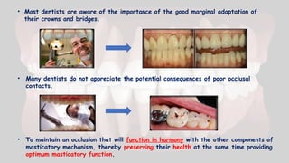
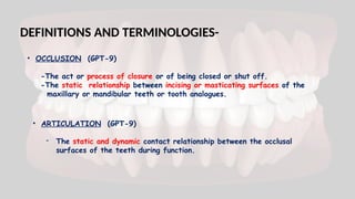
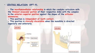
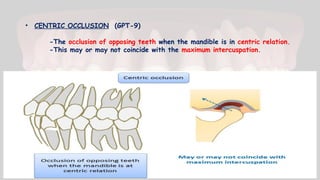
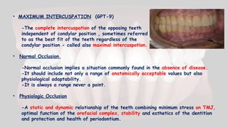
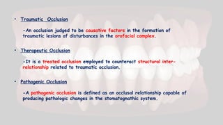
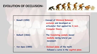
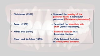
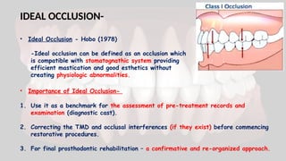
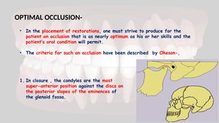
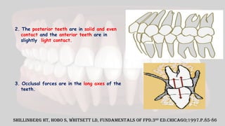
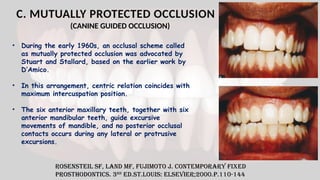
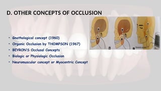
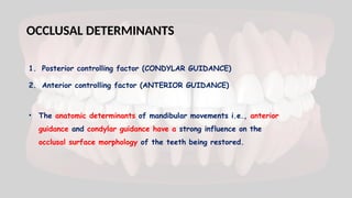
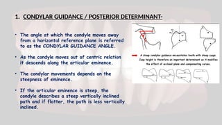
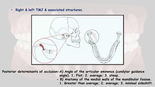
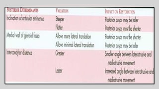
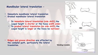
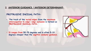
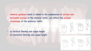
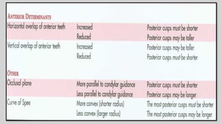
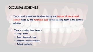
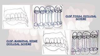
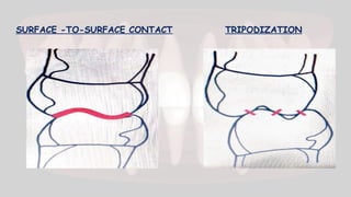
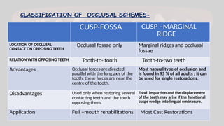
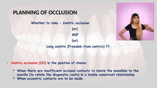
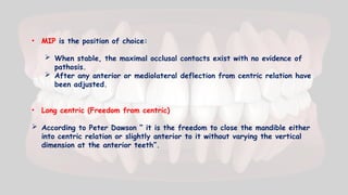
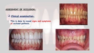
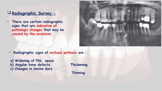
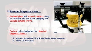
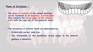
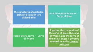
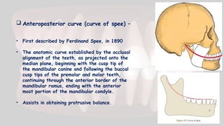
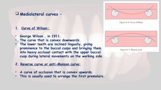
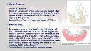
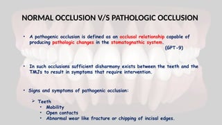
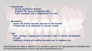
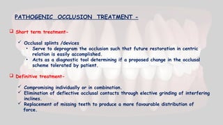
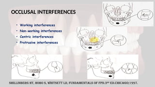
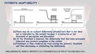
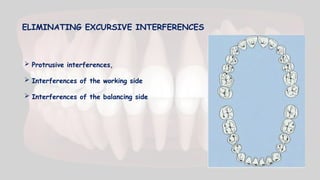
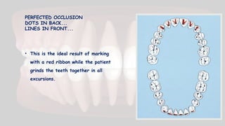
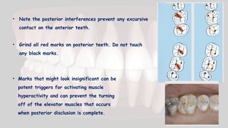
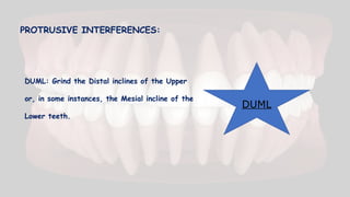
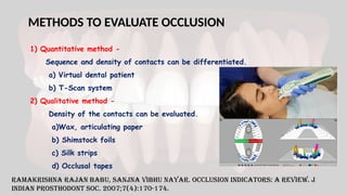
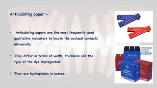
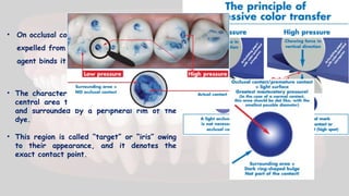
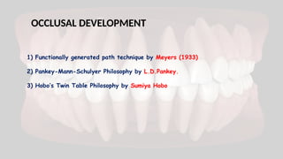
OCCLUSION IN FIXED PARTIAL
DENTURES
PRESENTER:
DR. KRUTIKA CHIKORDE.
2nd
YEAR POSTGRADUATE
DEPT. OF PROSTHODONTICS,
AND CROWN AND BRIDGE
COLLEGE OF DENTAL SCIENCES,
DAVANGERE .
STAFF INCHARGE:
DR. VEENA PRAKASH MAM.
PROFESSOR,
DEPT. OF PROSTHODONTICS
AND CROWN AND BRIDGE,
COLLEGE OF DENTAL SCIENCES,
DAVANGERE .
CONTENTS :
• Introduction
• Definitions and Terminologies
• Evolution of Occlusion
• Ideal occlusion
• Optimal occlusion
• Organization of occlusion
• Occlusal determinants
• Occlusal schemes
• Planning of Occlusion
• Methods to evaluate occlusal relationship.
• Normal versus pathogenic occlusion
• Occlusal interferences
• Occlusion development by different techniques
• Summary
• Conclusion
• References
INTRODUCTION-
• Long- term success of a restoration is dependent on maintenance
of occlusal harmony between the various components.
• In health, the occlusal anatomy of the teeth functions in harmony
with structures controlling the movements patterns of the mandible.
• Stomatognathic system is designed to dissipate the forces of
mastication to the supporting structures.
• Requirements of ideal occlusal contact relationships-
a) It should be within the adaptive capacity of the patient.
b) To restore and maintain heath and function of the
stomatognathic system.
c) Simultaneous bilateral contact of opposing posterior teeth must
occur in centric occlusion.
• Most dentists are aware of the importance of the good marginal adaptation of
their crowns and bridges.
• Many dentists do not appreciate the potential consequences of poor occlusal
contacts.
• To maintain an occlusion that will function in harmony with the other components of
masticatory mechanism, thereby preserving their health at the same time providing
optimum masticatory function.
DEFINITIONS AND TERMINOLOGIES-
• OCCLUSION (GPT-9)
-The act or process of closure or of being closed or shut off.
-The static relationship between incising or masticating surfaces of the
maxillary or mandibular teeth or tooth analogues.
• ARTICULATION (GPT-9)
- The static and dynamic contact relationship between the occlusal
surfaces of the teeth during function.
• CENTRIC RELATION (GPT-9)
- The maxillomandibular relationship in which the condyles articulate with
the thinnest avascular portion of their respective disks with the complex
in the anterior-superior position against the slopes of the articular
eminencies.
- This position is independent of tooth contact.
- This position is clinically discernible when the mandible is directed
superiorly and anteriorly.
• CENTRIC OCCLUSION (GPT-9)
-The occlusion of opposing teeth when the mandible is in centric relation.
-This may or may not coincide with the maximum intercuspation.
• MAXIMUM INTERCUSPATION (GPT-9)
-The complete intercuspation of the opposing teeth
independent of condylar position , sometimes referred
to as the best fit of the teeth regardless of the
condylar position - called also maximal intercuspation.
• Normal Occlusion
-Normal occlusion implies a situation commonly found in the absence of disease.
-It should include not only a range of anatomically acceptable values but also
physiological adaptability.
-It is always a range never a point.
• Physiologic Occlusion
-A static and dynamic relationship of the teeth combining minimum stress on TMJ,
optimal function of the orofacial complex, stability and esthetics of the dentition
and protection and health of periodontium.
• Traumatic Occlusion
-An occlusion judged to be causative factors in the formation of
traumatic lesions of disturbances in the orofacial complex.
• Therapeutic Occlusion
-It is a treated occlusion employed to counteract structural inter-
relationship related to traumatic occlusion.
• Pathogenic Occlusion
-A pathogenic occlusion is defined as an occlusal relationship capable of
producing pathologic changes in the stomatognathic system.
EVOLUTION OF OCCLUSION-
• Bonwill (1858) - Concept of Bilateral Balanced
occlusion and developed an
articulator that applied his 4-inch
triangular theory.
• Balkwill (1866) - The translating condyle moved
medially during lateral jaw
movement.
• Von Spee (1890) - Occlusal plane of the teeth
followed a curve in the sagittal plane.
• Christensen (1901) - Observed the opening of the
posterior teeth in mandibular
protrusion (Christensen phenomenon).
• Bennet (1908) - Described the immediate side
shift (Bennet movement).
• Alfred Gysi (1927) - Balanced occlusion on a
Removable Denture.
• Stuart and McCollum (1955) - Fully Balanced Occlusion
on fixed partial dentures.
• Mann and Pankey (1960) - Group function occlusion
• D’Amico (1961) - Cuspid Guidance
• Gutierrez MF, - Preservation of Natural
Cavada G, Valenzuela S (2010) Occurring Occlusion.
• Frank Spear (2013) - Occlusion defined by
Patient’s Physiologic
Capability of Compensation
IDEAL OCCLUSION-
• Ideal Occlusion - Hobo (1978)
-Ideal occlusion can be defined as an occlusion which
is compatible with stomatognathic system providing
efficient mastication and good esthetics without
creating physiologic abnormalities.
• Importance of Ideal Occlusion-
1. Use it as a benchmark for the assessment of pre-treatment records and
examination (diagnostic cast).
2. Correcting the TMD and occlusal interferences (if they exist) before commencing
restorative procedures.
3. For final prosthodontic rehabilitation – a confirmative and re-organized approach.
OPTIMAL OCCLUSION-
• In the placement of restorations, one must strive to produce for the
patient an occlusion that is as nearly optimum as his or her skills and the
patient’s oral condition will permit.
• The criteria for such an occlusion have been described by Okeson-,
1. In closure , the condyles are the most
super-anterior position against the discs on
the posterior slopes of the eminences of
the glenoid fossa.
2. The posterior teeth are in solid and even
contact and the anterior teeth are in
slightly light contact.
3. Occlusal forces are in the long axes of the
teeth.
Shillinberg HT, Hobo S, Whitsett LD, Fundamentals of FPD.3rd
ed.Chicago;1997.p.85-86
4. In the lateral excursions, working side contacts (preferably on canines)
disocclude or separate the non- working teeth instantly.
5. In protrusive excursions,
anterior tooth contacts will
disocclude the posterior teeth.
6. In an upright posture,
posterior
teeth contact more heavily than
do anterior teeth.
ORGANIZATION OF OCCLUSION-
Three recognized concepts-
A. Bilateral Balanced Occlusion
B. Unilateral Balanced Occlusion
C. Mutually protected Occlusion
Rosenstiel, Land, Fujimoto .Contemporary Fixed prosthodontics.5th
ed.
• However, since restorative treatment requirements vary, the clinician should
understand possible combinations of occlusal schemes and their advantages, dis
advantages and indications.
A. BILATERAL BALANCED OCCLUSION
• In 1935 ,Schuyler developed the first detailed technique for occlusal
adjustment. By 1953 he began to observe failure of natural dentition
restored with balance. His observations and suggestions effectively
signaled the end of BALANCE as an acceptable treatment approach for
the dentulous patient.
• Stuart and Stallard (1960) noted that balanced occlusion in reconstructed
natural dentitions
1.Often required injudicious increase in occlusal vertical dimension to achieve
balance.
2. Often led to instability of occlusion.
3. Frequently showed increased wear of teeth and restorations
• Thus the concept of a unilateral balanced occlusion (group
function) evolved.
B. UNILATERAL BALANCED OCCLUSION
• Multiple contact relations between the maxillary and
mandibular teeth in lateral movements on the working-
side whereby simultaneous contact of several teeth
acts as a group to distribute occlusal forces.
• Here excursive contact occurs between all opposing
posterior teeth on the laterotrusive (working) side only.
On the mediotrusive (non-working) side, no contact
occurs until the mandible has reached centric relation.
Glossary of Prosthodontic Terms, Edition Nine, J Prosthet Dent 2017;117 (5S):e1-e105
(GROUP FUNCTION OCCLUSION)
ADVANTAGES:
• Group function of the teeth on the working
side distributes the occlusal load.
• The absence of contact on the nonworking
side prevents those from getting subjected
to destructive ,obliquely directed forces
found in nonworking interferences.
• It also saves centric holding cusps that is
mandibular buccal cusps and maxillary
palatal cusps from excessive wear.
• In the presence of anterior bone loss or
missing canines, mouth should be restored
to group function.
C. MUTUALLY PROTECTED OCCLUSION
(CANINE GUIDED OCCLUSION)
Rosensteil SF, Land MF, Fujimoto J. Contemporary fixed
prosthodontics. 3rd
ed.St.Louis: Elsevier;2000.p.110-144
• During the early 1960s, an occlusal scheme called
as mutually protected occlusion was advocated by
Stuart and Stallard, based on the earlier work by
D’Amico.
• In this arrangement, centric relation coincides with
maximum intercuspation position.
• The six anterior maxillary teeth, together with six
anterior mandibular teeth, guide excursive
movements of mandible, and no posterior occlusal
contacts occurs during any lateral or protrusive
excursions.
 Advantages-
• Patients tolerance
• Ease of construction
Disadvantages-
• Periodontally weak anterior teeth
• Missing canine
• Class II and class III situations
• Cross-bite situations.
 Features of mutually protected occlusion-
• Uniform contact of all teeth around the arch when the mandibular condylar
processes are in their most superior position.
• Stable posterior tooth contacts with vertically directed resultant forces.
• Centric relation coincident with maximum intercuspation (intercuspal postion).
• No contact of posterior teeth in lateral or protrusive movements.
• Anterior tooth contacts harmonizing with functional jaw movements.
 Anterior teeth are suited for guidance by the
virtue of –
• Canines having longest, strongest roots.
• Good crown to root ratio
• The load being reduced by distance from the fulcrum as
the lever arm increases.
• Location is far from the TMJ thus receiving less stress.
• The proprioceptive threshold and concomitant reflexes reducing the load.
• Surrounded by dense compact bone which tolerates forces better.
• It has many receptors in the periodontal ligament so it controls lateral
pressure by directing vertical masticatory movements.
Dawson P. Evaluation and Diagnosis of Occlusion.St.Louis:Mosby Elsevier;2007
The farther anterior a tooth is
located, the less the influence of the
TMJ and the greater the influence of
the anterior guidance (AG).
D. OTHER CONCEPTS OF OCCLUSION
• Gnathological concept (1960)
• Organic Occlusion by THOMPSON (1967)
• BEYRON’S Occlusal Concepts
• Biologic or Physiologic Occlusion
• Neuromuscular concept or Myocentric Concept
OCCLUSAL DETERMINANTS
1. Posterior controlling factor (CONDYLAR GUIDANCE)
2. Anterior controlling factor (ANTERIOR GUIDANCE)
• The anatomic determinants of mandibular movements i.e., anterior
guidance and condylar guidance have a strong influence on the
occlusal surface morphology of the teeth being restored.
1. CONDYLAR GUIDANCE / POSTERIOR DETERMINANT-
• The angle at which the condyle moves away
from a horizontal reference plane is referred
to as the CONDYLAR GUIDANCE ANGLE.
• As the condyle moves out of centric relation
it descends along the articular eminence.
• The condylar movements depends on the
steepness of eminence.
• If the articular eminence is steep, the
condyle describes a steep vertically inclined
path and if flatter, the path is less vertically
inclined.
• Right & left TMJ & associated structures.
Posterior determinants of occlusion-A) Angle of the articular eminence (condylar guidance
angle). 1. Flat; 2. average; 3. steep.
- B) Anatomy of the medial walls of the mandibular fossae.
1. Greater than average; 2. average; 3. minimal sideshift.
Mandibular lateral translation -
• Immediate mandibular lateral translation
• Gradual mandibular lateral translation
• In immediate lateral translation (side shift),the
cuspal height is shorter or the fossa width wider.
• In gradual lateral translation (no side shift),the
cuspal height is longer or the fossa be narrower.
• Ridges and groove direction are affected by
the condylar path, particularly the lateral
translation. Working Condyle
2. ANTERIOR GUIDANCE / ANTERIOR DETERMINANT-
PROTRUSIVE INCISAL PATH-
• The track of the incisal edges from the maximum
intercuspation to edge- edge occlusion is termed as
PROTRUSIVE INCISAL PATH.
• It ranges from 50-70 degrees and is often 5-10
degrees steeper than the sagittal condylar guidance.
• Anterior guidance which is linked to the combination of vertical and
horizontal overlap of the anterior teeth, can affect the occlusal
morphology of the posterior teeth.
• a) Vertical Overlap and cuspal height
• b) Horizontal Overlap and cuspal height
• The occlusal scheme can be classified by the location of the occlusal
contact made by the functional cusp on the opposing tooth in the centric
relation.
• They are mainly four types -
 Cusp- fossa
 Cusp –Marginal ridge
 Surface-surface contact
 Tripod contacts
OCCLUSAL SCHEMES
CUSP FOSSA OCCLUSAL
SCHEME
CUSP-MARGINAL RIDGE
OCCLUSAL SCHEME
SURFACE -TO-SURFACE CONTACT TRIPODIZATION
CLASSIFICATION OF OCCLUSAL SCHEMES-
CUSP-FOSSA CUSP –MARGINAL
RIDGE
LOCATION OF OCCLUSAL
CONTACT ON OPPOSING TEETH
Occlusal fossae only Marginal ridges and occlusal
fossae
RELATION WITH OPPOSING TEETH Tooth-to- tooth Tooth-to-two teeth
Advantages Occlusal forces are directed
parallel with the long axis of the
tooth; these forces are near the
centre of the tooth.
Most natural type of occlusion and
is found in 95 % of all adults ; it can
be used for single restorations.
Disadvantages Used only when restoring several
contacting teeth and the tooth
opposing them.
Food impaction and the displacement
of the teeth may arise if the functional
cusps wedge into lingual embrasure.
Application Full –mouth rehabilitations Most Cast Restorations
PLANNING OF OCCLUSION
Whether to take - Centric occlusion
(or)
MIP
(or)
Long centric (Freedom from centric) ??
• Centric occlusion (CO) is the position of choice:
 When there are insufficient occlusal contacts to relate the mandible to the
maxilla (to relate the diagnostic casts) in a stable consistent relationship.
 When eccentric contacts are to be made.
• MIP is the position of choice:
 When stable, the maximal occlusal contacts exist with no evidence of
pathosis.
 After any anterior or mediolateral deflection from centric relation have
been adjusted.
• Long centric (Freedom from centric)
 According to Peter Dawson “ it is the freedom to close the mandible either
into centric relation or slightly anterior to it without varying the vertical
dimension at the anterior teeth”.
ASSESSMENT OF OCCLUSION -
 Clinical examination-
• This is done to reveal signs and symptoms
of occlusal pathosis.
 Radiographic Survey -
• There are certain radiographic
signs that are indicative of
pathologic changes that may be
caused by the occlusion.
• Radiographic signs of occlusal pathosis are :
a) Widening of PDL space
b) Angular bony defects Thickening
c) Changes in lamina dura
Thinning
 Mounted Diagnostic casts -
• Occlusal plane and occlusal contacts study
to facilitate and aid in the designing the
Occlusal scheme of FPD.
• Factors to be studied on the Mounted
diagnostic Casts -
1. Centric occlusion(CO),MIP and initial tooth contacts.
2. Plane Of Occlusion.
Plane of Occlusion -
• The plane of occlusion of the natural dentition
can be visualized as an imaginary curved plane
that connects the incisal edges of the anterior
teeth with the cusp tips of the posterior teeth.
• The curvatures of anterior teeth are determined by:
 Esthetically correct smile line.
 The relationship of the mandibular incisal edges to the anterior
guidance & phonetics.
The curvatures of posterior
plane of occlusion are
divided into:
an Anteroposterior curve 
Curve of Spee
Mediolateral curve  Curve
of Wilson
Together, the composite of
the curve of Spee, the curve
of Wilson, and the curve of
the incisal edges is properly
referred to as the curve of
occlusion.
 Anteroposterior curve (curve of spee) –
• First described by Ferdinand Spee, in 1890
• The anatomic curve established by the occlusal
alignment of the teeth, as projected onto the
median plane, beginning with the cusp tip of
the mandibular canine and following the buccal
cusp tips of the premolar and molar teeth,
continuing through the anterior border of the
mandibular ramus, ending with the anterior
most portion of the mandibular condyle.
• Assists in obtaining protrusive balance.
 Mediolateral curves –
1. Curve of Wilson:
• George Wilson , in 1911.
• The curve that is convex downwards.
• The lower teeth are inclined lingually, giving
prominence to the buccal cusps and bringing them
into heavy occlusal contact with the upper buccal
cusp during lateral movements on the working side.
2. Reverse curve or anti-Monson curve:
• A curve of occlusion that is convex upwards.
• This is usually used to arrange the first premolars.
3. Curve of monson:
• George S. Monson
• Curve of occlusion in which each cusp and incisal edge
touches or conforms to a segment of the surface of a
sphere 8 inches in diameter with its centre in the
region of the glabella.
• Combination of ‘curve of spee and curve of Wilson.
4. Pleasure curve:
• In excessive wear of the teeth, the obliteration of
the cusps and formation of either flat or cupped-out
occlusal surfaces, associated with the reversal of the
occlusal plane of the premolar, first and second molar
teeth, (the third molars being generally unaffected),
whereby the occlusal surfaces of the mandibular teeth
slope facially instead of lingually and those of the
maxillary teeth incline lingually.
• Combination of monson and anti-monson curves
NORMAL OCCLUSION V/S PATHOLOGIC OCCLUSION
• A pathogenic occlusion is defined as an occlusal relationship capable of
producing pathologic changes in the stomatognathic system.
(GPT-9)
• In such occlusions sufficient disharmony exists between the teeth and the
TMJs to result in symptoms that require intervention.
• Signs and symptoms of pathogenic occlusion:
 Teeth
• Mobility
• Open contacts
• Abnormal wear like fracture or chipping of incisal edges.
 Periodontium
• Chronic periodontal diseases
• Widened PDL space (radiographically)
• Tooth movement and a compromised C:R ratio.
 Musculature
• Acute and chronic muscular pain due to the bruxism
• Trismus due to no relaxation of elevator muscle
 TMJ
• Pain, clicking or popping due to muscular origin or internal derangement
of joint.
• Unilateral clicking with midline deviation due to displaced disc
Shillingburg HT, Hobo S, Whitsett LD, Jacobi R, Brakett SE. Shillinburg's Fundamentals
Of Fixed Prosthodontics.4th ed. Chicago: quintessence;2012
 Short term treatment-
 Occlusal splints /devices
• Serve to deprogram the occlusion such that future restoration in centric
relation is easily accomplished.
• Acts as a diagnostic tool determining if a proposed change in the occlusal
scheme tolerated by patient.
 Definitive treatment-
 Compromising individually or in combination.
 Elimination of deflective occlusal contacts through elective grinding of interfering
inclines.
 Replacement of missing teeth to produce a more favourable distribution of
force.
PATHOGENIC OCCLUSION TREATMENT -
OCCLUSAL INTERFERENCES
• Working interferences
• Non-working interferences
• Centric interferences
• Protrusive interferences
Shillinberg HT, Hobo S, Whitsett LD, Fundamentals of FPD.3rd
ed.Chicago;1997.
(a)There may be an occlusal disharmony (shaded bar) that is not ideal
but is tolerated by the patient because it is below his or her
threshold of perception and discomfort.
(b) If the threshold is lowered, the disharmony that had been previously
tolerated may produce symptoms in the patient.
(c)Treatment is then rendered by first raising the patient’s threshold
and then decreasing or eliminating the disharmony.
Shillinberg HT, Hobo S, Whitsett LD, Fundamentals of FPD.3rd
ed.Chicago;1997.
PATIENTS ADAPTABILITY -
EQUILIBRATION PROCEDURES -
Reduction of
interferences in
Centric Relation
Selective
reduction of
interferences in
lateral
excursions
Elimination of all
posterior
interferences
during protrusive
excursions
Harmonization
of anterior
guidance
Dawson E.P. Functional Occlusion From TMJ To Smile Design.1st Ed. St. Louis:
Elsevier;2009. p. 189-198
Selective Grinding for Occlusal Equilibration
CENTRIC RELATION INTERFERENCES CAN
BE DIFFERENTIATED INTO TWO TYPES:
1. Interference to the arc of closure
2. Interference to the line of closure
1.Interference to the arc of closure -
As the condyles rotate on their
centric relation axis, each lower
tooth follows an arc of closure
Any interference  condyles
displaced downward and forward 
anterior slide
Basic grinding rule to correct anterior slide -
MUDL
2.Interference to the line of closure -
Primary interferences that cause the mandible
to deviate to left or right from first point of
contact in CR to most closed position.
1. If the interfering incline causes the
mandible to deviate off the line of closure
toward the cheek, grind the
Basic grinding rules-
BULL
Basic grinding rules-
2. If the interfering incline causes the
mandible to deviate off the line of
closure toward the tongue, grind the
LUBL
LATERAL EXCURSION INTERFERENCES:
 Dictated by two determinants:
1. The border movements of the condyles
2. The anterior guidance
• When lateral excursions are being equilibrated, the
mandible must be guided with firm upward pressure
through the condyles to ensure that all
interferences are recorded and eliminated through
the uppermost ranges of motion that can occur at
true border paths for both the condyles and the
anterior guidance.
ELIMINATING EXCURSIVE INTERFERENCES
 Protrusive interferences,
 Interferences of the working side
 Interferences of the balancing side
PERFECTED OCCLUSION
DOTS IN BACK...
LINES IN FRONT...
• This is the ideal result of marking
with a red ribbon while the patient
grinds the teeth together in all
excursions.
• Note the posterior interferences prevent any excursive
contact on the anterior teeth.
• Grind all red marks on posterior teeth. Do not touch
any black marks.
• Marks that might look insignificant can be
potent triggers for activating muscle
hyperactivity and can prevent the turning
off of the elevator muscles that occurs
when posterior disclusion is complete.
DUML: Grind the Distal inclines of the Upper
or, in some instances, the Mesial incline of the
Lower teeth.
PROTRUSIVE INTERFERENCES:
DUML
VERIFICATION OF COMPLETION -
Clench Test
Anterior
Deprogramming
Splint
Ask the patient to clench the teeth together
and squeeze firmly (empty mouth). If the
patient can feel any discomfort in any tooth,
the equilibration is not complete.
To confirm whether the problem is or is not
related to occluso-muscle pain. If the anterior
splint completely separates all the posterior
teeth, all discomfort will dissipate if the cause
of the discomfort is totally related to
occlusion.
METHODS TO EVALUATE OCCLUSION
1) Quantitative method -
Sequence and density of contacts can be differentiated.
a) Virtual dental patient
b) T-Scan system
2) Qualitative method -
Density of the contacts can be evaluated.
a)Wax, articulating paper
b) Shimstock foils
c) Silk strips
d) Occlusal tapes
Ramakrishna Rajan Babu, Sanjna Vibhu Nayar. Occlusion indicators: A review. J
Indian Prosthodont Soc. 2007;7(4):170-174.
Articulating paper -
• Articulating papers are the most frequently used
qualitative indicators to locate the occlusal contacts
intraorally.
• They differ in terms of width, thickness and the
type of the dye impregnated.
• They are hydrophobic in nature.
• On occlusal contact, the coloring agent is
expelled from the film and the bonding
agent binds it on to the tooth surface.
• The characteristic marking is observed as a
central area that is devoid of the colorant
and surrounded by a peripheral rim of the
dye.
• This region is called “target” or “iris” owing
to their appearance, and it denotes the
exact contact point.
OCCLUSAL DEVELOPMENT
1) Functionally generated path technique by Meyers (1933)
2) Pankey-Mann-Schulyer Philosophy by L.D.Pankey.
3) Hobo’s Twin Table Philosophy by Sumiya Hobo
Pankey –Mann Schyuler Philosophy by LD Pankey -
Shetty Et al . PHILOSOPHIES IN FULL MOUTH REHABILITATION – A SYSTEMATIC REVIEW.
IJDCR 2013; 3(3): 30-39.
Hobo’s Twin Table philosophy by Sumiya Hobo -
Shetty Et al . PHILOSOPHIES IN FULL MOUTH REHABILITATION – A SYSTEMATIC REVIEW.
IJDCR 2013; 3(3): 30-39.
Shetty Et al . PHILOSOPHIES IN FULL MOUTH REHABILITATION – A SYSTEMATIC REVIEW.
IJDCR 2013; 3(3): 30-39.
DIAGNOSTIC WAX-UP
PRINCIPLE - The diagnostic wax-up is the
process of converting a programmed treatment
plan into a three-dimensional visualization.
Steps in the diagnostic wax-up -
Step 1: Mount upper and lower casts with centric relation
bite record and facebow.
Step 2: Verify the accuracy of the mounting.
Step 3: Examine the occlusal relationship on the casts.
Step 4: Lock the centric latch when observing the casts and
determine the correct vertical dimension
Step 5: Return the condyles to centric relation and lock the
centric lock.
Step 6: Observe the teeth that were reshaped, Remove
unsavable teeth from the casts.
Step 7: Equilibration is the first treatment option to explore.
Step 8: Examine the plane of occlusion.
Step 9: Establish stable holding contacts on the anterior teeth.
Step 10: Correct lower incisal edges if needed.
Step 11: Re-evaluate the total occlusion with the upper cast to
see how it can be adapted to occlude with the lower arch.
SUMMARY
RESTORING DIFFERENT COMBINATIONS
PROSTHESIS POSITION
ICP /CR
ARTICULATOR
AND RECORDS
OCCLUSAL
MORPHOLOGY
Single Crown ICP Simple Hinge Conform to
occlusal
morphology
FPD-one
Quadrant
ICP Semi adjustable
/anterior
guidance
Conform to
occlusal
morphology
Several
Quadrants
Long
Centric
Fully adjustable
/anterior guidance
and condylar
guidance
Group function is
desired /cusp to
fossa
CONCLUSION
• Occlusion is the integrated relationship of the tooth, periodontium ,TMJ and neuro
musculature, and not merely the interdigitation of the tooth.
• There is a complex interaction of many components of masticatory system.
• Changes in one component would affect the entire system.
• One who practices the restorative dentistry should appreciate their significance for
the long term successful restorations by maintaining the occlusal integrity.
• There is no one answer to occlusal problems, the dentist should use the philosophy
that works best in his own hands and at the same time do the most good, or better
yet, the least harm to the patient.”
 Dawson; Functional occlusion From TMJ To Smile Design – 1st
edt.
 Dawson; Evaluation, diagnosis & treatment of occlusal problems
 Okeson; Management of temporomandibular disorders and occlusion – 8th
edt.
 Shillingburg, Hobo, Whitsett, Jacobi, Brackett; Fundamentals Of Fixed
Prosthodontics – 3rd
edt.
 Takayama and hobo; Oral rehabilitation: Clinical determination of occlusion.
 Rosensteil, Land, Fujimoto; Contemporary Fixed Prosthodontics – 5th
edt.
REFRENCES
TEXTBOOKS -
SCIENTIFIC JOURNALS-
 Ogawa, Ogimoto. Pattern Of Occlusal Contacts In Lateral Position: JPD 1998;80:67-
74.
 Schuyler. Factors of Occlusion As Applicable To Restorative Dentistry; JPD 1953;3:
772-15.
 Pokorny, Wiens, Litvak. Occlusion For Fixed Prosthodontics – A Historical Perspective
of the Gnathological Influence; JPD 2008;99:299-313.
 Johnson. Variations in Organic Occlusion ; JPD 1979;41:625-629.
 Clark JR, Evans RD. Functional Occlusion: A Review; JO 2001;28(1):76-81.
 Babu RR, Nayar SV. Occlusion indicators: A review. J Indian Prosthodont Soc.
2007;7(4):170-174.
THANK YOU

OCCLUSION IN FIXED PARTIAL DENTURE .pptx

  • 2.
    OCCLUSION IN FIXEDPARTIAL DENTURES PRESENTER: DR. KRUTIKA CHIKORDE. 2nd YEAR POSTGRADUATE DEPT. OF PROSTHODONTICS, AND CROWN AND BRIDGE COLLEGE OF DENTAL SCIENCES, DAVANGERE . STAFF INCHARGE: DR. VEENA PRAKASH MAM. PROFESSOR, DEPT. OF PROSTHODONTICS AND CROWN AND BRIDGE, COLLEGE OF DENTAL SCIENCES, DAVANGERE .
  • 3.
    CONTENTS : • Introduction •Definitions and Terminologies • Evolution of Occlusion • Ideal occlusion • Optimal occlusion • Organization of occlusion • Occlusal determinants • Occlusal schemes
  • 4.
    • Planning ofOcclusion • Methods to evaluate occlusal relationship. • Normal versus pathogenic occlusion • Occlusal interferences • Occlusion development by different techniques • Summary • Conclusion • References
  • 5.
    INTRODUCTION- • Long- termsuccess of a restoration is dependent on maintenance of occlusal harmony between the various components. • In health, the occlusal anatomy of the teeth functions in harmony with structures controlling the movements patterns of the mandible. • Stomatognathic system is designed to dissipate the forces of mastication to the supporting structures. • Requirements of ideal occlusal contact relationships- a) It should be within the adaptive capacity of the patient. b) To restore and maintain heath and function of the stomatognathic system. c) Simultaneous bilateral contact of opposing posterior teeth must occur in centric occlusion.
  • 6.
    • Most dentistsare aware of the importance of the good marginal adaptation of their crowns and bridges. • Many dentists do not appreciate the potential consequences of poor occlusal contacts. • To maintain an occlusion that will function in harmony with the other components of masticatory mechanism, thereby preserving their health at the same time providing optimum masticatory function.
  • 7.
    DEFINITIONS AND TERMINOLOGIES- •OCCLUSION (GPT-9) -The act or process of closure or of being closed or shut off. -The static relationship between incising or masticating surfaces of the maxillary or mandibular teeth or tooth analogues. • ARTICULATION (GPT-9) - The static and dynamic contact relationship between the occlusal surfaces of the teeth during function.
  • 8.
    • CENTRIC RELATION(GPT-9) - The maxillomandibular relationship in which the condyles articulate with the thinnest avascular portion of their respective disks with the complex in the anterior-superior position against the slopes of the articular eminencies. - This position is independent of tooth contact. - This position is clinically discernible when the mandible is directed superiorly and anteriorly.
  • 9.
    • CENTRIC OCCLUSION(GPT-9) -The occlusion of opposing teeth when the mandible is in centric relation. -This may or may not coincide with the maximum intercuspation.
  • 10.
    • MAXIMUM INTERCUSPATION(GPT-9) -The complete intercuspation of the opposing teeth independent of condylar position , sometimes referred to as the best fit of the teeth regardless of the condylar position - called also maximal intercuspation. • Normal Occlusion -Normal occlusion implies a situation commonly found in the absence of disease. -It should include not only a range of anatomically acceptable values but also physiological adaptability. -It is always a range never a point. • Physiologic Occlusion -A static and dynamic relationship of the teeth combining minimum stress on TMJ, optimal function of the orofacial complex, stability and esthetics of the dentition and protection and health of periodontium.
  • 11.
    • Traumatic Occlusion -Anocclusion judged to be causative factors in the formation of traumatic lesions of disturbances in the orofacial complex. • Therapeutic Occlusion -It is a treated occlusion employed to counteract structural inter- relationship related to traumatic occlusion. • Pathogenic Occlusion -A pathogenic occlusion is defined as an occlusal relationship capable of producing pathologic changes in the stomatognathic system.
  • 12.
    EVOLUTION OF OCCLUSION- •Bonwill (1858) - Concept of Bilateral Balanced occlusion and developed an articulator that applied his 4-inch triangular theory. • Balkwill (1866) - The translating condyle moved medially during lateral jaw movement. • Von Spee (1890) - Occlusal plane of the teeth followed a curve in the sagittal plane.
  • 13.
    • Christensen (1901)- Observed the opening of the posterior teeth in mandibular protrusion (Christensen phenomenon). • Bennet (1908) - Described the immediate side shift (Bennet movement). • Alfred Gysi (1927) - Balanced occlusion on a Removable Denture. • Stuart and McCollum (1955) - Fully Balanced Occlusion on fixed partial dentures.
  • 14.
    • Mann andPankey (1960) - Group function occlusion • D’Amico (1961) - Cuspid Guidance • Gutierrez MF, - Preservation of Natural Cavada G, Valenzuela S (2010) Occurring Occlusion. • Frank Spear (2013) - Occlusion defined by Patient’s Physiologic Capability of Compensation
  • 15.
    IDEAL OCCLUSION- • IdealOcclusion - Hobo (1978) -Ideal occlusion can be defined as an occlusion which is compatible with stomatognathic system providing efficient mastication and good esthetics without creating physiologic abnormalities. • Importance of Ideal Occlusion- 1. Use it as a benchmark for the assessment of pre-treatment records and examination (diagnostic cast). 2. Correcting the TMD and occlusal interferences (if they exist) before commencing restorative procedures. 3. For final prosthodontic rehabilitation – a confirmative and re-organized approach.
  • 16.
    OPTIMAL OCCLUSION- • Inthe placement of restorations, one must strive to produce for the patient an occlusion that is as nearly optimum as his or her skills and the patient’s oral condition will permit. • The criteria for such an occlusion have been described by Okeson-, 1. In closure , the condyles are the most super-anterior position against the discs on the posterior slopes of the eminences of the glenoid fossa.
  • 17.
    2. The posteriorteeth are in solid and even contact and the anterior teeth are in slightly light contact. 3. Occlusal forces are in the long axes of the teeth. Shillinberg HT, Hobo S, Whitsett LD, Fundamentals of FPD.3rd ed.Chicago;1997.p.85-86
  • 18.
    4. In thelateral excursions, working side contacts (preferably on canines) disocclude or separate the non- working teeth instantly. 5. In protrusive excursions, anterior tooth contacts will disocclude the posterior teeth. 6. In an upright posture, posterior teeth contact more heavily than do anterior teeth.
  • 19.
    ORGANIZATION OF OCCLUSION- Threerecognized concepts- A. Bilateral Balanced Occlusion B. Unilateral Balanced Occlusion C. Mutually protected Occlusion Rosenstiel, Land, Fujimoto .Contemporary Fixed prosthodontics.5th ed. • However, since restorative treatment requirements vary, the clinician should understand possible combinations of occlusal schemes and their advantages, dis advantages and indications.
  • 20.
    A. BILATERAL BALANCEDOCCLUSION • In 1935 ,Schuyler developed the first detailed technique for occlusal adjustment. By 1953 he began to observe failure of natural dentition restored with balance. His observations and suggestions effectively signaled the end of BALANCE as an acceptable treatment approach for the dentulous patient. • Stuart and Stallard (1960) noted that balanced occlusion in reconstructed natural dentitions 1.Often required injudicious increase in occlusal vertical dimension to achieve balance. 2. Often led to instability of occlusion. 3. Frequently showed increased wear of teeth and restorations • Thus the concept of a unilateral balanced occlusion (group function) evolved.
  • 21.
    B. UNILATERAL BALANCEDOCCLUSION • Multiple contact relations between the maxillary and mandibular teeth in lateral movements on the working- side whereby simultaneous contact of several teeth acts as a group to distribute occlusal forces. • Here excursive contact occurs between all opposing posterior teeth on the laterotrusive (working) side only. On the mediotrusive (non-working) side, no contact occurs until the mandible has reached centric relation. Glossary of Prosthodontic Terms, Edition Nine, J Prosthet Dent 2017;117 (5S):e1-e105 (GROUP FUNCTION OCCLUSION)
  • 22.
    ADVANTAGES: • Group functionof the teeth on the working side distributes the occlusal load. • The absence of contact on the nonworking side prevents those from getting subjected to destructive ,obliquely directed forces found in nonworking interferences. • It also saves centric holding cusps that is mandibular buccal cusps and maxillary palatal cusps from excessive wear. • In the presence of anterior bone loss or missing canines, mouth should be restored to group function.
  • 23.
    C. MUTUALLY PROTECTEDOCCLUSION (CANINE GUIDED OCCLUSION) Rosensteil SF, Land MF, Fujimoto J. Contemporary fixed prosthodontics. 3rd ed.St.Louis: Elsevier;2000.p.110-144 • During the early 1960s, an occlusal scheme called as mutually protected occlusion was advocated by Stuart and Stallard, based on the earlier work by D’Amico. • In this arrangement, centric relation coincides with maximum intercuspation position. • The six anterior maxillary teeth, together with six anterior mandibular teeth, guide excursive movements of mandible, and no posterior occlusal contacts occurs during any lateral or protrusive excursions.
  • 24.
     Advantages- • Patientstolerance • Ease of construction Disadvantages- • Periodontally weak anterior teeth • Missing canine • Class II and class III situations • Cross-bite situations.
  • 25.
     Features ofmutually protected occlusion- • Uniform contact of all teeth around the arch when the mandibular condylar processes are in their most superior position. • Stable posterior tooth contacts with vertically directed resultant forces. • Centric relation coincident with maximum intercuspation (intercuspal postion). • No contact of posterior teeth in lateral or protrusive movements. • Anterior tooth contacts harmonizing with functional jaw movements.
  • 26.
     Anterior teethare suited for guidance by the virtue of – • Canines having longest, strongest roots. • Good crown to root ratio • The load being reduced by distance from the fulcrum as the lever arm increases. • Location is far from the TMJ thus receiving less stress. • The proprioceptive threshold and concomitant reflexes reducing the load. • Surrounded by dense compact bone which tolerates forces better. • It has many receptors in the periodontal ligament so it controls lateral pressure by directing vertical masticatory movements. Dawson P. Evaluation and Diagnosis of Occlusion.St.Louis:Mosby Elsevier;2007 The farther anterior a tooth is located, the less the influence of the TMJ and the greater the influence of the anterior guidance (AG).
  • 27.
    D. OTHER CONCEPTSOF OCCLUSION • Gnathological concept (1960) • Organic Occlusion by THOMPSON (1967) • BEYRON’S Occlusal Concepts • Biologic or Physiologic Occlusion • Neuromuscular concept or Myocentric Concept
  • 28.
    OCCLUSAL DETERMINANTS 1. Posteriorcontrolling factor (CONDYLAR GUIDANCE) 2. Anterior controlling factor (ANTERIOR GUIDANCE) • The anatomic determinants of mandibular movements i.e., anterior guidance and condylar guidance have a strong influence on the occlusal surface morphology of the teeth being restored.
  • 29.
    1. CONDYLAR GUIDANCE/ POSTERIOR DETERMINANT- • The angle at which the condyle moves away from a horizontal reference plane is referred to as the CONDYLAR GUIDANCE ANGLE. • As the condyle moves out of centric relation it descends along the articular eminence. • The condylar movements depends on the steepness of eminence. • If the articular eminence is steep, the condyle describes a steep vertically inclined path and if flatter, the path is less vertically inclined.
  • 30.
    • Right &left TMJ & associated structures. Posterior determinants of occlusion-A) Angle of the articular eminence (condylar guidance angle). 1. Flat; 2. average; 3. steep. - B) Anatomy of the medial walls of the mandibular fossae. 1. Greater than average; 2. average; 3. minimal sideshift.
  • 32.
    Mandibular lateral translation- • Immediate mandibular lateral translation • Gradual mandibular lateral translation • In immediate lateral translation (side shift),the cuspal height is shorter or the fossa width wider. • In gradual lateral translation (no side shift),the cuspal height is longer or the fossa be narrower. • Ridges and groove direction are affected by the condylar path, particularly the lateral translation. Working Condyle
  • 33.
    2. ANTERIOR GUIDANCE/ ANTERIOR DETERMINANT- PROTRUSIVE INCISAL PATH- • The track of the incisal edges from the maximum intercuspation to edge- edge occlusion is termed as PROTRUSIVE INCISAL PATH. • It ranges from 50-70 degrees and is often 5-10 degrees steeper than the sagittal condylar guidance.
  • 34.
    • Anterior guidancewhich is linked to the combination of vertical and horizontal overlap of the anterior teeth, can affect the occlusal morphology of the posterior teeth. • a) Vertical Overlap and cuspal height • b) Horizontal Overlap and cuspal height
  • 36.
    • The occlusalscheme can be classified by the location of the occlusal contact made by the functional cusp on the opposing tooth in the centric relation. • They are mainly four types -  Cusp- fossa  Cusp –Marginal ridge  Surface-surface contact  Tripod contacts OCCLUSAL SCHEMES
  • 37.
  • 38.
  • 39.
    CLASSIFICATION OF OCCLUSALSCHEMES- CUSP-FOSSA CUSP –MARGINAL RIDGE LOCATION OF OCCLUSAL CONTACT ON OPPOSING TEETH Occlusal fossae only Marginal ridges and occlusal fossae RELATION WITH OPPOSING TEETH Tooth-to- tooth Tooth-to-two teeth Advantages Occlusal forces are directed parallel with the long axis of the tooth; these forces are near the centre of the tooth. Most natural type of occlusion and is found in 95 % of all adults ; it can be used for single restorations. Disadvantages Used only when restoring several contacting teeth and the tooth opposing them. Food impaction and the displacement of the teeth may arise if the functional cusps wedge into lingual embrasure. Application Full –mouth rehabilitations Most Cast Restorations
  • 40.
    PLANNING OF OCCLUSION Whetherto take - Centric occlusion (or) MIP (or) Long centric (Freedom from centric) ?? • Centric occlusion (CO) is the position of choice:  When there are insufficient occlusal contacts to relate the mandible to the maxilla (to relate the diagnostic casts) in a stable consistent relationship.  When eccentric contacts are to be made.
  • 41.
    • MIP isthe position of choice:  When stable, the maximal occlusal contacts exist with no evidence of pathosis.  After any anterior or mediolateral deflection from centric relation have been adjusted. • Long centric (Freedom from centric)  According to Peter Dawson “ it is the freedom to close the mandible either into centric relation or slightly anterior to it without varying the vertical dimension at the anterior teeth”.
  • 42.
    ASSESSMENT OF OCCLUSION-  Clinical examination- • This is done to reveal signs and symptoms of occlusal pathosis.
  • 43.
     Radiographic Survey- • There are certain radiographic signs that are indicative of pathologic changes that may be caused by the occlusion. • Radiographic signs of occlusal pathosis are : a) Widening of PDL space b) Angular bony defects Thickening c) Changes in lamina dura Thinning
  • 44.
     Mounted Diagnosticcasts - • Occlusal plane and occlusal contacts study to facilitate and aid in the designing the Occlusal scheme of FPD. • Factors to be studied on the Mounted diagnostic Casts - 1. Centric occlusion(CO),MIP and initial tooth contacts. 2. Plane Of Occlusion.
  • 45.
    Plane of Occlusion- • The plane of occlusion of the natural dentition can be visualized as an imaginary curved plane that connects the incisal edges of the anterior teeth with the cusp tips of the posterior teeth. • The curvatures of anterior teeth are determined by:  Esthetically correct smile line.  The relationship of the mandibular incisal edges to the anterior guidance & phonetics.
  • 46.
    The curvatures ofposterior plane of occlusion are divided into: an Anteroposterior curve  Curve of Spee Mediolateral curve  Curve of Wilson Together, the composite of the curve of Spee, the curve of Wilson, and the curve of the incisal edges is properly referred to as the curve of occlusion.
  • 47.
     Anteroposterior curve(curve of spee) – • First described by Ferdinand Spee, in 1890 • The anatomic curve established by the occlusal alignment of the teeth, as projected onto the median plane, beginning with the cusp tip of the mandibular canine and following the buccal cusp tips of the premolar and molar teeth, continuing through the anterior border of the mandibular ramus, ending with the anterior most portion of the mandibular condyle. • Assists in obtaining protrusive balance.
  • 48.
     Mediolateral curves– 1. Curve of Wilson: • George Wilson , in 1911. • The curve that is convex downwards. • The lower teeth are inclined lingually, giving prominence to the buccal cusps and bringing them into heavy occlusal contact with the upper buccal cusp during lateral movements on the working side. 2. Reverse curve or anti-Monson curve: • A curve of occlusion that is convex upwards. • This is usually used to arrange the first premolars.
  • 49.
    3. Curve ofmonson: • George S. Monson • Curve of occlusion in which each cusp and incisal edge touches or conforms to a segment of the surface of a sphere 8 inches in diameter with its centre in the region of the glabella. • Combination of ‘curve of spee and curve of Wilson. 4. Pleasure curve: • In excessive wear of the teeth, the obliteration of the cusps and formation of either flat or cupped-out occlusal surfaces, associated with the reversal of the occlusal plane of the premolar, first and second molar teeth, (the third molars being generally unaffected), whereby the occlusal surfaces of the mandibular teeth slope facially instead of lingually and those of the maxillary teeth incline lingually. • Combination of monson and anti-monson curves
  • 50.
    NORMAL OCCLUSION V/SPATHOLOGIC OCCLUSION • A pathogenic occlusion is defined as an occlusal relationship capable of producing pathologic changes in the stomatognathic system. (GPT-9) • In such occlusions sufficient disharmony exists between the teeth and the TMJs to result in symptoms that require intervention. • Signs and symptoms of pathogenic occlusion:  Teeth • Mobility • Open contacts • Abnormal wear like fracture or chipping of incisal edges.
  • 51.
     Periodontium • Chronicperiodontal diseases • Widened PDL space (radiographically) • Tooth movement and a compromised C:R ratio.  Musculature • Acute and chronic muscular pain due to the bruxism • Trismus due to no relaxation of elevator muscle  TMJ • Pain, clicking or popping due to muscular origin or internal derangement of joint. • Unilateral clicking with midline deviation due to displaced disc Shillingburg HT, Hobo S, Whitsett LD, Jacobi R, Brakett SE. Shillinburg's Fundamentals Of Fixed Prosthodontics.4th ed. Chicago: quintessence;2012
  • 52.
     Short termtreatment-  Occlusal splints /devices • Serve to deprogram the occlusion such that future restoration in centric relation is easily accomplished. • Acts as a diagnostic tool determining if a proposed change in the occlusal scheme tolerated by patient.  Definitive treatment-  Compromising individually or in combination.  Elimination of deflective occlusal contacts through elective grinding of interfering inclines.  Replacement of missing teeth to produce a more favourable distribution of force. PATHOGENIC OCCLUSION TREATMENT -
  • 53.
    OCCLUSAL INTERFERENCES • Workinginterferences • Non-working interferences • Centric interferences • Protrusive interferences Shillinberg HT, Hobo S, Whitsett LD, Fundamentals of FPD.3rd ed.Chicago;1997.
  • 54.
    (a)There may bean occlusal disharmony (shaded bar) that is not ideal but is tolerated by the patient because it is below his or her threshold of perception and discomfort. (b) If the threshold is lowered, the disharmony that had been previously tolerated may produce symptoms in the patient. (c)Treatment is then rendered by first raising the patient’s threshold and then decreasing or eliminating the disharmony. Shillinberg HT, Hobo S, Whitsett LD, Fundamentals of FPD.3rd ed.Chicago;1997. PATIENTS ADAPTABILITY -
  • 55.
    EQUILIBRATION PROCEDURES - Reductionof interferences in Centric Relation Selective reduction of interferences in lateral excursions Elimination of all posterior interferences during protrusive excursions Harmonization of anterior guidance
  • 56.
    Dawson E.P. FunctionalOcclusion From TMJ To Smile Design.1st Ed. St. Louis: Elsevier;2009. p. 189-198 Selective Grinding for Occlusal Equilibration
  • 57.
    CENTRIC RELATION INTERFERENCESCAN BE DIFFERENTIATED INTO TWO TYPES: 1. Interference to the arc of closure 2. Interference to the line of closure 1.Interference to the arc of closure - As the condyles rotate on their centric relation axis, each lower tooth follows an arc of closure Any interference  condyles displaced downward and forward  anterior slide
  • 58.
    Basic grinding ruleto correct anterior slide - MUDL
  • 59.
    2.Interference to theline of closure - Primary interferences that cause the mandible to deviate to left or right from first point of contact in CR to most closed position.
  • 60.
    1. If theinterfering incline causes the mandible to deviate off the line of closure toward the cheek, grind the Basic grinding rules- BULL
  • 61.
    Basic grinding rules- 2.If the interfering incline causes the mandible to deviate off the line of closure toward the tongue, grind the LUBL
  • 62.
    LATERAL EXCURSION INTERFERENCES: Dictated by two determinants: 1. The border movements of the condyles 2. The anterior guidance • When lateral excursions are being equilibrated, the mandible must be guided with firm upward pressure through the condyles to ensure that all interferences are recorded and eliminated through the uppermost ranges of motion that can occur at true border paths for both the condyles and the anterior guidance.
  • 63.
    ELIMINATING EXCURSIVE INTERFERENCES Protrusive interferences,  Interferences of the working side  Interferences of the balancing side
  • 64.
    PERFECTED OCCLUSION DOTS INBACK... LINES IN FRONT... • This is the ideal result of marking with a red ribbon while the patient grinds the teeth together in all excursions.
  • 65.
    • Note theposterior interferences prevent any excursive contact on the anterior teeth. • Grind all red marks on posterior teeth. Do not touch any black marks. • Marks that might look insignificant can be potent triggers for activating muscle hyperactivity and can prevent the turning off of the elevator muscles that occurs when posterior disclusion is complete.
  • 66.
    DUML: Grind theDistal inclines of the Upper or, in some instances, the Mesial incline of the Lower teeth. PROTRUSIVE INTERFERENCES: DUML
  • 67.
    VERIFICATION OF COMPLETION- Clench Test Anterior Deprogramming Splint Ask the patient to clench the teeth together and squeeze firmly (empty mouth). If the patient can feel any discomfort in any tooth, the equilibration is not complete. To confirm whether the problem is or is not related to occluso-muscle pain. If the anterior splint completely separates all the posterior teeth, all discomfort will dissipate if the cause of the discomfort is totally related to occlusion.
  • 68.
    METHODS TO EVALUATEOCCLUSION 1) Quantitative method - Sequence and density of contacts can be differentiated. a) Virtual dental patient b) T-Scan system 2) Qualitative method - Density of the contacts can be evaluated. a)Wax, articulating paper b) Shimstock foils c) Silk strips d) Occlusal tapes Ramakrishna Rajan Babu, Sanjna Vibhu Nayar. Occlusion indicators: A review. J Indian Prosthodont Soc. 2007;7(4):170-174.
  • 69.
    Articulating paper - •Articulating papers are the most frequently used qualitative indicators to locate the occlusal contacts intraorally. • They differ in terms of width, thickness and the type of the dye impregnated. • They are hydrophobic in nature.
  • 70.
    • On occlusalcontact, the coloring agent is expelled from the film and the bonding agent binds it on to the tooth surface. • The characteristic marking is observed as a central area that is devoid of the colorant and surrounded by a peripheral rim of the dye. • This region is called “target” or “iris” owing to their appearance, and it denotes the exact contact point.
  • 72.
    OCCLUSAL DEVELOPMENT 1) Functionallygenerated path technique by Meyers (1933) 2) Pankey-Mann-Schulyer Philosophy by L.D.Pankey. 3) Hobo’s Twin Table Philosophy by Sumiya Hobo
  • 73.
    Pankey –Mann SchyulerPhilosophy by LD Pankey - Shetty Et al . PHILOSOPHIES IN FULL MOUTH REHABILITATION – A SYSTEMATIC REVIEW. IJDCR 2013; 3(3): 30-39.
  • 74.
    Hobo’s Twin Tablephilosophy by Sumiya Hobo - Shetty Et al . PHILOSOPHIES IN FULL MOUTH REHABILITATION – A SYSTEMATIC REVIEW. IJDCR 2013; 3(3): 30-39.
  • 75.
    Shetty Et al. PHILOSOPHIES IN FULL MOUTH REHABILITATION – A SYSTEMATIC REVIEW. IJDCR 2013; 3(3): 30-39.
  • 76.
    DIAGNOSTIC WAX-UP PRINCIPLE -The diagnostic wax-up is the process of converting a programmed treatment plan into a three-dimensional visualization. Steps in the diagnostic wax-up - Step 1: Mount upper and lower casts with centric relation bite record and facebow. Step 2: Verify the accuracy of the mounting. Step 3: Examine the occlusal relationship on the casts.
  • 77.
    Step 4: Lockthe centric latch when observing the casts and determine the correct vertical dimension Step 5: Return the condyles to centric relation and lock the centric lock. Step 6: Observe the teeth that were reshaped, Remove unsavable teeth from the casts. Step 7: Equilibration is the first treatment option to explore. Step 8: Examine the plane of occlusion. Step 9: Establish stable holding contacts on the anterior teeth. Step 10: Correct lower incisal edges if needed. Step 11: Re-evaluate the total occlusion with the upper cast to see how it can be adapted to occlude with the lower arch.
  • 78.
    SUMMARY RESTORING DIFFERENT COMBINATIONS PROSTHESISPOSITION ICP /CR ARTICULATOR AND RECORDS OCCLUSAL MORPHOLOGY Single Crown ICP Simple Hinge Conform to occlusal morphology FPD-one Quadrant ICP Semi adjustable /anterior guidance Conform to occlusal morphology Several Quadrants Long Centric Fully adjustable /anterior guidance and condylar guidance Group function is desired /cusp to fossa
  • 79.
    CONCLUSION • Occlusion isthe integrated relationship of the tooth, periodontium ,TMJ and neuro musculature, and not merely the interdigitation of the tooth. • There is a complex interaction of many components of masticatory system. • Changes in one component would affect the entire system. • One who practices the restorative dentistry should appreciate their significance for the long term successful restorations by maintaining the occlusal integrity. • There is no one answer to occlusal problems, the dentist should use the philosophy that works best in his own hands and at the same time do the most good, or better yet, the least harm to the patient.”
  • 80.
     Dawson; Functionalocclusion From TMJ To Smile Design – 1st edt.  Dawson; Evaluation, diagnosis & treatment of occlusal problems  Okeson; Management of temporomandibular disorders and occlusion – 8th edt.  Shillingburg, Hobo, Whitsett, Jacobi, Brackett; Fundamentals Of Fixed Prosthodontics – 3rd edt.  Takayama and hobo; Oral rehabilitation: Clinical determination of occlusion.  Rosensteil, Land, Fujimoto; Contemporary Fixed Prosthodontics – 5th edt. REFRENCES TEXTBOOKS -
  • 81.
    SCIENTIFIC JOURNALS-  Ogawa,Ogimoto. Pattern Of Occlusal Contacts In Lateral Position: JPD 1998;80:67- 74.  Schuyler. Factors of Occlusion As Applicable To Restorative Dentistry; JPD 1953;3: 772-15.  Pokorny, Wiens, Litvak. Occlusion For Fixed Prosthodontics – A Historical Perspective of the Gnathological Influence; JPD 2008;99:299-313.  Johnson. Variations in Organic Occlusion ; JPD 1979;41:625-629.  Clark JR, Evans RD. Functional Occlusion: A Review; JO 2001;28(1):76-81.  Babu RR, Nayar SV. Occlusion indicators: A review. J Indian Prosthodont Soc. 2007;7(4):170-174.
  • 82.

Editor's Notes

  • #21 Most desirable group function consists of canine, premolar and mesiobuccal cusp of first molar. Indications – where the relationship doesn’t allow the ant. Guidance to do its job - Like class 1 with extreme overjet, Class 3, End to end bites, Ant open bite
  • #22 Due to these factors, this concept has had broad support from PANKEY , MANN and SCHYLUER (1960) RAMJFORD, ASH(1966), POSSELT(1968), and LAURITZEN(1974). It has been adapted by PANKEY and MANN for complete mouth rehabilitation
  • #23 Stallard found that anterior teeth protect posterior teeth and that the posterior teeth protect the anterior teeth.
  • #29 Condylar guidance is a fixed factor and can not be changed Steeper angle of eminence allows for the steeper post. Cusps
  • #32 The angle between the working (W) and nonworking (NW) paths is greater on teeth located farther from the condyle.
  • #37 The buccal cusp tip of the lower bicuspid is received by mesial fossa of the upper and the palatal cusp of the upper bicuspid is received by the distal of the lower bicuspid and thus one balances the other. In case of molars, the three lower buccal cusps opposes the fossa and the upper palatal cusp opposes the central and distal fossa of the lower molars. This design lends to stability and receprocation of forces of occlusion. Cusp marginal ridge- the functional cusp contacts the opposing occlusal surface on the marginal ridges of the opposing Pair of the teeth or in the fossae.
  • #38 Dawson has referred it to as- “mashed-potato occlusion.” It is the form that results if the articulator is simply closed together when the wax on the dies is soft. A- between the upper buccal triangular ridge and the central ridge of the lower buccal cusps. B- between the triangular ridges of the upper lingual and the lower buccal and it is common contact between the stamp cusps. C- between the central ridge of the upper lingual cusp and lower lingual triangular ridge. Buccolingual stability results from this contacts being present.
  • #47 Anteroposterior curvature of occlusal surfaces, begins at the tip of lower canine and following the buccal cusp tips of the biscuspids and molars and continuing to the anterior border of the ramus. If the curved line continued further back, it would ideally follow an arc through the condyle. The curvature of the arc would relate, on an average, to part of a circle with a 4-inch radius.
  • #50 Hypermobility – due to the premature contact in CR Open contacts – due to the toothn migration and unstable occlusion Abnorma wear due to parafunctional habits
  • #51 Widening due to the premature contact Adv perio diseases – bone loss, rapid tooth migration due to the occlusal discrepancies
  • #53 occlusal interference - any tooth contact that inhibits the remaining occluding surfaces from achieving stable and harmonious contacts or any undesirable occlusal contact. A working interference may occur when there is contact between the maxillary and mandibular posterior teeth on the same side of the arches as the direction in which the mandible has moved . If that contact is heavy enough to disocclude anterior teeth, it is an interference. A nonworking interference is an occlusal contact between maxillary and mandibular teeth on the side of the arches opposite the direction in which the mandible has moved in a lateral excursion. The centric interference - is a premature contact that occurs when the mandible closes with the condyles in their optimum position in the glenoid fossae. It will cause deflection of the mandible in a posterior, anterior, and/or lateral direction. A protrusive interference is a premature contact occurring between the mesial aspects of mandibular posterior teeth and the distal aspects of maxillary posterior teeth
  • #58 Mesial inclines of maxillary teeth OR distal inclines of mandibular teeth
  • #60 If the interfering incline causes the mandible to deviate off the line of closure toward the cheek, grind the buccal incline of maxillary or lingual incline of mandibular or both
  • #61 If the interfering incline causes the mandible to deviate off the line of closure toward the tongue, grind the lingual incline of maxillary or buccal incline of mandibular or both Refers to inclines and not cusp tips
  • #62 When lateral excursions are being equilibrated, the mandible must be guided with firm upward pressure through the condyles to ensure that all interferences are recorded and eliminated through the uppermost ranges of motion that can occur at true border paths for both the condyles and the anterior guidance.
  • #63 Can be marked and adjusted without concern for whether the interference is in protrusive, lateral working side, or balancing side.
  • #64 All teeth touch in centric relation. Only the anterior teeth contact in excursion.
  • #65 armamentarium : A small diamond wheel stone, 12-sided football-shaped finishing bur work well for precise reduction and reshaping. Red and black marking ribbons are held in Miller ribbon holders. Such interferences can easily be eliminated, and must be, for a predictably successful result.
  • #66 slide forward and back, forward and back.” from centric The patient should do the sliding, but we should maintain a firm hold on the mandible to make sure the condyles are staying up.
  • #69 The natural perception of occlusal thickness usually ranges from 12.5 to 100 micrometer.
  • #70 The density of these markings does not denote the force of the contact; instead, heavier contact tends to spread the mark peripheral to the actual location of the occlusal contact. Only the central portion is heavy contact areas indicates the interference requiring correction.
  • #73  One of the most practical philosophies for developing occlusion that was originally organized into a workable concept by Dr.L.D.Pankey utilizing the principles of occlusion proposed by Dr.Clyde Schuyler. 1. Pro op photographs, 2. broadricks occlusal plane analysis, 3. tooth prep of lower ant., 4. provisionalization of lower ant. 5. transfer of cusp to fossa relation, 6. fabrication of fossa guide, 7. wax prep of mandi. Post. Using fossa guide 8. re-establishment of occlusal plane.
  • #74 Twin table concept which developed anterior guidance to create a predetermined, harmonious disclusion with the condylar path. The technique utilizes 2 different customized incisal guide tables. The first incisal table is termed incisal table without disclusion to achieve uniform contacts in the posterior restorations during eccentric movements. The other incisal table is made when the articulator can simulate border movements by placing 3 mm plastic separators behind the condylar elements. This is termed the incisal guidance with disclusion. A. Recording of interocclusal centric relation using Aluwax, B. Mounting of the prepared models using facebow transfer and interocclusal record C. Condylar insert of 3 mm placed behind the condylar elements to achieve disclusion of posterior teeth. D. Disclusion of 1 mm achieved on the nonworking side
  • #75 A) Condylar inserts inserted behind condylar elements B) Preparation of wax patterns C) Disclusion achieved in lateral excursive movement D) Post operative photograph of the completed full mouth rehabilitation
  • #76 Objective: To achieve centric relation contact on all teeth. However, one should start by determining what must be done to achieve contact of the anterior teeth.
  • #77 Objective: Occlusal interferences should be eliminated by selective grinding on the casts until the incisal pin contacts the guide plate. At that point, the original vertical dimension will have been reestablished in centric relation. If a change in VDO is needed to fulfill requirements for stability, it can be determined now.