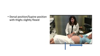
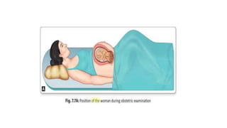
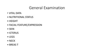
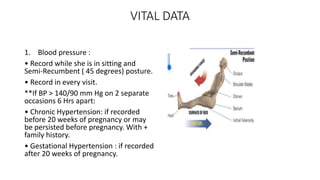
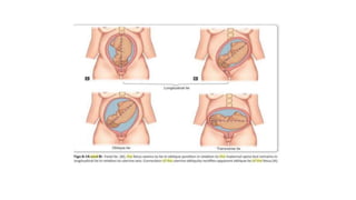
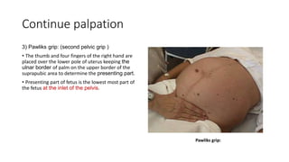
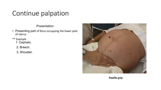
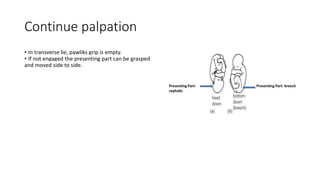
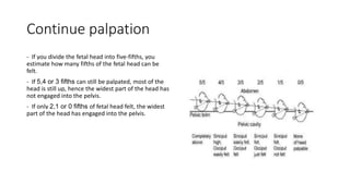
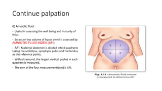
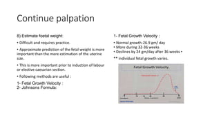
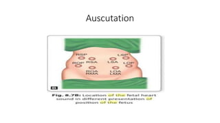
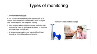
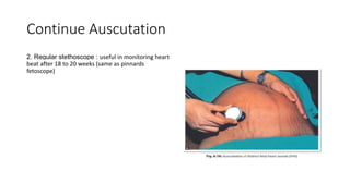
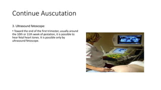
Obstetric Examination During
Pregnancy
Lecture by: Prof.Srey Sopha
Prepared by: DES.Deng Makara
Content
1. Keywords Before Examination
2. General physical examination
3. Obstetrical Examination:
A- Abdominal.
B- Vaginal & Cervical.
Keywords Before Examination
• Before examination, explain to the patient the need and the nature of
the proposed examination.
• Obtain a verbal consent.
• The examiner (either male or female) should be accompanied by
another female.
• Respect her privacy and examine in a private room.
Keywords Before Examination
• Expose only relevant parts of her anatomy for examination .
• Ensure the patient is comfortable and warm.
• Ask patient to empty the bladder .
• Patient should lie in the dorsal position with thighs slightly flexed.
• Stand right to her.
Keywords Before Examination
• She is slightly rolled to the left side to prevent compression of the
inferior vena cava by the enlarged uterus (inferior vena caval
syndrome or supine hypotensive syndrome).
• Ask for any tender area before palpating the abdomen.
• Dorsal position/Supine position
with thighs slightly flexed
General Examination
• VITAL DATA
• NUTRITIONAL STATUS
• HEIGHT
• FACIAL FEATURE/EXPRESSION
• SKIN
• ICTERUS
• LEGS
• NECK
• BREAS T
VITAL DATA
1. Blood pressure :
• Record while she is in sitting and
Semi-Recumbent ( 45 degrees) posture.
• Record in every visit.
**If BP > 140/90 mm Hg on 2 separate
occasions 6 Hrs apart:
• Chronic Hypertension: if recorded
before 20 weeks of pregnancy or may
be persisted before pregnancy. With +
family history.
• Gestational Hypertension : if recorded
after 20 weeks of pregnancy.
VITAL DATA
2.Pulse rate: slightly increase
3. Respiratory rate: feels shortness
of breath with slight exertion due
to elevated diaphragm.
3.Temperature
NUTRITIONAL STATUS
• Nails- white spots in zinc
deficiency, brittle nails in
magnesium deficiency.
• Tongue- May be Large in iodine or
niacin deficiency. May be pallor in
Fe++ deficiency.
• WEIGHT- The abnormal nutritional
status can be described as obesity
and emaciation.
• Check weight in every visit.
• Parameter-Body mass index (BMI)
• Parameters helps in early
intervention of preeclampsia ( in
obese ) and IUGR of fetus ( in
under weight ).
+HEIGHT
-Short stature women are mostly to
suffer with small pelvis.
+ FACIAL FEATURE/EXPRESSION
-Some facial appearances are
pathognomonic of disease.
- Here the patient may be having
thyrotoxicosis.
- The appearance of the patient’s face
may also provide information regarding
psychological makeup: is the person
happy, sad, angry
+SKIN : Extreme pigmentation around
neck, face, forehead. Common in
pregnancy
• Palmar erythema– due to high
estrogen
• Hirsutism– mild common, if more–
Cushing syndrome .
+ICTERUS- Bulbar conjunctiva, under
surface of tongue, Hard palate- to rule
out any LIVER pathology
+LEGS-EDEMA–common-physiological
• Other causes- Preeclampsia, Cardiac
Failure, Nephrotic Syndrome
ABDOMINAL EXAMINATION
• Can be examined in three parts
1- INSPECTION
2- PALPATION
3- AUSCULTATION
Inspection
- Size of the uterus:
• If the length & breadth are both increased multiple pregnancies,
polyhydramnios
• If the length is increased only large baby- Shape of the uterus:
• Length should be larger than broad this indicates longitudinal lie. But
if the uterus is low and broad indicates transverse fetus lie.
• Pendulous abdomen- in primigravidae is sign of inlet contraction.
Inspection
• If there is lateral implantation of
the placenta then the uterus
enlargement will be
asymmetrical- piskacek’s sign.
- Look for fetal movements. (More
prominently seen in 3rd
trimester / Less in
oligohydramnios)
- Look for scars.
- Herniations.
• CUTANEOUS SIGNS- Linea nigra,
Striae gravidarum, Striae
albicans, Umbilicus flat or
everted, Superficial veins.
• SKIN CONDITIONS
- Ringworm/Scabies
Palpation
• Palpation of fetal parts
• Active fetal movements
• Height of the uterus (symphysis-
fundal height)
• Gestational age
• Foetal poles
• Foetal lie
• Presentation part- cephalic(head),
breech,etc
• Attitude
• Level of engagement of presenting
part.
• Uterine contractions.
• Estimate fetal weight.
• Amniotic fluid.
• Any cephalo-pelvic disproportion
Of the above parameters
• To assess FETAL POLE, FETAL LIE,
FETAL PRESENTING PART, ATTITUDE
AND ENGAGEMENT OF FETAL HEAD-
LEOPOLD’S MANOUEVRE IS
FOLLOWED
Palpation
1 ) Palpation of fetal parts
- Distinctly felt after 20th week
- Usually done to estimate the fetal pole/presenting part.
2 ) Active fetal movements
- Gives positive evidence of pregnancy.
- Felt at intervals by placing the hand over the uterus as early as 20th
week. Indicates live fetus.
- Intensity more in last trimester.
Palpation
3) Height of the uterus (Symphysis-Fundal Height):
• The distance from the symphysis pubis to the uterine fundus (top of
the uterus)- size of the uterus directly related to the size of the fetus.
**Technique:
• Place ulnar border of the left hand on the highest part of the uterus
(fundus).
• Mark this point with a pen after obtaining her permission.
• The distance between the upper border of the symphysis pubis upto
the marked point is measured by tape.
• This corresponds to gestational age
Palpation
4) Gestational age :
• The distance from the symphysis pubis to the uterine fundus (top of the uterus) corresponds to the
gestational age/duration of pregnancy.
• After 24 weeks of pregnancy, the distance measured in cm normally corresponds to the period of gestation in
weeks.
5) Fetal Pole, Lie , Presenting Part , Engagement And Attitude Of Fetal Head are assessed by LEOPOLD’S
• MANOUEVRE.
• LEOPOLD’S MANOUEVRE: Done by four
• obstetric grips
1- Fundal grip - To assess fetal pole
2- Lateral grip - To assess fetal lie
3- Pawliks grip - To assess presenting part
4- Deep pelvic grip – To assess engagement and attitude of fetal head.
Palpation
• 1) Fundal grip:
• Both hands placed over the fundus and the
contents of the fundus determined.
• A hard smooth, round pole indicates a fetal head.
• Broad, soft and irregular mass suggestive of
breech.
• In transverse lie no parts are palpated.
Palpation
• 2) Lateral Grip or umbilical grip:
• Move both hands in a downward direction from
the fundus along the sides of the uterus to
determine the "lie" of the fetus.
• "Lie" is the relationship btw the longitudinal axis of
the fetus and the longitudinal axis of the mother.
• The "lie" is usually longitudinal, hence baby is lying
length-wise in the same direction as mother's
longitudinal axis.
Continue palpation
• Other "lies" are :
• Transverse Lie: fetus lies across the longitudinal
axis of mother and
• oblique lie: fetus lies at an oblique angle to the
mother's longitudinal axis.
• Can also determine which side the foetal back is
situated by feeling the firm regular surface of the
foetal back on one side and the irregular, lumpy
surface as the foetal limbs on the other side.
Continue palpation
3) Pawliks grip: (second pelvic grip )
• The thumb and four fingers of the right hand are
placed over the lower pole of uterus keeping the
ulnar border of palm on the upper border of the
suprapubic area to determine the presenting part.
• Presenting part of fetus is the lowest most part of
the fetus at the inlet of the pelvis.
Continue palpation
Presentation:
• Presenting part of fetus occupying the lower pole
of uterus
** Example
1. Cephalic.
2. Breech.
3. Shoulder.
Continue palpation
• In transverse lie, pawliks grip is empty.
• If not engaged the presenting part can be grasped
and moved side to side.
Continue palpation
4) Deep pelvic grip: ( first pelvic grip )
• Determines two points about the fetus
1) The attitude of the fetal head
2) Engagement of the fetal head
1) The attitude of the fetal head :
The examiner turns around to face patients
feet.
• Each hand placed on either side of the fetal
trunk lower down.
• The hands moved downwards towards the
fetal head.
• Note made as to which hand first touches
the fetal head (This point called cephalic
prominence).
• Cephalic prominence helps determine the
attitude (i.e. flexion, deflexed or extended) of
fetal head.
Continue Palpation
• If cephalic prominence (sinciput) is on the
opposite side of fetal back, fetal head is well
flexed (Normal Position).
• If cephalic prominence (occiput) on the same side
as fetal back, fetal head is extended (abnormal
position).
• If examiners hands reach the fetal head equally on
both sides (both sinciput and occiput), fetal head
is deflexed (Military position, indicating mal-
position)
Continue palpation
2)Engagement of the fetal head:
- Engagement of the fetal head defined as having
occurred once the widest transverse diameter of the
fetal head (bi-parietal diameter) has passed through
the pelvic inlet into the true pelvis.
- Procedure: Continue moving both hands down
around the fetal head, determine how far around the
head you can get.
- Examiner should be able to palpate part of fetal
head still in the lower abdomen (also called the 'false'
pelvis but cannot palpate the part of fetal head in the
true pelvis).
Continue palpation
- If you divide the fetal head into five-fifths, you
estimate how many fifths of the fetal head can be
felt.
- If 5,4 or 3 fifths can still be palpated, most of the
head is still up, hence the widest part of the head has
not engaged into the pelvis.
- If only 2,1 or 0 fifths of fetal head felt, the widest
part of the head has engaged into the pelvis.
Continue palpation
6) Amniotic fluid :
- Useful in assessing the well being and maturity of
fetus
- Excess or less volume of liquor amnii is assessed by
AMNIOTIC FLUID INDEX (AFI)
- AFI: Maternal abdomen is divided into 4 quadrants
taking the umbilicus, symphysis pubis and the fundus
as the reference points.
- With ultrasound, the largest vertical pocket in each
quadrant is measured.
- The sum of the four measurements(cm) is AFI.
Continue palpation
• AFI helps to diagnose the clinical conditions called
oligohydramnios and polyhydramnios.
• Normal level of amniotic fluid at Term- 40 weeks
is 600-800 ml.
• Other values:
• at 12 weeks: 50 ml,
• at 20 weeks: 400 ml,
• at 36-38 weeks: 1 liter.
• There is gradual decrease in levels after 38 weeks
7)Uterine contractions:
Braxton-Hicks:
• Felt bimanually.
• During early months of pregnancy- usually in 2nd
trimester begin.
• Irregular,Infrequent,Spasmodic,Painless
• Increases by near term.
• Elicited by rubbing the uterus.
• Absent in abdominal pregnancy.
Continue Palpation
• PalmerSign
• In early weeks of pregnancy palmer sign is
• elicited to diagnose the pregnancy.
• This method is done to note the uterine
contractions.
• Done by- cupping uterus between internal fingers
and external fingers for about 2-3 mins.
• During contraction- uterus is firm and well defined.
• During relaxation – soft and ill defined
Continue palpation
8) Estimate foetal weight:
• Difficult and requires practice.
• Approximate prediction of the fetal weight is more
important than the mere estimation of the uterine
size.
• This is more important prior to induction of labour
or elective caesarian section.
• Following methods are useful :
1- Fetal Growth Velocity :
2- Johnsons Formula:
1- Fetal Growth Velocity :
• Normal growth-26.9 gm/ day
• More during 32-36 weeks
• Declines by 24 gm/day after 36 weeks •
** individual fetal growth varies.
Continue palpation
2- Johnson's formula:
• Applicable only in vertex presentation
• Fundal height (cm) noted above the pubic
symphysis
• Fundal height (cm)- 12 (if Vertex above Ischial
Spine ) × 155 = weight
• Fundal height (cm)- 11 (if vertex below Ischial
Spine) × 155 = weight
• This will be fetal weight in grams.
• • • e.g., 32 (Fundal height)-12(constant) x155(
constant) => 20 x 155=3100gms.
8) Cephalo-pelvic disproportion:
- State were the normal proportion between fetal size
and size of the pelvis is disturbed.
• Two methods:
1. Abdominal method.
2. Abdomino-vaginal method. (explained
in vaginal examination)
Abdominal method
• Patient is placed in dorsal position with the thighs slightly flexed and separated.
• The head is grasped by the left head
• Two fingers, index and middle fingers, of the right hand are placed above the symphysis pubis keeping the
inner surface of the fingers in line with the anterior surface of the symphysis pubis to note the degree of
overlapping, if any, when the head is pushed downwards and backwards.
• No disproportion- if the head can be pushed down in the pelvis without pelvis overlapping of the parietal
bone on the symphysis pubis.
• Disproportion- if the head cannot be pushed down and instead the parietal bone overhangs the symphysis
pubis displacing the fingers.
• Abdominal method is difficult to elicit in deflexed head.
• It can be used as screening method.
Auscutation
• Importance: for monitoring FETAL HEART
SOUNDS
• Helps in diagnosis of live baby but its location of
maximum intensity can resolve doubt about the
presentation of the fetus.
• FHS are best audible through back in vertex and
breech presentation where the convex portion of the
back is in contact with the uterine wall.
• How ever in face presentation, FHS are heard
through fetal chest.
• FHS is maximum below the umbilicus in cephalic
presentation and
• FHS is maximum around the umbilicus in breech.
• Location of FHS depends on the position of the
head and degree of decent of the head even in
cephalic presentation.
• In Occipito anterior position, FHS is heard in
middle of the spino-umbilical line.
• In occipito-posterior –> towards the mother flank
on same side
• In occipito-lateral -> towards laterally .
• In left occipito-posterior position –> FHS is most
difficult to locate.
Auscutation
Types of monitoring
1. Pinnard stethoscope:
• The heartbeat of the baby may be checked by a
simple instrument which looks like a short trumpet
that is held against the pregnant tummy.
• This is called a Pinnard stethoscope (or fetoscope)
and can be used by a midwife or doctor to listen to
the heartbeat periodically.
• A fetoscope can detect and transmit fetal heart
sounds at 18 to 20 weeks and beyond.
Continue Auscutation
2. Regular stethoscope : useful in monitoring heart
beat after 18 to 20 weeks (same as pinnards
fetoscope)
Continue Auscutation
3. Ultrasound fetoscope:
• Toward the end of the first trimester, usually around
the 10th or 11th week of gestation, it is possible to
hear fetal heart tones. It is possible only by
ultrasound fetoscope.
Continue Auscutation
4. Doppler: Doptone machine
• Doppler machines may be very simple and report
only the rate and rhythm of the beat, but more
sophisticated models will provide additional
information about blood flow in the umbilical artery.
vaginal examination
• A vaginal examination (speculum or digital
examination) is not part of a routine obstetric
examination but may be indicated to diagnose
the pregnancy, to see any rupture of
membranes, onset of labour by checking
cervix, cephalopelvic disproportion.
• Can be done bimanually by hands and by speculum.
Continue vaginal examination
• Technique of vaginal examination:
Mother in Supine, Hips Flexed And Abducted,Knees
Flexed.
Aseptic technique as much as possible.
 Note: In Placenta previae & Abruptio
placentae- usually vaginal examination is avoided.
Only vulval examination done.
Continue vaginal examination
• Premature rupture of membranes:
- Check the collected fluid in posterior fornix (vaginal pool).
• Cephalopelvic disproportion:
- Done by Muller-MunroKerr method.
- It is a bimanual examination
• It is superficial to abdominal method
• Two fingers are introduced into vagina with the finger tips
placed over the ischial spines and thumb is placed over the
symphysis pubis.
• The head is grasped by the left hand and is pushed in a
downward and backward direction into the pelvis.
• No disproportion- if the head can be pushed down up to the
level of ischial spines and there is no overlapping of the parietal
bone on the symphysis pubis.
• Disproportion- if the head cannot be pushed down and
instead the parietal bone overhangs the symphysis pubis
displacing the thumb.
Continue vaginal examination
Cervical examination
-Done simultaneously in vaginal examination
- Helps in diagnosing the pregnancy in early weeks-
Goodells sign- (soft cervix-6th week)
- To check the dilatation of cervix, effacement of
cervix in labour.
- Hegars sign: Gently done- Bimanual examination-
two fingers in the anterior fornix and two abdominal
fingers behind the uterus. +Ve sign- cervix is firm.
References
• TEXT BOOK OF OBSTETRICS- D.C DUTTA, sixth
• edition-2004.
• D.C. DUTTA’S TEXBOOK OF OBSTETRICS, 8th edition-2015- Google eBook
• MUDALIAR AND MENONS CLINICAL OBSTETRICS- 9TH edition.
• OXFORD HANDBOOK OF CLINICAL EXAMINATION AND PRACTICAL SKILLS, 1st edition (vishal).
• GOOGLEIMAGES
• UpToDate App
• AMBOSS App

Obstetric Examination during pregnancy.pptx

  • 1.
    Obstetric Examination During Pregnancy Lectureby: Prof.Srey Sopha Prepared by: DES.Deng Makara
  • 2.
    Content 1. Keywords BeforeExamination 2. General physical examination 3. Obstetrical Examination: A- Abdominal. B- Vaginal & Cervical.
  • 3.
    Keywords Before Examination •Before examination, explain to the patient the need and the nature of the proposed examination. • Obtain a verbal consent. • The examiner (either male or female) should be accompanied by another female. • Respect her privacy and examine in a private room.
  • 4.
    Keywords Before Examination •Expose only relevant parts of her anatomy for examination . • Ensure the patient is comfortable and warm. • Ask patient to empty the bladder . • Patient should lie in the dorsal position with thighs slightly flexed. • Stand right to her.
  • 5.
    Keywords Before Examination •She is slightly rolled to the left side to prevent compression of the inferior vena cava by the enlarged uterus (inferior vena caval syndrome or supine hypotensive syndrome). • Ask for any tender area before palpating the abdomen.
  • 6.
    • Dorsal position/Supineposition with thighs slightly flexed
  • 8.
    General Examination • VITALDATA • NUTRITIONAL STATUS • HEIGHT • FACIAL FEATURE/EXPRESSION • SKIN • ICTERUS • LEGS • NECK • BREAS T
  • 9.
    VITAL DATA 1. Bloodpressure : • Record while she is in sitting and Semi-Recumbent ( 45 degrees) posture. • Record in every visit. **If BP > 140/90 mm Hg on 2 separate occasions 6 Hrs apart: • Chronic Hypertension: if recorded before 20 weeks of pregnancy or may be persisted before pregnancy. With + family history. • Gestational Hypertension : if recorded after 20 weeks of pregnancy.
  • 10.
    VITAL DATA 2.Pulse rate:slightly increase 3. Respiratory rate: feels shortness of breath with slight exertion due to elevated diaphragm. 3.Temperature
  • 11.
    NUTRITIONAL STATUS • Nails-white spots in zinc deficiency, brittle nails in magnesium deficiency. • Tongue- May be Large in iodine or niacin deficiency. May be pallor in Fe++ deficiency. • WEIGHT- The abnormal nutritional status can be described as obesity and emaciation. • Check weight in every visit. • Parameter-Body mass index (BMI) • Parameters helps in early intervention of preeclampsia ( in obese ) and IUGR of fetus ( in under weight ).
  • 12.
    +HEIGHT -Short stature womenare mostly to suffer with small pelvis. + FACIAL FEATURE/EXPRESSION -Some facial appearances are pathognomonic of disease. - Here the patient may be having thyrotoxicosis. - The appearance of the patient’s face may also provide information regarding psychological makeup: is the person happy, sad, angry +SKIN : Extreme pigmentation around neck, face, forehead. Common in pregnancy • Palmar erythema– due to high estrogen • Hirsutism– mild common, if more– Cushing syndrome . +ICTERUS- Bulbar conjunctiva, under surface of tongue, Hard palate- to rule out any LIVER pathology +LEGS-EDEMA–common-physiological • Other causes- Preeclampsia, Cardiac Failure, Nephrotic Syndrome
  • 15.
    ABDOMINAL EXAMINATION • Canbe examined in three parts 1- INSPECTION 2- PALPATION 3- AUSCULTATION
  • 16.
    Inspection - Size ofthe uterus: • If the length & breadth are both increased multiple pregnancies, polyhydramnios • If the length is increased only large baby- Shape of the uterus: • Length should be larger than broad this indicates longitudinal lie. But if the uterus is low and broad indicates transverse fetus lie. • Pendulous abdomen- in primigravidae is sign of inlet contraction.
  • 17.
    Inspection • If thereis lateral implantation of the placenta then the uterus enlargement will be asymmetrical- piskacek’s sign. - Look for fetal movements. (More prominently seen in 3rd trimester / Less in oligohydramnios) - Look for scars. - Herniations. • CUTANEOUS SIGNS- Linea nigra, Striae gravidarum, Striae albicans, Umbilicus flat or everted, Superficial veins. • SKIN CONDITIONS - Ringworm/Scabies
  • 19.
    Palpation • Palpation offetal parts • Active fetal movements • Height of the uterus (symphysis- fundal height) • Gestational age • Foetal poles • Foetal lie • Presentation part- cephalic(head), breech,etc • Attitude • Level of engagement of presenting part. • Uterine contractions. • Estimate fetal weight. • Amniotic fluid. • Any cephalo-pelvic disproportion Of the above parameters • To assess FETAL POLE, FETAL LIE, FETAL PRESENTING PART, ATTITUDE AND ENGAGEMENT OF FETAL HEAD- LEOPOLD’S MANOUEVRE IS FOLLOWED
  • 20.
    Palpation 1 ) Palpationof fetal parts - Distinctly felt after 20th week - Usually done to estimate the fetal pole/presenting part. 2 ) Active fetal movements - Gives positive evidence of pregnancy. - Felt at intervals by placing the hand over the uterus as early as 20th week. Indicates live fetus. - Intensity more in last trimester.
  • 21.
    Palpation 3) Height ofthe uterus (Symphysis-Fundal Height): • The distance from the symphysis pubis to the uterine fundus (top of the uterus)- size of the uterus directly related to the size of the fetus. **Technique: • Place ulnar border of the left hand on the highest part of the uterus (fundus). • Mark this point with a pen after obtaining her permission. • The distance between the upper border of the symphysis pubis upto the marked point is measured by tape. • This corresponds to gestational age
  • 23.
    Palpation 4) Gestational age: • The distance from the symphysis pubis to the uterine fundus (top of the uterus) corresponds to the gestational age/duration of pregnancy. • After 24 weeks of pregnancy, the distance measured in cm normally corresponds to the period of gestation in weeks. 5) Fetal Pole, Lie , Presenting Part , Engagement And Attitude Of Fetal Head are assessed by LEOPOLD’S • MANOUEVRE. • LEOPOLD’S MANOUEVRE: Done by four • obstetric grips 1- Fundal grip - To assess fetal pole 2- Lateral grip - To assess fetal lie 3- Pawliks grip - To assess presenting part 4- Deep pelvic grip – To assess engagement and attitude of fetal head.
  • 24.
    Palpation • 1) Fundalgrip: • Both hands placed over the fundus and the contents of the fundus determined. • A hard smooth, round pole indicates a fetal head. • Broad, soft and irregular mass suggestive of breech. • In transverse lie no parts are palpated.
  • 25.
    Palpation • 2) LateralGrip or umbilical grip: • Move both hands in a downward direction from the fundus along the sides of the uterus to determine the "lie" of the fetus. • "Lie" is the relationship btw the longitudinal axis of the fetus and the longitudinal axis of the mother. • The "lie" is usually longitudinal, hence baby is lying length-wise in the same direction as mother's longitudinal axis.
  • 26.
    Continue palpation • Other"lies" are : • Transverse Lie: fetus lies across the longitudinal axis of mother and • oblique lie: fetus lies at an oblique angle to the mother's longitudinal axis. • Can also determine which side the foetal back is situated by feeling the firm regular surface of the foetal back on one side and the irregular, lumpy surface as the foetal limbs on the other side.
  • 28.
    Continue palpation 3) Pawliksgrip: (second pelvic grip ) • The thumb and four fingers of the right hand are placed over the lower pole of uterus keeping the ulnar border of palm on the upper border of the suprapubic area to determine the presenting part. • Presenting part of fetus is the lowest most part of the fetus at the inlet of the pelvis.
  • 29.
    Continue palpation Presentation: • Presentingpart of fetus occupying the lower pole of uterus ** Example 1. Cephalic. 2. Breech. 3. Shoulder.
  • 30.
    Continue palpation • Intransverse lie, pawliks grip is empty. • If not engaged the presenting part can be grasped and moved side to side.
  • 32.
    Continue palpation 4) Deeppelvic grip: ( first pelvic grip ) • Determines two points about the fetus 1) The attitude of the fetal head 2) Engagement of the fetal head 1) The attitude of the fetal head : The examiner turns around to face patients feet. • Each hand placed on either side of the fetal trunk lower down. • The hands moved downwards towards the fetal head. • Note made as to which hand first touches the fetal head (This point called cephalic prominence). • Cephalic prominence helps determine the attitude (i.e. flexion, deflexed or extended) of fetal head.
  • 33.
    Continue Palpation • Ifcephalic prominence (sinciput) is on the opposite side of fetal back, fetal head is well flexed (Normal Position). • If cephalic prominence (occiput) on the same side as fetal back, fetal head is extended (abnormal position). • If examiners hands reach the fetal head equally on both sides (both sinciput and occiput), fetal head is deflexed (Military position, indicating mal- position)
  • 34.
    Continue palpation 2)Engagement ofthe fetal head: - Engagement of the fetal head defined as having occurred once the widest transverse diameter of the fetal head (bi-parietal diameter) has passed through the pelvic inlet into the true pelvis. - Procedure: Continue moving both hands down around the fetal head, determine how far around the head you can get. - Examiner should be able to palpate part of fetal head still in the lower abdomen (also called the 'false' pelvis but cannot palpate the part of fetal head in the true pelvis).
  • 35.
    Continue palpation - Ifyou divide the fetal head into five-fifths, you estimate how many fifths of the fetal head can be felt. - If 5,4 or 3 fifths can still be palpated, most of the head is still up, hence the widest part of the head has not engaged into the pelvis. - If only 2,1 or 0 fifths of fetal head felt, the widest part of the head has engaged into the pelvis.
  • 36.
    Continue palpation 6) Amnioticfluid : - Useful in assessing the well being and maturity of fetus - Excess or less volume of liquor amnii is assessed by AMNIOTIC FLUID INDEX (AFI) - AFI: Maternal abdomen is divided into 4 quadrants taking the umbilicus, symphysis pubis and the fundus as the reference points. - With ultrasound, the largest vertical pocket in each quadrant is measured. - The sum of the four measurements(cm) is AFI.
  • 37.
    Continue palpation • AFIhelps to diagnose the clinical conditions called oligohydramnios and polyhydramnios. • Normal level of amniotic fluid at Term- 40 weeks is 600-800 ml. • Other values: • at 12 weeks: 50 ml, • at 20 weeks: 400 ml, • at 36-38 weeks: 1 liter. • There is gradual decrease in levels after 38 weeks 7)Uterine contractions: Braxton-Hicks: • Felt bimanually. • During early months of pregnancy- usually in 2nd trimester begin. • Irregular,Infrequent,Spasmodic,Painless • Increases by near term. • Elicited by rubbing the uterus. • Absent in abdominal pregnancy.
  • 38.
    Continue Palpation • PalmerSign •In early weeks of pregnancy palmer sign is • elicited to diagnose the pregnancy. • This method is done to note the uterine contractions. • Done by- cupping uterus between internal fingers and external fingers for about 2-3 mins. • During contraction- uterus is firm and well defined. • During relaxation – soft and ill defined
  • 39.
    Continue palpation 8) Estimatefoetal weight: • Difficult and requires practice. • Approximate prediction of the fetal weight is more important than the mere estimation of the uterine size. • This is more important prior to induction of labour or elective caesarian section. • Following methods are useful : 1- Fetal Growth Velocity : 2- Johnsons Formula: 1- Fetal Growth Velocity : • Normal growth-26.9 gm/ day • More during 32-36 weeks • Declines by 24 gm/day after 36 weeks • ** individual fetal growth varies.
  • 40.
    Continue palpation 2- Johnson'sformula: • Applicable only in vertex presentation • Fundal height (cm) noted above the pubic symphysis • Fundal height (cm)- 12 (if Vertex above Ischial Spine ) × 155 = weight • Fundal height (cm)- 11 (if vertex below Ischial Spine) × 155 = weight • This will be fetal weight in grams. • • • e.g., 32 (Fundal height)-12(constant) x155( constant) => 20 x 155=3100gms. 8) Cephalo-pelvic disproportion: - State were the normal proportion between fetal size and size of the pelvis is disturbed. • Two methods: 1. Abdominal method. 2. Abdomino-vaginal method. (explained in vaginal examination)
  • 41.
    Abdominal method • Patientis placed in dorsal position with the thighs slightly flexed and separated. • The head is grasped by the left head • Two fingers, index and middle fingers, of the right hand are placed above the symphysis pubis keeping the inner surface of the fingers in line with the anterior surface of the symphysis pubis to note the degree of overlapping, if any, when the head is pushed downwards and backwards. • No disproportion- if the head can be pushed down in the pelvis without pelvis overlapping of the parietal bone on the symphysis pubis. • Disproportion- if the head cannot be pushed down and instead the parietal bone overhangs the symphysis pubis displacing the fingers. • Abdominal method is difficult to elicit in deflexed head. • It can be used as screening method.
  • 42.
    Auscutation • Importance: formonitoring FETAL HEART SOUNDS • Helps in diagnosis of live baby but its location of maximum intensity can resolve doubt about the presentation of the fetus. • FHS are best audible through back in vertex and breech presentation where the convex portion of the back is in contact with the uterine wall. • How ever in face presentation, FHS are heard through fetal chest. • FHS is maximum below the umbilicus in cephalic presentation and • FHS is maximum around the umbilicus in breech. • Location of FHS depends on the position of the head and degree of decent of the head even in cephalic presentation. • In Occipito anterior position, FHS is heard in middle of the spino-umbilical line. • In occipito-posterior –> towards the mother flank on same side • In occipito-lateral -> towards laterally . • In left occipito-posterior position –> FHS is most difficult to locate.
  • 43.
  • 44.
    Types of monitoring 1.Pinnard stethoscope: • The heartbeat of the baby may be checked by a simple instrument which looks like a short trumpet that is held against the pregnant tummy. • This is called a Pinnard stethoscope (or fetoscope) and can be used by a midwife or doctor to listen to the heartbeat periodically. • A fetoscope can detect and transmit fetal heart sounds at 18 to 20 weeks and beyond.
  • 45.
    Continue Auscutation 2. Regularstethoscope : useful in monitoring heart beat after 18 to 20 weeks (same as pinnards fetoscope)
  • 46.
    Continue Auscutation 3. Ultrasoundfetoscope: • Toward the end of the first trimester, usually around the 10th or 11th week of gestation, it is possible to hear fetal heart tones. It is possible only by ultrasound fetoscope.
  • 47.
    Continue Auscutation 4. Doppler:Doptone machine • Doppler machines may be very simple and report only the rate and rhythm of the beat, but more sophisticated models will provide additional information about blood flow in the umbilical artery.
  • 48.
    vaginal examination • Avaginal examination (speculum or digital examination) is not part of a routine obstetric examination but may be indicated to diagnose the pregnancy, to see any rupture of membranes, onset of labour by checking cervix, cephalopelvic disproportion. • Can be done bimanually by hands and by speculum.
  • 49.
    Continue vaginal examination •Technique of vaginal examination: Mother in Supine, Hips Flexed And Abducted,Knees Flexed. Aseptic technique as much as possible.  Note: In Placenta previae & Abruptio placentae- usually vaginal examination is avoided. Only vulval examination done.
  • 50.
    Continue vaginal examination •Premature rupture of membranes: - Check the collected fluid in posterior fornix (vaginal pool). • Cephalopelvic disproportion: - Done by Muller-MunroKerr method. - It is a bimanual examination • It is superficial to abdominal method • Two fingers are introduced into vagina with the finger tips placed over the ischial spines and thumb is placed over the symphysis pubis. • The head is grasped by the left hand and is pushed in a downward and backward direction into the pelvis. • No disproportion- if the head can be pushed down up to the level of ischial spines and there is no overlapping of the parietal bone on the symphysis pubis. • Disproportion- if the head cannot be pushed down and instead the parietal bone overhangs the symphysis pubis displacing the thumb.
  • 51.
  • 52.
    Cervical examination -Done simultaneouslyin vaginal examination - Helps in diagnosing the pregnancy in early weeks- Goodells sign- (soft cervix-6th week) - To check the dilatation of cervix, effacement of cervix in labour. - Hegars sign: Gently done- Bimanual examination- two fingers in the anterior fornix and two abdominal fingers behind the uterus. +Ve sign- cervix is firm.
  • 54.
    References • TEXT BOOKOF OBSTETRICS- D.C DUTTA, sixth • edition-2004. • D.C. DUTTA’S TEXBOOK OF OBSTETRICS, 8th edition-2015- Google eBook • MUDALIAR AND MENONS CLINICAL OBSTETRICS- 9TH edition. • OXFORD HANDBOOK OF CLINICAL EXAMINATION AND PRACTICAL SKILLS, 1st edition (vishal). • GOOGLEIMAGES • UpToDate App • AMBOSS App