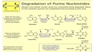
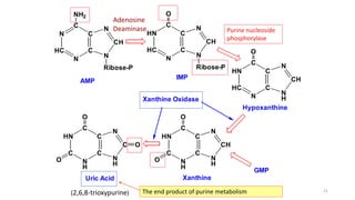
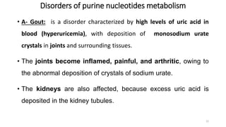
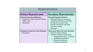
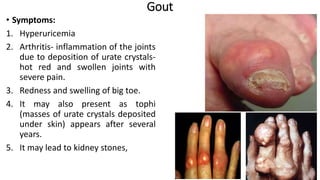
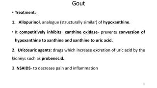
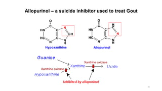
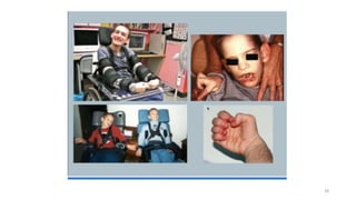
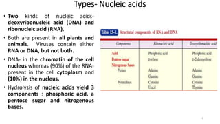
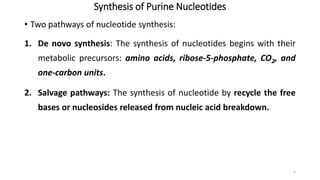
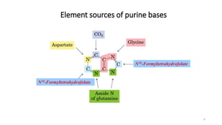
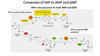
Nucleotide Metabolism
Noor Ullah
M.Phil Biochemistry& Molecular Biology
Lecturer IPMS, KMU
Tuesday, March 12, 2024 1
Nucleic acids- Introduction
• Nucleotides are energy rich compounds that drive metabolic processes (esp.,
biosynthetic) in all cells.
• They also serve as chemical signals, key links in cellular systems that respond to
hormones and other extracellular stimuli, and are structural components of a
number of enzyme cofactors and metabolic intermediates.
• The nucleic acids (DNA and RNA) are the molecular storehouses for genetic
information and are jointly referred to as the ‘molecules of heredity’.
• The structure of every protein, and ultimately of every cell constituent, is a
product of information programmed into the nucleotide sequence of a cell’s
nucleic acids.
2
Definitions- Nucleic acids
• The nucleic acids are the hereditary determinants of living organisms.
• They are the macromolecules present in most living cells either in the free
state or protein bound- nucleoproteins.
• Just as amino acids are the repeating units of proteins, mononucleotide
as their repeating units of nucleic acids.
• Elemental composition- the nucleic acids contain carbon, hydrogen,
oxygen, nitrogen and, strangely enough, phosphorus ; the percentage of
the last two elements being about 15 and 10, respectively.
3
Types- Nucleic acids
• Two kinds of nucleic acids-
deoxyribonucleic acid (DNA) and
ribonucleic acid (RNA).
• Both are present in all plants and
animals. Viruses contain either
RNA or DNA, but not both.
• DNA- in the chromatin of the cell
nucleus whereas (90%) of the RNA-
present in the cell cytoplasm and
(10%) in the nucleus.
• Hydrolysis of nucleic acids yield 3
components : phosphoric acid, a
pentose sugar and nitrogenous
bases.
4
Synthesis of Purine Nucleotides
• Two pathways of nucleotide synthesis:
1. De novo synthesis: The synthesis of nucleotides begins with their
metabolic precursors: amino acids, ribose-5-phosphate, CO2, and
one-carbon units.
2. Salvage pathways: The synthesis of nucleotide by recycle the free
bases or nucleosides released from nucleic acid breakdown.
5
Synthesis of Purine Nucleotides
• De Novo synthesis:
• Site:
• in cytosol of liver, small intestine and thymus
• Characteristics:
a. Purines are synthesized using ribose-5-phosphate (R-5-P) as the
starting material step by step.
b. PRPP(5-phosphoribosyl-1-pyrophosphate) is active donor of R-
5-P
c. AMP and GMP are synthesized from IMP(Inosine-5'-
Monophosphate)
6
Synthesis of Inosine Monophosphate (IMP)
• Ribose phosphate is formed in pentose-phosphate pathway from glucose
• Basic pathway for biosynthesis of purine ribonucleotides starts from ribose-5-
phosphate (R-5-P)
• Purine ring is synthesized on ribose-5-phosphate by the way of gradual adding
of nitrogen and carbon atoms and cyclization.
• The pathway ends with the formation of a purine nucleotide called Inosine
monophosphate (IMP) which is the precursor of AMP and GMP which then
converted into ATP and GTP, respectively
• Requires 11 steps.
• Occurs primarily in the liver
7
N10-Formyltetrahydrofolate
Element sources of purine bases
8
Conversion of IMP to AMP and GMP
GTP is used for AMP synthesis
ATP is used for GMP
synthesis
IMP is the precursor for both AMP and GMP.
9
kinase
ADP
kinase
ADP
ATP
ATP ADP
AMP
ATP
kinase
GDP
kinase
ADP
GTP
ATP ADP
GMP
ATP
ADP, ATP, GDP and GTP biosynthesis
10
Regulation of de novo synthesis
• The significance of regulation:
1. Meet the need of the body, without wasting.
2. AMP and GMP control their respective synthesis from IMP by a
feedback mechanism, [GTP]=[ATP]
11
Regulation of de novo synthesis
• Amidophosphoribosyl transferase catalyses the rate limiting step of
the pathway which is activated by activated by PRPP.
• So PRPP is an activator of the pathway. Increased PRPP leads to
overproduction of purine nucleotides.
• Inhibitors of the amidotransferase:
• The enzyme is inhibited by the final products of the pathway (IMP,
AMP and GMP).
12
13
Salvage pathway of purines- Resynthesis of purine nucleotides
• De novo biosynthesis occurs in liver due to the presence of enzymes.
• Other tissues can’t do de novo synthesis.
• In these organs, free purine bases (guanine, hypoxanthine and
adenine) reacts with PRPP again to resynthesize purine nucleotides.
• These free purine bases are obtained from diet or by degradation of
RNA or DNA
14
Salvage pathway of purines- Resynthesis of purine nucleotides
• The significance of salvage pathway :
• Save the fuel
• Some tissues and organs such as brain and bone marrow are only
capable of synthesizing nucleotides by salvage pathway.
• Two phosphoribosyl transferases are involved:
• APRT (adenine phosphoribosyl transferase) for adenine.
• HGPRT (hypoxanthine guanine phosphoribosyl transferase) for
guanine or hypoxanthine.
• Both enzymes use PRPP as the source of ribose-5-phosphate
15
Purine Salvage Pathway
N
N
N
N
NH2
O
Guanine
N
N N
O
N
Hypoxanthine
O
OH
HO
2-O3POH2C
N
N N
O
N
IMP
O
OH
HO
2-O3POH2C
N
N
N
N
NH2
O
GMP
.
.
Adenine AMP
PRPP PPi
adenine
phosphoribosyl transferase
PRPP PPi
hypoxanthine-guanine
phosphoribosyl transferase
(HGPRT)
Absence of activity of HGPRT leads to Lesch-Nyhan syndrome. 16
Nucleoproteins (nucleic
acids + proteins)
Pepsin, gastricsin, HCl
Nucleic acids Histones, protamines
Nucleases (DNA-ases, RNA-ases)
Oligonucleotides
Mononucleotides
Phosphodiesterases
Nuclesides Phosphoric acid
+
+
Phosphatases
Nitrogenous
bases
+ Pentose
Nucleosidases
Decomposition of nucleic acids in intestine and tissue
17
Phosphoric acid
Nitrogenous
bases
Pentoses
phosphorylation;
ATP synthesis;
synthesis of phospholipids;
buffer systems;
constituent of bones, cartilages
oxidation with energy formation;
synthesis of nucleotides, hexoses,
coenzymes
oxidation to the end products
Destiny of nitrogenous bases, pentoses and phosphoric acids in the organism
18
Catabolism of purine nucleotides-Uric acid
• Uric acid is the end product of purine metabolism in primates, birds and
terrestrial reptiles
• AMP or GMP is metabolized to give hypoxanthine which is then converted into
xanthine and finally into uric acid.
• Most of uric acid is excreted by the kidneys. The rate of uric acid excretion by the
normal adult human is about 0.6 g/24 h, arising in part from ingested purines
and in part from the turnover of the purine nucleotides of nucleic acids.
• The normal serum uric acid concentration in adults is in the range of 3-7 mg/dl.
• But under certain circumstances, the body produces too much uric acid or
removes too little.
• In either case, concentrations of uric acid increase in the blood. This condition is
known as hyperuricemia.
19
20
N
HC
N
C
C
C
N
CH
N
NH2
Ribose-P
AMP
HN
HC
N
C
C
C
N
CH
N
O
Ribose-P
IMP
HN
HC
N
C
C
C
N
H
CH
N
O
HN
C
N
H
C
C
C
N
H
CH
N
O
O
HN
C
N
H
C
C
C
N
H
C
N
O
O
O
GMP
Hypoxanthine
Uric Acid Xanthine
Xanthine Oxidase
(2,6,8-trioxypurine)
Adenosine
Deaminase
The end product of purine metabolism 21
Purine nucleoside
phosphorylase
Disorders of purine nucleotides metabolism
• A- Gout: is a disorder characterized by high levels of uric acid in
blood (hyperuricemia), with deposition of monosodium urate
crystals in joints and surrounding tissues.
• The joints become inflamed, painful, and arthritic, owing to
the abnormal deposition of crystals of sodium urate.
• The kidneys are also affected, because excess uric acid is
deposited in the kidney tubules.
22
23
Gout
• Symptoms:
1. Hyperuricemia
2. Arthritis- inflammation of the joints
due to deposition of urate crystals-
hot red and swollen joints with
severe pain.
3. Redness and swelling of big toe.
4. It may also present as tophi
(masses of urate crystals deposited
under skin) appears after several
years.
5. It may lead to kidney stones,
24
Gout
• Treatment:
1. Allopurinol, analogue (structurally similar) of hypoxanthine.
• It competitively inhibits xanthine oxidase- prevents conversion of
hypoxanthine to xanthine and xanthine to uric acid.
2. Uricosuric agents: drugs which increase excretion of uric acid by the
kidneys such as probenecid.
3. NSAIDS- to decrease pain and inflammation
25
HN
HC
N
C
C
C
N
H
CH
N
O
Hypoxanthine
HN
HC
N
C
C
C
N
H
N
H
C
O
Allopurinol
Allopurinol – a suicide inhibitor used to treat Gout
Xanthine oxidase
Xanthine oxidase
26
Lesch- Nyhan syndrome
• First described by Michael Lesch and William L. Nyhan in 1964
• There is a defect or lack in the HGPRT enzyme
• Sex-linked metabolic disorder: only males
• The rate of purine synthesis is increased about 200-fold
• Loss of HGPRT → block (inhibit) salvage pathway of guanine and hypoxanthine → ↓ use of
PRPP in salvage pathway and ↑ de novo purine synthesis leading to overproduction of
purine nucleotides which by catabolism, will give increased levels of uric acid
Symptoms: appear at age 3-6 months. The first symptom is orange colored crystals in the diaper of
the baby.
1- Hyperuricemia: in aggressive way than in gout.
2- urate kidney stones:
Some symptoms of unknown mechanism are:
3- mental retardation
4- involuntary movements of legs and arms
5- lack of muscle coordination
6- self mutilation (biting of fingers and lips leading to lip lesions). 27
28

Nucleotide Metabolism.biochemistryblecture.pptx

  • 1.
    Nucleotide Metabolism Noor Ullah M.PhilBiochemistry& Molecular Biology Lecturer IPMS, KMU Tuesday, March 12, 2024 1
  • 2.
    Nucleic acids- Introduction •Nucleotides are energy rich compounds that drive metabolic processes (esp., biosynthetic) in all cells. • They also serve as chemical signals, key links in cellular systems that respond to hormones and other extracellular stimuli, and are structural components of a number of enzyme cofactors and metabolic intermediates. • The nucleic acids (DNA and RNA) are the molecular storehouses for genetic information and are jointly referred to as the ‘molecules of heredity’. • The structure of every protein, and ultimately of every cell constituent, is a product of information programmed into the nucleotide sequence of a cell’s nucleic acids. 2
  • 3.
    Definitions- Nucleic acids •The nucleic acids are the hereditary determinants of living organisms. • They are the macromolecules present in most living cells either in the free state or protein bound- nucleoproteins. • Just as amino acids are the repeating units of proteins, mononucleotide as their repeating units of nucleic acids. • Elemental composition- the nucleic acids contain carbon, hydrogen, oxygen, nitrogen and, strangely enough, phosphorus ; the percentage of the last two elements being about 15 and 10, respectively. 3
  • 4.
    Types- Nucleic acids •Two kinds of nucleic acids- deoxyribonucleic acid (DNA) and ribonucleic acid (RNA). • Both are present in all plants and animals. Viruses contain either RNA or DNA, but not both. • DNA- in the chromatin of the cell nucleus whereas (90%) of the RNA- present in the cell cytoplasm and (10%) in the nucleus. • Hydrolysis of nucleic acids yield 3 components : phosphoric acid, a pentose sugar and nitrogenous bases. 4
  • 5.
    Synthesis of PurineNucleotides • Two pathways of nucleotide synthesis: 1. De novo synthesis: The synthesis of nucleotides begins with their metabolic precursors: amino acids, ribose-5-phosphate, CO2, and one-carbon units. 2. Salvage pathways: The synthesis of nucleotide by recycle the free bases or nucleosides released from nucleic acid breakdown. 5
  • 6.
    Synthesis of PurineNucleotides • De Novo synthesis: • Site: • in cytosol of liver, small intestine and thymus • Characteristics: a. Purines are synthesized using ribose-5-phosphate (R-5-P) as the starting material step by step. b. PRPP(5-phosphoribosyl-1-pyrophosphate) is active donor of R- 5-P c. AMP and GMP are synthesized from IMP(Inosine-5'- Monophosphate) 6
  • 7.
    Synthesis of InosineMonophosphate (IMP) • Ribose phosphate is formed in pentose-phosphate pathway from glucose • Basic pathway for biosynthesis of purine ribonucleotides starts from ribose-5- phosphate (R-5-P) • Purine ring is synthesized on ribose-5-phosphate by the way of gradual adding of nitrogen and carbon atoms and cyclization. • The pathway ends with the formation of a purine nucleotide called Inosine monophosphate (IMP) which is the precursor of AMP and GMP which then converted into ATP and GTP, respectively • Requires 11 steps. • Occurs primarily in the liver 7
  • 8.
  • 9.
    Conversion of IMPto AMP and GMP GTP is used for AMP synthesis ATP is used for GMP synthesis IMP is the precursor for both AMP and GMP. 9
  • 10.
  • 11.
    Regulation of denovo synthesis • The significance of regulation: 1. Meet the need of the body, without wasting. 2. AMP and GMP control their respective synthesis from IMP by a feedback mechanism, [GTP]=[ATP] 11
  • 12.
    Regulation of denovo synthesis • Amidophosphoribosyl transferase catalyses the rate limiting step of the pathway which is activated by activated by PRPP. • So PRPP is an activator of the pathway. Increased PRPP leads to overproduction of purine nucleotides. • Inhibitors of the amidotransferase: • The enzyme is inhibited by the final products of the pathway (IMP, AMP and GMP). 12
  • 13.
  • 14.
    Salvage pathway ofpurines- Resynthesis of purine nucleotides • De novo biosynthesis occurs in liver due to the presence of enzymes. • Other tissues can’t do de novo synthesis. • In these organs, free purine bases (guanine, hypoxanthine and adenine) reacts with PRPP again to resynthesize purine nucleotides. • These free purine bases are obtained from diet or by degradation of RNA or DNA 14
  • 15.
    Salvage pathway ofpurines- Resynthesis of purine nucleotides • The significance of salvage pathway : • Save the fuel • Some tissues and organs such as brain and bone marrow are only capable of synthesizing nucleotides by salvage pathway. • Two phosphoribosyl transferases are involved: • APRT (adenine phosphoribosyl transferase) for adenine. • HGPRT (hypoxanthine guanine phosphoribosyl transferase) for guanine or hypoxanthine. • Both enzymes use PRPP as the source of ribose-5-phosphate 15
  • 16.
    Purine Salvage Pathway N N N N NH2 O Guanine N NN O N Hypoxanthine O OH HO 2-O3POH2C N N N O N IMP O OH HO 2-O3POH2C N N N N NH2 O GMP . . Adenine AMP PRPP PPi adenine phosphoribosyl transferase PRPP PPi hypoxanthine-guanine phosphoribosyl transferase (HGPRT) Absence of activity of HGPRT leads to Lesch-Nyhan syndrome. 16
  • 17.
    Nucleoproteins (nucleic acids +proteins) Pepsin, gastricsin, HCl Nucleic acids Histones, protamines Nucleases (DNA-ases, RNA-ases) Oligonucleotides Mononucleotides Phosphodiesterases Nuclesides Phosphoric acid + + Phosphatases Nitrogenous bases + Pentose Nucleosidases Decomposition of nucleic acids in intestine and tissue 17
  • 18.
    Phosphoric acid Nitrogenous bases Pentoses phosphorylation; ATP synthesis; synthesisof phospholipids; buffer systems; constituent of bones, cartilages oxidation with energy formation; synthesis of nucleotides, hexoses, coenzymes oxidation to the end products Destiny of nitrogenous bases, pentoses and phosphoric acids in the organism 18
  • 19.
    Catabolism of purinenucleotides-Uric acid • Uric acid is the end product of purine metabolism in primates, birds and terrestrial reptiles • AMP or GMP is metabolized to give hypoxanthine which is then converted into xanthine and finally into uric acid. • Most of uric acid is excreted by the kidneys. The rate of uric acid excretion by the normal adult human is about 0.6 g/24 h, arising in part from ingested purines and in part from the turnover of the purine nucleotides of nucleic acids. • The normal serum uric acid concentration in adults is in the range of 3-7 mg/dl. • But under certain circumstances, the body produces too much uric acid or removes too little. • In either case, concentrations of uric acid increase in the blood. This condition is known as hyperuricemia. 19
  • 20.
  • 21.
  • 22.
    Disorders of purinenucleotides metabolism • A- Gout: is a disorder characterized by high levels of uric acid in blood (hyperuricemia), with deposition of monosodium urate crystals in joints and surrounding tissues. • The joints become inflamed, painful, and arthritic, owing to the abnormal deposition of crystals of sodium urate. • The kidneys are also affected, because excess uric acid is deposited in the kidney tubules. 22
  • 23.
  • 24.
    Gout • Symptoms: 1. Hyperuricemia 2.Arthritis- inflammation of the joints due to deposition of urate crystals- hot red and swollen joints with severe pain. 3. Redness and swelling of big toe. 4. It may also present as tophi (masses of urate crystals deposited under skin) appears after several years. 5. It may lead to kidney stones, 24
  • 25.
    Gout • Treatment: 1. Allopurinol,analogue (structurally similar) of hypoxanthine. • It competitively inhibits xanthine oxidase- prevents conversion of hypoxanthine to xanthine and xanthine to uric acid. 2. Uricosuric agents: drugs which increase excretion of uric acid by the kidneys such as probenecid. 3. NSAIDS- to decrease pain and inflammation 25
  • 26.
    HN HC N C C C N H CH N O Hypoxanthine HN HC N C C C N H N H C O Allopurinol Allopurinol – asuicide inhibitor used to treat Gout Xanthine oxidase Xanthine oxidase 26
  • 27.
    Lesch- Nyhan syndrome •First described by Michael Lesch and William L. Nyhan in 1964 • There is a defect or lack in the HGPRT enzyme • Sex-linked metabolic disorder: only males • The rate of purine synthesis is increased about 200-fold • Loss of HGPRT → block (inhibit) salvage pathway of guanine and hypoxanthine → ↓ use of PRPP in salvage pathway and ↑ de novo purine synthesis leading to overproduction of purine nucleotides which by catabolism, will give increased levels of uric acid Symptoms: appear at age 3-6 months. The first symptom is orange colored crystals in the diaper of the baby. 1- Hyperuricemia: in aggressive way than in gout. 2- urate kidney stones: Some symptoms of unknown mechanism are: 3- mental retardation 4- involuntary movements of legs and arms 5- lack of muscle coordination 6- self mutilation (biting of fingers and lips leading to lip lesions). 27
  • 28.