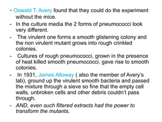
The document outlines the historical development of our understanding of nucleic acids, starting from the discovery of nuclein by Johann Friedrich Miescher in 1869 to the establishment of DNA as the genetic material through various experiments. Key experiments mentioned include Frederick Griffith's bacterial transformation, Alfred Hershey and Martha Chase's studies on viral DNA, and the Watson-Crick model of DNA structure. The document also emphasizes the central dogma of molecular biology, detailing the flow of genetic information from DNA to RNA to proteins.


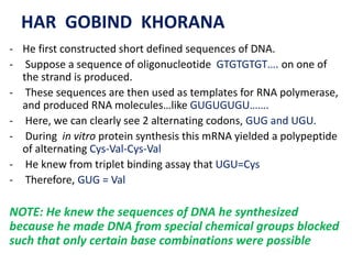
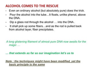
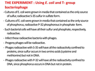
![It all begins with a thin soup of
bacterial cells
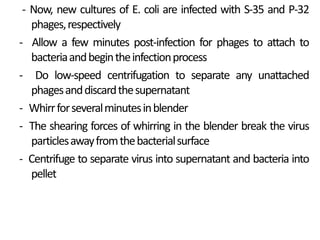
- Millions of bacterial cells are suspended in a broth of nutrient
chemicals, doubling their number in about every half an hour.
- Let‟s concentrate these cells a bit.
- The test tube full of cloudy soup is whirled round at about
8000 revolutions per minute in a centrifuge.
[ The soup is cloudy because it reflects light]
- The cells are then forced down to the bottom of the test tube.
- After 30 minutes or so, the broth has separated into an
absolutely clear liquid above a small pellet, dingy and greyish
yellow.](https://image.slidesharecdn.com/nucleicacids-ahistoricalpesrpective-190213044413/85/Nucleic-acids-a-historical-pesrpective-3-320.jpg)
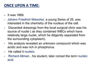


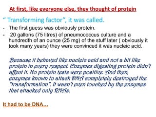
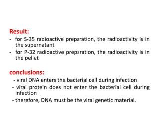
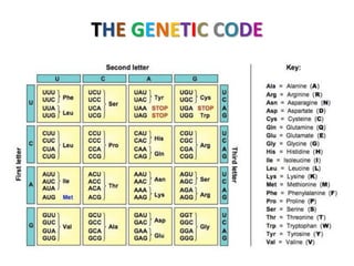
![CRICK and BRENNER, from these evidences, and their other
works deduced 3 fundamental properties of the code:
1. The code is made of triplets (called a ‘codon’) i.e. 1
amino acid is represented by 3 nucleotides in the
mRNA.
2. Because there are now 64 codons, and only 20
amino acids, the code is degenerate i.e. any amino
acid can correspond to more than one codon.
3. The code is sequential and non overlapping.
[ Later, exceptions to the third point was found…one being
the bacteriophage φX174 which had overlapping
genes]](https://image.slidesharecdn.com/nucleicacids-ahistoricalpesrpective-190213044413/85/Nucleic-acids-a-historical-pesrpective-37-320.jpg)
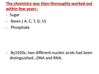
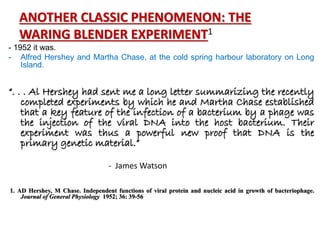
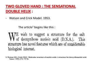
![- Then they tried to synthesize more complex RNA
molecules.
- Let‟s imagine a mix of 3:1 U to G. So, the synthetic RNA
would be a poly UG [ which might have UUU, UUG and
other codons…and was guessed on the basis of
probability].
- The protein thus synthesized from poly UG, 3:1 in vitro
were found to be Valine, Leucine and Cysteine present
about 1-3rd as often as Phenylalanine.
{ Phe was present 2-3rd because the amount of U to G
was 3:1, hence more chance of forming UUU codon}.
- It also suggested that Val, Leu and Cys must have
codon that have 2 Us and 1 G…](https://image.slidesharecdn.com/nucleicacids-ahistoricalpesrpective-190213044413/85/Nucleic-acids-a-historical-pesrpective-40-320.jpg)