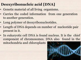
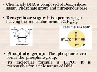
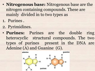
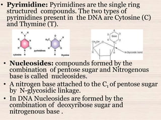
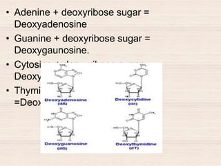
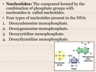
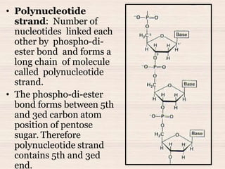
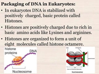
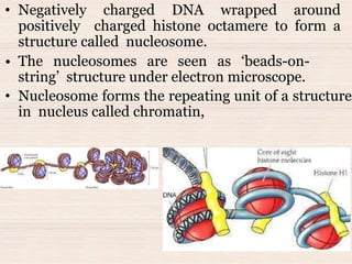
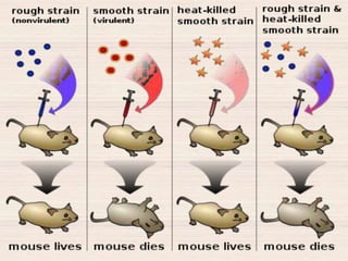
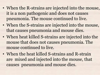
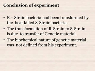
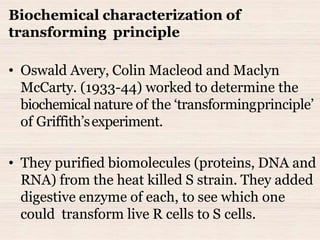
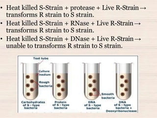
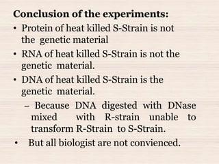
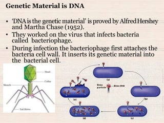
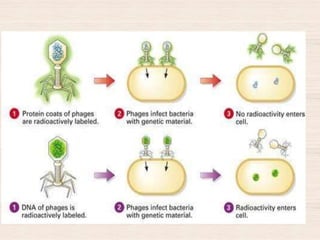
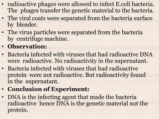
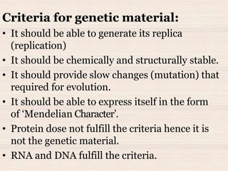
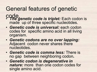
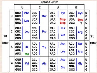
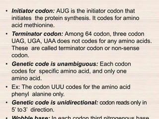
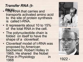
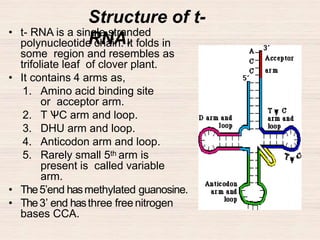
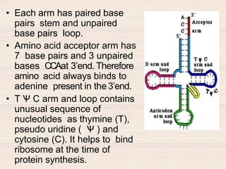
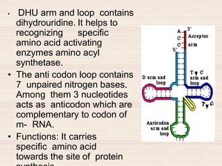
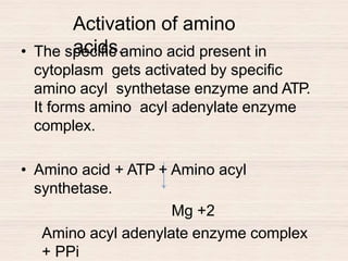
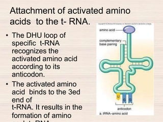
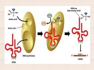
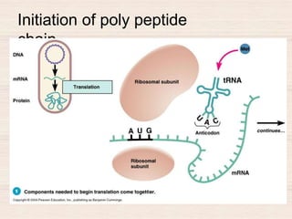
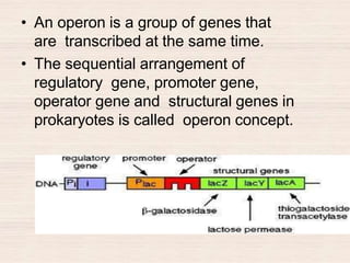
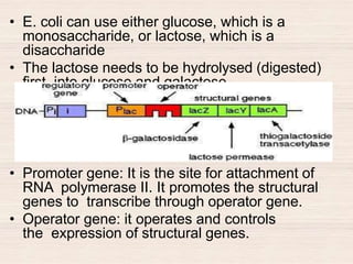
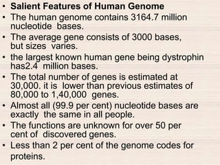
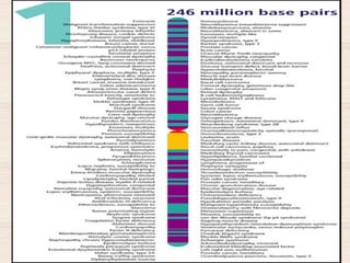
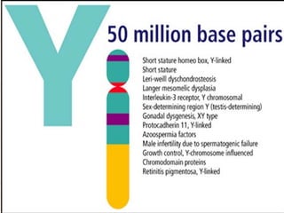
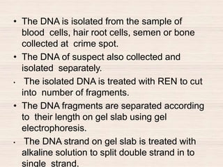
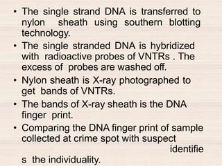
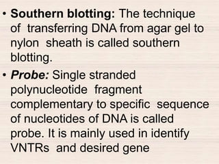
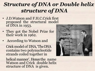
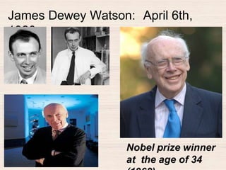
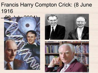
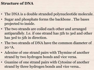
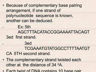
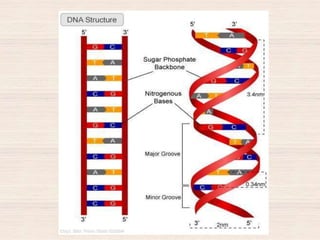
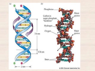
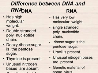
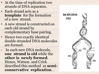
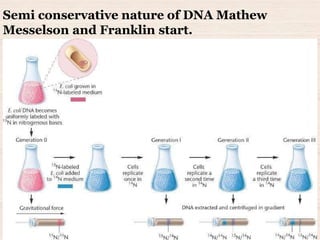
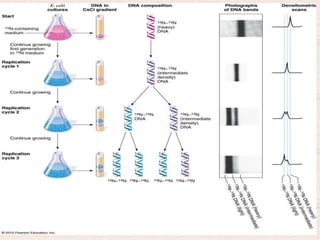
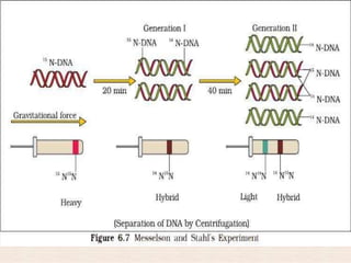
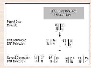
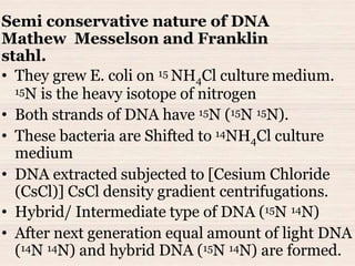
This document discusses the structure and replication of DNA. It provides details on:
1. The key components of DNA including deoxyribose sugar, phosphate groups, and nitrogenous bases. It also describes how nucleotides and polynucleotide strands are formed.
2. The double helix structure of DNA proposed by Watson and Crick, including how the strands are antiparallel and connected through complementary base pairing.
3. Semiconservative replication of DNA, where each old DNA strand acts as a template for a new complementary strand, resulting in two identical double-stranded DNA molecules after replication.
4. Experiments by Meselson and Stahl using isotopes that provided evidence supporting the
3. • Nucleic acids are the
macromolecules present in
all living cell.
• Freidrich Miescher was the
first person isolated the
nucleic acids from the pus
cells. He called it as nuclein.
• As it has an acidic nature,
hence Altmann called it as
nucleic acids.
• Nucleic acids are the
macromolecules present in
all living cell.
Nucleic acids
Freidrich Miescher
5. • Genetic material of all living organisms.
• Carries the coded information from one generation
to another generation.
• Long polymer of deoxyribonucleotides.
• Length of DNA depends on number of nucleotide pair
present in it.
• In eukaryotic cell DNA is found nucleus. It is the chief
component of chromosomes. DNA also found in the
mitochondria and chloroplasts
Deoxyribonucleic acid [DNA]
6. • Chemically DNA is composed of Deoxyribose
sugar, Phosphate group and nitrogenous base .
• Deoxyribose sugar: It is a pentose sugar
heaving the molecular formula C5H10O4.
• Phosphate group: The phosphoric acid
forms the phosphate group.
• its molecular formula is H3PO4. It is
responsible for acidic nature of DNA.
7. • Nitrogenous base: Nitrogenous base are the
nitrogen containing compounds. These are
mainly divided in to two types as
1. Purines .
2. Pyrimidines.
• Purines: Purines are the double ring
heterocyclic structural compounds. The two
types of purines present in the DNA are
Adenine (A) and Guanine (G).
8. • Pyrimidine: Pyrimidines are the single ring
structured compounds. The two types of
pyrimidines present in the DNA are Cytosine (C)
and Thymine (T).
• Nucleosides: compounds formed by the
combination of pentose sugar and Nitrogenous
base is called nucleosides.
• A nitrogen base attached to the C1 of pentose sugar
by N-glycosidic linkage.
• In DNA Nucleosides are formed by the
combination of deoxyribose sugar and
nitrogenous base .
10. • Nucleotides: The compound formed by the
combination of phosphate groups with
nucleosides is called nucleotides.
• Four types of nucleotides present in the DNA:
1. Deoxyadenosine monophosphate.
2. Deoxygaunosine monophosphate.
3. Deoxycytidine monophosphate.
4. Deoxythymidine monophosphate.
11. • Polynucleotide
strand: Number of
nucleotides linked each
other by phospho-di-
ester bond and forms a
long chain of molecule
called polynucleotide
strand.
• The phospho-di-ester
bond forms between 5th
and 3ed carbon atom
position of pentose
sugar. Therefore
polynucleotide strand
contains 5th and 3ed
end.
12. Structure of DNA or Double helix
structure of DNA
• J.D.Watson and F.H.C.Crick first
proposed the structural model
of DNA in 1953.
• They got the Nobel Prize for
their work in 1962.
• According to Watson and
Crickmodel of DNA,‘TheDNA
contains two polynucleotide
strands coiled together in
helical manner’. Hencethe name
Watson and Crick double helix
structure of DNA is given.
15. Structure of DNA
• The DNA is a double stranded polynucleotide molecule.
• Sugar and phosphate forms the backbone . The bases
projected to inside.
• The two strands are coiled each other and arranged
antiparallely. I.e. if one strand has 5th to 3ed and other
has 3ed to 5th in direction.
• The two strands of DNA have the common diameter of
20 0A.
• Adenine of one strand pairs with Thymine of another
strand by two hydrogen bonds and vice versa.
• Guanine of one strand pairs with Cytosine of another
strand by three hydrogen bonds and vice versa..
16. • Because of complementary base pairing
arrangement, if one strand of
polynucleotide sequence is known,
another can be deduced.
• Ex: 5th
AGCTTTACATACCGGAAAATTACAGT
3ed first strand.
• 3ed
TCGAAATGTATGGCCTTTTAATGT
CA 5TH second strand.
• The complementary strand twisted each
other at the distance of 34 0A.
17.
18.
19. Central dogma
• One way flow of genetic information from
DNA to protein is called central dogma.
• DNA→ RNA→Protein.
20. Packaging of DNA in prokaryotes:
• Prokaryotes do not have definite nucleus.
• The DNA is not scattered throughout the cell.
• It is held together with some proteins in a
region is called‘nucleoid’.
• The DNA in nucleoid is organized in large loops
held be proteins.
21. Packaging of DNA in Eukaryotes:
• In eukaryotes DNA is stabilized with
positively charged, basic protein called
Histones.
• Histones are positively charged due to rich in
basic amino acids like Lysines and arginines.
• Histones are organized to form a unit of
eight molecules called histone octamere.
22. • Negatively charged DNA wrapped around
positively charged histone octamere to form a
structure called nucleosome.
• The nucleosomes are seen as ‘beads-on-
string’ structure under electron microscope.
• Nucleosome forms the repeating unit of a structure
in nucleus called chromatin,
23. • The chromatin is packaged to form chromatin
fibers. These are further coiled and condensed
at metaphase stage to form chromosome.
• euchromatin: In the nucleus some loosely
coiled regions of chromatin (light stained) is
called euchromatin.
• Heterochromatin: In the nucleus more densely
packed regions of chromatin (stains dark) are
called Heterochromatin.
• Euchromatin is transcriptionally active than
heterochromatin.
24. Transforming principle
• Frederich Griffith conducted experiments to
show Transforming principle in bacteria
• He conducted an experiment on mice and
pneumonia bacteria streptococcus pneumoniae.
These bacteria are found in two strains, as
• 1 virulent (smooth strain)
• 2 non-virulent (rough strain).
• The S-strain bacteria produce capsule and
is pathogenic.
• The R-strain lacks capsule and is non
pathogenic.
25.
26. • When the R-strains are injected into the mouse,
it is a non pathogenic and does not causes
pneumonia. The mouse continued to live.
• When the S-strains are injected into the mouse,
that causes pneumonia and mouse dies.
• When heat killed S-strains are injected into the
mouse that does not causes pneumonia. The
mouse continued to live.
• When the heat killed S-strains and R-strain
are mixed and injected into the mouse, that
causes pneumonia and mouse dies.
27. Conclusion of experiment
• R –Strain bacteria had been transformed by
the heat killed S-Strain bacteria.
• The transformation of R-Strain to S-Strain
is due to transfer of Genetic material.
• The biochemical nature of genetic material
was not defined from his experiment.
28. Biochemical characterization of
transforming principle
• Oswald Avery, Colin Macleod and Maclyn
McCarty. (1933-44) worked to determine the
biochemical nature of the ‘transformingprinciple’
of Griffith’sexperiment.
• They purified biomolecules (proteins, DNA and
RNA) from the heat killed S strain. They added
digestive enzyme of each, to see which one
could transform live R cells to S cells.
29. • Heat killed S-Strain + protease + Live R-Strain→
transforms R strain to S strain.
• Heat killed S-Strain + RNase + Live R-Strain→
transforms R strain to S strain.
• Heat killed S-Strain + DNase + Live R-Strain→
unable to transforms R strain to S strain.
30. Conclusion of the experiments:
• Protein of heat killed S-Strain is not
the genetic material
• RNA of heat killed S-Strain is not the
genetic material.
• DNA of heat killed S-Strain is the
genetic material.
– Because DNA digested with DNase
mixed with R-strain unable to
transform R-Strain to S-Strain.
• But all biologist are not convienced.
31. Genetic Material is DNA
• ‘DNAisthe genetic material’ isproved by AlfredHershey
and Martha Chase (1952).
• They worked on the virus that infects bacteria
called bacteriophage.
• During infection the bacteriophage first attaches the
bacteria cell wall. It inserts its genetic material into
the bacterial cell.
32. • The viral genetic material became part of the
bacterial genome. It manufactures more
virus particle using host content.
• Hershey and Chase worked to discover whether it
was protein or DNA from the viruses that entered
the bacteria.
• Experiment :( blenders experiment)
• They grew some viruses on a medium having
radioactive phosphorus . Some others on
medium having radioactive sulfur.
• Viruses grown in radioactive Phosphorus have
radioactive DNA but not radioactive protein.
Because Phosphorus present in DNA not in
protein.
• Viruses grown in radioactive sulfur have
radioactive protein not radioactive DNA. Because
sulfur present
33.
34. • radioactive phages were allowed to infect E.coli bacteria.
The phages transfer the genetic material to the bacteria.
• The viral coats were separated from the bacteria surface
by blender.
• The virus particles were separated from the bacteria
by centrifuge machine.
• Observation:
• Bacteria infected with viruses that had radioactive DNA
were radioactive. No radioactivity in the supernatant.
• Bacteria infected with viruses that had radioactive
protein were not radioactive. But radioactivity found
in the supernatant.
• Conclusion of Experiment:
• DNA is the infecting agent that made the bacteria
radioactive hence DNA is the genetic material not the
protein.
35. Criteria for genetic material:
• It should be able to generate its replica
(replication)
• It should be chemically and structurally stable.
• It should provide slow changes (mutation) that
required for evolution.
• It should be able to express itself in the form
of ‘Mendelian Character’.
• Protein dose not fulfill the criteria hence it is
not the genetic material.
• RNA and DNA fulfill the criteria.
36. Difference between DNA and
RNADNA
• Has high
molecular
weight.
• Double stranded
poly nucleotide
chain.
• Deoxy ribose sugar
is the pentose
sugar.
• Thymine is present.
• Unusual nitrogen
bases are absent
RNA
• Has very low
molecular weight.
• single stranded
poly nucleotide
chain.
• ribose sugar is the
pentose sugar.
• Uracil is present.
• Unusual nitrogen bases
are present.
• Genetic material of
37. RNA World
• According to the RNA world hypothesis,
ribonucleic acid is the first genetic material where
all genetic information was stored and first life
arose from it.
• It is a self-replicating molecule.
• In simple words, RNA is the precursor to the every
life form that exists today on the earth.
• It is believed that modern cells arose from them
and every essential process that occurs in living
organisms evolved around RNA.
• RNA world hypothesis was widely accepted by the
scientific community.
38. RNA World
• RNA’s was also considered as a catalyst for certain
biochemical reactions in the primitive cells.
• Presence of 2-OH group made them more reactive
and thus a suitable biocatalyst.
• This reactive nature made them labile to
degradation and hence unstable.
• This instability gave a wide scope of mutation
required for evolution. But being a hereditary
material it should have been stable in both
chemically and structurally.
• Eventually, these unstable molecules have been
replaced by more stable hereditary molecules.
39. RNA World
• During this evolutionary period, DNA and protein
molecules came into the picture. They replaced the
role of RNA as the genetic material and structural
component.
• RNA was a single-stranded hereditary molecule which
stored and expressed genetic information.
• But their unstable and degrading nature led to the
evolution of double-stranded DNA- genetic material
which was more stable both chemically and
structurally.
• However, RNA is not completely eliminated. They still
serve as genetic material in some organisms and they
catalyze few essential biochemical reactions in the
cells.
• Also, the complex machinery of protein synthesis from
DNA is still proceeding through RNA.
40. Replication of DNA
• “It isthe processby which DNA produces theexact
copies of the original DNA."
• In eukaryotes, DNA is double stranded. The two
strands are complementary to each other because of
their base sequences.
Semi-conservative method of DNA replication
• It is the most common method of DNA replication.
• It takes place in the nucleus where the DNA
is present.
• Replication takes place in the S-phase of cell cycle.
• Deoxyribose nucleotides needed for formation of
new DNA strands are present in nucleoplasm.
41. • At the time of replication two
strands of DNA separates.
• Each strand acts as a
template for the formation
of a new strand.
• A new strand is constructed on
each old strand by
complementary base pairing.
• Hence two exactly identical
double stranded DNA molecules
are formed.
• In each new DNA molecule,
one strand is old while the
other is newly formed.
Hence, Watson and Crick
described this method as semi-
conservative replication. .
46. Semi conservative nature of DNA
Mathew Messelson and Franklin
stahl.
• They grew E. coli on 15 NH4Cl culture medium.
15N is the heavy isotope of nitrogen
• Both strands of DNA have 15N (15N 15N).
• These bacteria are Shifted to 14NH4Cl culture
medium
• DNA extracted subjected to [Cesium Chloride
(CsCl)] CsCl density gradient centrifugations.
• Hybrid/ Intermediate type of DNA (15N 14N)
• After next generation equal amount of light DNA
(14N 14N) and hybrid DNA (15N 14N) are formed.
47. • Mechanism of DNA replication:
• The process of DNA replication takes
place by number of substance, enzymes
and proteins. They are,
• Substance: Deoxyribonucleotides.
• Enzymes: DNA Helicase.
DNA Polymarase III,
II, I. RNA Primase.
DNA ligase.
• Protein: SSB [ single strand binding
protein]
48.
49.
50. • Mechanism of replication starts at a specific
point of DNA molecule called point of ori.
• At origin, DNA strand unwinds by breaking
hydrogen bonds. This takes place with the help
of an enzyme DNA Helicases.
• At the point separation it appears like a fork or
a Y- shape. It is called replication fork.
• Each old DNA strand acts as a template
for the synthesis of new strand.
• SSB protein attaches to
un-winded strand and prevents
rejoining.
• the synthesis of new DNA strand on old strand
takes place by an enzymes DNA
polymerase III. It adds new nucleotides through
51. • DNA polymerase III always synthesis new
strand in 5 to 3 direction.
• The synthesis of new strand for the parent
templet strand heaving 3 to 5 end is
continuous. This strand is called leading
strand.
• The synthesis of new strand for the parent
templet strand heaving 5 to 3 end is
discontinuous. It forms by small segments of
DNA called Okazoki fragments. This strand is
called lagging strand .
• During the formation of okazoki fragment
RNA primase first synthesis RNA primer
52. • DNA polymarase III continuous the
synthesis of okazoki fragment in 5 to3
direction.
• At the end RAN primer are replaced by
deoxyribonucleotides with the help of
enzyme DNA polymarase I.
• The okazoki fragments joins together with
the help of an enzyme DNA ligase.
• DNA polymarase II once again checks
and repairs the errors occurred in
new strand.
53. Central
dogma.• One way flow of information from DNA to
m- RNA and from m-RNA to protein is
called central dogma.
54. Mechanism of
Transcription.• The processes of transcription takes
place by
1. DNA helicase.
2. RNA polymerase II.
3. Ribonucleotides.
• Ribonucleoside monophosphate
activates into ribonucleoside tri
phosphate by phosphorylase enzyme
and phosphoric acid. Hence ATP,GTP,
UTP, CTP forms.
55. • Transcription begins by uncoiling of DNA at
specific site called promoter region of cistron. It
takes place by DNA helicase.
• RNA polymerase II recognizes and binds to the
promoter sequence.
• RNA polymerase II synthesis RNA, always from 5’
to3’ direction.
• Hence among two strands of DNAone strand 3’ to 5’ end
act as a templet.
56. G C A G T A C A T G T C
C G T C A T G T A C A G
5'
3'
3'
5'
coding
strand
template
strand
transcription
RNAG C A G U A C A U G U C5' 3'
57.
58. • The complementary base for templet strand
pairs to transcribe m- RNA.
• When the RNA polymerase reaches the
terminator point, synthesis of RNA stops up.
• Newly synthesized m-RNA in eukaryotic is
called hnRNA (heterogeneous RNA). It
undergoes post transcriptional process by
splicing.
• The eukaryotic m-RNA contains exons and
introns. In splicing introns are removed
and exons are joined in a defined order.
59. • hnRNA undergoes additional processing
called as capping and tailing.
• In capping an unusual nucleotide methyl
guanosine triphosphate is added to the 5′-
end of hnRNA.
• In tailing, adenylate residues are added
at 3′- end as a template independent
manner.
• Fully processed hnRNA, now called
mRNA. It transported out of the nucleus
for translation
60. Messenger RNA (m-
RNA)
• It is the RNA that carries message from
DNA to the site of protein synthesis. It
represents about 5 to 10% of the total RNA
of cell.
• It carries the genetic information in the form
of triplet codons,
61. • m-RNA is a single stranded poly nucleotide
chain.
• The eukaryotic m-RNA contains a cap at 5th
end composed of 7 methyl gonosine. It is
absent in prokaryotic m-RNA.
• Next to the cap it has non coding region
called leader sequence.
• Next to it has an initiator codon. AUG. It
initiate the protein synthesis.
62. • Next to initiator codon, the coding sequence is
present. It codes for specific protein. It is called
exon.
• Next to this terminator codon, UAA, UGA or
UAG is present. It terminates the protein
synthesis.
• Next to this again non coding region is present.
• In eukaryotic m-RNApoly Atail is present at 3’end.
63. Functions of r- RNA:
1. It helps in binding of ribosomes to m-
RNA.
2. It acts as an enzyme(ribozymes),
helps in formation of peptide bond
during protein synthesis.
64. Note:
• RNA polymerase is also called DNA dependent
RNA polymerase.
• RNA polymerase I is found in nucleolus and helps
in synthesis of r- RNA. Hence nucleolus is called
ribosome factories of cell.
• RNA polymerase II helps in synthesis of m-RNA.
• RNA polymerase III helps in synthesis of t-RNA.
• Ribosomal RNA is the insoluble RNA.
• Pribnow box: It is a DNA sequence found
in the promoter region of genes in
prokaryotes. It has the sequence of TATAATG.
• TATAbox: The TATAbox (Goldberg-Hogness box) is
a DNA sequence found in the promoter region
of genes in eukaryotes. It has the sequence of
TATAAAT.
65. Concept of
gene
.
• Gene is the basic unit of inheritance that
express specific character.
• 1857 - Gregor Johann Mendel conducted
hybridization experiments with pea plants.
He called them as factors.
• 1909 - Danish botanist Johannsen
proposed
that each portion of a chromosome that
controls a phenotype is called a“gene”
(Greek: “to give birth to”).
• 1941 - George W. Beadle and Edward L.
Tatum discovered that genes control the
production of enzymes. They proposed
One gene one enzyme hypothesis.
66. • 1944 - Oswald T. Avery announced that
DNA
i
s the substance responsible for heredity.
• 1950’s– Watson and Crick discover
chemical structure of DNA,
• The gene is made up of a specific
sequence of DNA nucleotides.
• Symer Benzer proposed modern concept of
gene, while working on Ecoli and T4
bacteriophage.
• According to him gene is ,
1. Cistron.
2. Recon.
67. • Cistron: It is the structural gene. It is
functional unit of DNA molecule that codes
for specific protein.
• Recon: It is the unit of DNA that
undergoes recombination.
• Muton: It is the unit of DNA that
capable to undergo mutation.
• Replicon: It is the unit of DNA that
undergoes preplication.
68. • Genetic code: The three specific
nucleotide sequence that codes for
specific amino acid present in DNA is
called genetic code.
• Codon: The three specific nucleotide
sequence that codes for specific amino
acid present in m-RNA is called codon.
• Anti codon: The three specific
nucleotide sequence that codes for
specific amino acid present in
anticodon arm of t-RNA is called
anticodon.
69. General features of genetic
code.• The genetic code is triplet: Each codon is
made up of three specific nucleotides.
• Genetic code is universal: each codon
codes for specific amino acid in all living
organism.
• Genetic codons are no over lapping:
Adjacent codon never shares there
nucleotides.
• Genetic code is comma less: There is
no gap between neighboring codon.
• Genetic codon is degenerative in
nature: more than one codon codes for
single amino acid.
70.
71. • Initiator codon: AUG is the initiator codon that
initiates the protein synthesis. It codes for amino
acid methionine.
• Terminator codon: Among 64 codon, three codon
UAG, UGA, UAA does not codes for any amino acids.
These are called terminator codon or non-sense
codon.
• Genetic code is unambiguous: Each codon
codes for specific amino acid, and only one
amino acid.
• Ex: The codon UUU codes for the amino acid
phenyl alanine only.
• Genetic code is unidirectional: codon reads only in
5’ to3’ direction.
72. Transfer RNA (t-
RNA)• The RNA that carries and
transports activated amino acid
to the site of protein synthesis
is called t-RNA.
• It represents about 10 to 15%
of the total RNA in the cell.
• The polynucleotide chain is
folded on itself to have the
shape of a cloverleaf.
• Cloverleaf model of t-RNA was
proposed by American
biochemist Robert Holley in
1965. He shared the Nobel
Prize in Physiology in
1968 1922 -
73. Structure of t-
RNA.• t- RNA is a single stranded
polynucleotide chain. It folds in
some region and resembles as
trifoliate leaf of clover plant.
• It contains 4 arms as,
1. Amino acid binding site
or acceptor arm.
2. T ѰC arm and loop.
3. DHU arm and loop.
4. Anticodon arm and loop.
5. Rarely small 5th arm is
present is called variable
arm.
• The5’end hasmethylated guanosine.
• The3’ end hasthree freenitrogen
bases CCA.
74. • Each arm has paired base
pairs stem and unpaired
base pairs loop.
• Amino acid acceptor arm has
7 base pairs and 3 unpaired
bases CCAat 3’end. Therefore
amino acid always binds to
adenine present in the3’end.
• T Ѱ C arm and loop contains
unusual sequence of
nucleotides as thymine (T),
pseudo uridine ( Ѱ ) and
cytosine (C). It helps to bind
ribosome at the time of
protein synthesis.
75. • DHU arm and loop contains
dihydrouridine. It helps to
recognizing specific
amino acid activating
enzymes amino acyl
synthetase.
• The anti codon loop contains
7 unpaired nitrogen bases.
Among them 3 nucleotides
acts as anticodon which are
complementary to codon of
m- RNA.
• Functions: It carries
specific amino acid
towards the site of protein
76.
77.
78. Ribosomal RNA (r-RNA):
• r- RNA is the structural and functional
unit of ribosomes.
• It represents nearly 80% of the total RNA in
the cell.
79. • Translation:
• decoding the coded information carried by m- RNA into
protein is called translation. OR
• Synthesis of poly peptide chain on m-RNA strand with the
help of t-RNA and ribosomes is called translation.
• Translation takes place in cytoplasm.
• The required components for translation are,
1. m-RNA.
2. t-RNA.
3. Ribosomes.
4. Amino acids.
5. Amino acyl synthetase.
6. Peptidyle synthetase.
7. ATP.
8. GTP.
9. Mg +2
80. • The process of translation takes place
by 5 steps.
1. Activation of amino acids.
2. Attachment of activated amino acids to
the t- RNA.
3. Initiation of poly peptide chain.
4. Elongation of polypeptide chain.
5. Termination of polypeptide chain.
81. Activation of amino
acids.• The specific amino acid present in
cytoplasm gets activated by specific
amino acyl synthetase enzyme and ATP.
It forms amino acyl adenylate enzyme
complex.
• Amino acid + ATP + Amino acyl
synthetase.
Mg +2
Amino acyl adenylate enzyme complex
+ PPi
82. Attachment of activated amino
acids to the t- RNA.
• The DHU loop of
specific t-RNA
recognizes the
activated amino acid
according to its
anticodon.
• The activated amino
acid binds to the 3ed
end of
t-RNA. It results in the
formation of amino
85. Initiation of poly peptide
chain• The ribosome binds to the m-RNAat 5’ end.
• The ribosome recognizes the initiator codon
AUG on m-RNA.
• The t- RNA having anticodon UAC carries
formyl methionine acid to the site of initiator
codon.
• It initiates the poly peptide chain.
87. • The charged t-RNA that carries specific
amino acid enters the ribosome. It
attaches to m-
RNA next to initiator codon with the help of
anticodon.
• The peptide bond forms between these
two amino acids by peptidyle
synthetase.
• As the peptide bond forms the t-RNA
becomes uncharged. It leaves the
ribosome.
• The ribosome moves codon by codon in
the direction of 5’ to 3’ end.
• As the ribosome moves over m-RNA, the
88.
89.
90.
91.
92. Termination of polypeptide
chain• The termination of poly peptide chain takes
place due to the presence of terminator
codon UAA, UGA or UAG.
• when terminator codon comes, it does not
codes for any amino acid. It leads to
termination of polypeptide chain.
• The poly peptide chain synthesized
releases out from ribosome. It undergoes
folding to form specific protein.
• The ribosome leaves the m-RNA.
93.
94.
95.
96. Lac operon concept.
OR
Gene expression in
prokaryotes.
OR
Inducesable operon
concept.
Lac-operon concept
was first proposed by
French biologists
Jacob and Monad.
Experimentally
they
E.coli
.
17 June 1920 (age
92)
9 February 1910
-
31 May , 1976.
97. • An operon is a group of genes that
are transcribed at the same time.
• The sequential arrangement of
regulatory gene, promoter gene,
operator gene and structural genes in
prokaryotes is called operon concept.
98. • The lac operon consists of three
structural genes. Each involved in
processing the sugar lactose
• One of them is the gene for enzyme β-
galactosidase. This enzyme hydrolyses
lactose into glucose and galactose.
99. Cultural situation for
E.coli
1. When glucose is present and
lactose is absent the E. coli does not
produce β- galactosidase.
2. When glucose is absent and lactose is
present the E. coli produce β-
galactosidase.
100. • E. coli can use either glucose, which is a
monosaccharide, or lactose, which is a
disaccharide
• The lactose needs to be hydrolysed (digested)
first into glucose and galactose.
• Promoter gene: It is the site for attachment of
RNA polymerase II. It promotes the structural
genes to transcribe through operator gene.
• Operator gene: it operates and controls
the expression of structural genes.
101. • Regulator gene: It regulates the operator
gene incorporation with repressor
chemical.
• Structural genes: Three structural genes
are present next to operator gene. They
are,
– Lac Z : codes for enzyme β –
galactosidase.
– Lac Y: codes for lactose permease
103. • The E.coli bacteria that cultured in
absence of lactose media, it would not
produces the enzyme β –galactosidase.
That necessary for lactose metabolism.
104. • In this condition structural genes
are in switched off.
• In absence of lactose, the repressor
protein synthesized by regulator gene
binds to operator gene.
105. • This blocks the RNA polymerase II to
transcribe structural gene.
• Hence enzymes are not produced.
107. • The E.coli bacteria that are cultured in
presence of lactose media, it produces
the enzyme β –galactosidase. It is
necessary for lactose metabolism.
108. • In this condition structural genes are in
switched on.
• In presence of lactose, the repressor
protein synthesized by regulator gene
binds to lactose molecule. It forms
repressor inducer complex.
109. • The inactive repressor complex does not
binds to operator gene.
• The RNA polymerase II transcribes
structural genes to produce enzymes.
110.
111. • U.S. govt. started Human genome project in
1990 co-ordinated by the Department of
Energy and the National Institutes of Health.
• GENOME – The whole hereditary information of
an organism that is encoded in the DNA is called
genome.
• . Human Genome Project (HGP) was called a
mega project because,
1. Human genome have approximately 3 x 10⁹ bp.
The cost of sequencing required 3 US $ per
bp. Then total estimated cost of the project is 9
billion US dollars.
2. The obtained sequences were to be stored in
typed form in books. If each page of the book
contained 1000 letters. each
112. • Aims or goal of the project:
1. To identify the approximate 20,000-
25,000 genes in the human DNA.
2. To determine the sequences of the 3
billion bases that make up human
DNA.
3. To store this information in data bases.
4. To Improve tools for data analysis.
5. To address the ethical, legal, and social
issues that arise from genome research
113. • Methodologies :
• The methods involved two major approaches.
1. Identifying all the genes that expressed as
RNA (Expressed Sequence Tags - ESTs).
2. Blind approach of simply sequencing the
whole set of genome. That contained all the
coding and non- coding sequence. later
assigning different regions in the sequence
with functions (Sequence Annotation).
114. • Salient Features of Human Genome
• The human genome contains 3164.7 million
nucleotide bases.
• The average gene consists of 3000 bases,
but sizes varies.
• the largest known human gene being dystrophin
has2.4 million bases.
• The total number of genes is estimated at
30,000. it is lower than previous estimates of
80,000 to 1,40,000 genes.
• Almost all (99.9 per cent) nucleotide bases are
exactly the same in all people.
• The functions are unknown for over 50 per
cent of discovered genes.
• Less than 2 per cent of the genome codes for
proteins.
115.
116.
117. • Repeated sequences make up very large
portion of the human genome.
• Chromosome 1 has most genes (2968), and
the Y has the fewest (231).
• Scientists have identified about 1.4 million
locations where singlebase DNA differences
(SNPs – single nucleotide polymorphism -
‘snips.
• This information helps to finding
chromosomal locations for disease-
associated sequences and tracing human
history.
118.
119. DNA finger printing
technology• It is the technology
used for identification
of individual at genetic
level.
• This technology was
first developed by
alec Jeffreys, in 1985.
• DNA fingerprintinginvolves identifying
differences in some
specific regions in
DNA sequence
called as repetitive
DNA,
Born: 9
January
1950 (age 62)
Oxford,
United
Kingdom
120. • These repetitive DNA are separated from bulk
genomic DNA at different peaks during density
gradient centrifugation. The bulk DNA forms a
major peak and the other small peaks are
referred to as satellite DNA.
• These sequences show high degree of
polymorphism and form basis of DNA
fingerprinting.
• The inheritable mutation is observed in a
population at high frequency it is referred as DNA
polymorphism.
• The principleof DNA finger printing is based on
matching of VNTRs of DNA collected at crime spot
121. • VNTRs: Variable number of tandem repeats. It is
also called as mini satellites that shows very high
degree of polymorphism.
• VNTRs are very specific to individual and differs
from person to person. It shows some
similarities between family members.
• VNTRs of identical twins are same. Hence it is
not possible to identify individuality in identical
twins by DNA finger printing technology.
• Southern blotting: It is the technique of
transferring DNA from agar gel to nylon sheath.
• Probe: Single stranded polynucleotide fragment
complementary to specific sequence of
nucleotides of DNA is called probe. It is mainly
used in identify VNTRs and desired gene
124. • The DNA finger printing technique involved
Southern blot and hybridisation using
radiolabelled VNTR as a probe. It included
I. isolation of DNA,
II. digestion of DNA by restriction
endonucleases,
III. separation of DNA fragments by
electrophoresis,
IV. transferring (blotting) of separated DNA
fragments to nylon sheath.
V. hybridisation using labelled VNTR probe,
VI. detection of hybridised DNA fragments
125. Application of DNA finger
printing technology.
1. It is used to identify criminals and rapist.
2. To solve parental dispute.
3. To solve immigrant problems.
4. To identify dead bodies of soldiers died in
wars.
5. To identify dead bodies of person
died at accidents and bomb blast.
6. To identify racial groups.
7. To detect inheritable disorders.
8. To detect donor cell in case of
126. • The DNA is isolated from the sample of
blood cells, hair root cells, semen or bone
collected at crime spot.
• The DNA of suspect also collected and
isolated separately.
• The isolated DNA is treated with REN to cut
into number of fragments.
• The DNA fragments are separated according
to their length on gel slab using gel
electrophoresis.
• The DNA strand on gel slab is treated with
alkaline solution to split double strand in to
single strand.
127. • The single strand DNA is transferred to
nylon sheath using southern blotting
technology.
• The single stranded DNA is hybridized
with radioactive probes of VNTRs . The
excess of probes are washed off.
• Nylon sheath is X-ray photographed to
get bands of VNTRs.
• The bands of X-ray sheath is the DNA
finger print.
• Comparing the DNA finger print of sample
collected at crime spot with suspect
identifie
s the individuality.
128. • Southern blotting: The technique
of transferring DNA from agar gel to
nylon sheath is called southern
blotting.
• Probe: Single stranded
polynucleotide fragment
complementary to specific sequence
of nucleotides of DNA is called
probe. It is mainly used in identify
VNTRs and desired gene