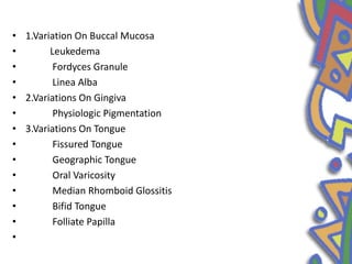
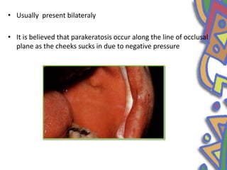
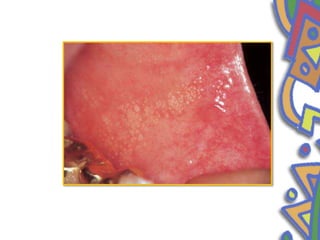
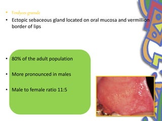
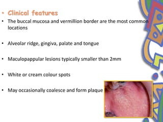
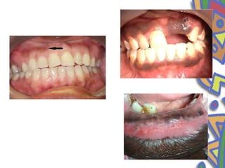
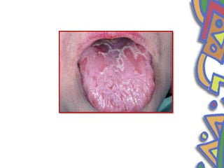
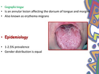
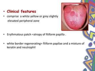
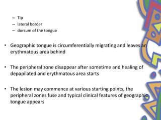
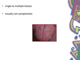
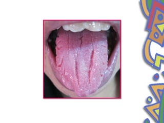
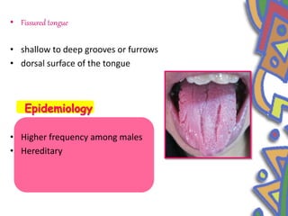
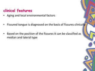
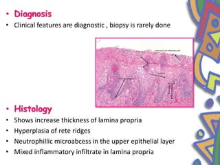
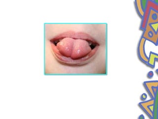
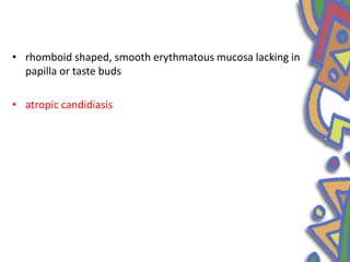
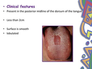
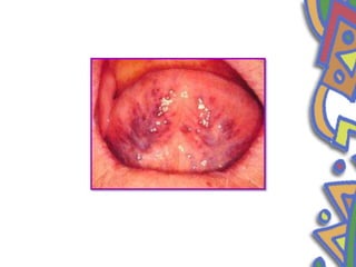
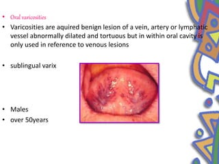
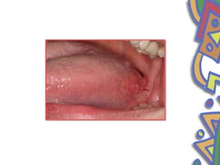
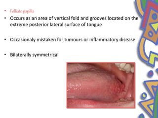
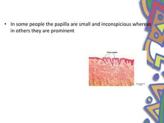
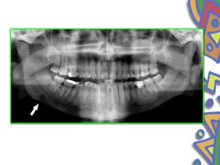
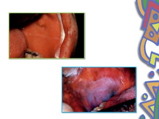
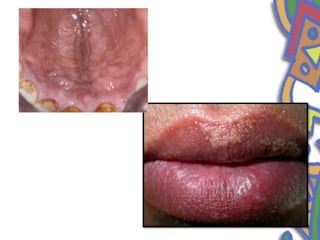
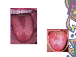
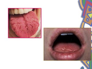
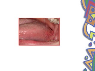
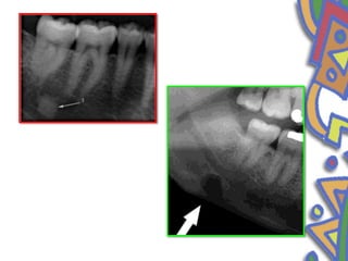
3. ā¢ Introduction
ā¢ Fundamental to diagnosing oral pathologic conditions is the
ability to recognize the spectrum of clinical findings that
represents variation of normal within the population.
ā¢ Some are purely developmental, while others have a clear
inflammatory or traumatic etiology.
4. ā¢ 1.Variation On Buccal Mucosa
ā¢ Leukedema
ā¢ Fordyces Granule
ā¢ Linea Alba
ā¢ 2.Variations On Gingiva
ā¢ Physiologic Pigmentation
ā¢ 3.Variations On Tongue
ā¢ Fissured Tongue
ā¢ Geographic Tongue
ā¢ Oral Varicosity
ā¢ Median Rhomboid Glossitis
ā¢ Bifid Tongue
ā¢ Folliate Papilla
ā¢
5. ā¢ 4.Variations On Lips
ā¢ Commissural Lip Pit
ā¢ Paramedian Lip Pit
ā¢ 5.Radiological Variations
ā¢ Idiopathic Osteosclerosis
ā¢ Focal Osteoporotic Bone Defect
ā¢ Stafne Bone Defect
6.
7. ā¢ Leukedema
ā¢ Common developmental alteration of the oral mucosa which
present most typically as an asymptomatic, bilateral, whitish, grey
semitransparent macule of the buccal mucosa
ā¢ Epidemology
ā¢ Distribution between genders has
been found to be equal
ā¢ The incidence and intensity
increases with age
8. ā¢ Clinical features
ā¢ White and wheel like alteration of oral mucosa
ā¢ bilaterally on buccal mucosa
ā¢ Less clinically evident after stretching of the mucosa but
reappear after this manipulation is discontinued
9. ā¢ Asymptomatic
ā¢ No malignant transformation
ā¢ Differential diagnosis
ā¢ Clinical examination readily differentiate leukedema from
leukoplakia since there is no loss of pliability or flexibilty of
involved tissue
ā¢ In addition tissue affected by leukedema manifest an
edematous state
12. ā¢ Linea alba
ā¢ Normal variations which appear as a white line extending from the
corner of the mouth to posterior region at the level of occlusal
plane bilaterally
ā¢ Frictional keratosis
ā¢ Trauma from facial surface of teeth.
13. ā¢ Usually present bilateraly
ā¢ It is believed that parakeratosis occur along the line of occlusal
plane as the cheeks sucks in due to negative pressure
14.
15. ā¢ Fordyces granule
ā¢ Ectopic sebaceous gland located on oral mucosa and vermillion
border of lips
ā¢ 80% of the adult population
ā¢ More pronounced in males
ā¢ Male to female ratio 11:5
16. ā¢ Clinical features
ā¢ The buccal mucosa and vermillion border are the most common
locations
ā¢ Alveolar ridge, gingiva, palate and tongue
ā¢ Maculopappular lesions typically smaller than 2mm
ā¢ White or cream colour spots
ā¢ May occasionally coalesce and form plaque
17. ā¢ Treatment
ā¢ Not required
ā¢ Chemical peel
ā¢ Laser
ā¢ Surgical diathermy or cryotherapy
18.
19. ā¢ Physiological pigmentation
ā¢ increased melanotic activity rather than an increase in number of
melanocytes
ā¢
ā¢ Africans, Asians and Mediterranean
ā¢ both gender
ā¢ 1st two decades
Epidemiology
Epidemiology
20. ā¢ Clinical features
ā¢ Attached gingiva
ā¢ light to dark brown
ā¢ It appears as bilateral, well demarcated, ribbon like dark brown
band
ā¢ The buccal mucosa, hard palate, lips and tongue may also be
affected
ā¢ symmetrically distributed
21.
22. ā¢ Disease that may be confused with melanin pigmentation are
ā¢ Addisons disease ,
ā¢ Oral melanotic macule.
ā¢ Oral mucosal melanoma,
ā¢ Drug induced pigmentation
23.
24. ā¢ Geographic tongue
ā¢ Is an annular lesion affecting the dorsum of tongue and margins
ā¢ Also known as erythema migrans
ā¢ Epidemiology
ā¢ 1-2.5% prevalence
ā¢ Gender distribution is equal
25. ā¢ Clinical features
ā¢ comprise a white yellow or grey slightly
elevated peripheral zone
ā¢ Erythmatous patch =atropy of filiform papilla .
ā¢ white border regenerating= filiform papilae and a mixture of
keratin and neutrophil
26. ā Tip
ā lateral border
ā dorsum of the tongue
ā¢ Geographic tongue is circumferentially migrating and leaves an
erythmatous area behind
ā¢ The peripheral zone disappear after sometime and healing of
depapilated and erythmatous area starts
ā¢ The lesion may commence at various starting points, the
peripheral zones fuse and typical clinical features of geographic
tongue appears
27. ā¢ single to multiple lesions
ā¢ Usually non symptomatic
28.
29. ā¢ Fissured tongue
ā¢ shallow to deep grooves or furrows
ā¢ dorsal surface of the tongue
Epidemiology
ā¢ Higher frequency among males
ā¢ Hereditary
30. clinical features
ā¢ Aging and local environmental factors
ā¢ Fissured tongue is diagnosed on the basis of fissures clinically
ā¢ Based on the position of the fissures it can be classified as
median and lateral type
31. ā¢ Diagnosis
ā¢ Clinical features are diagnostic , biopsy is rarely done
ā¢ Histology
ā¢ Shows increase thickness of lamina propria
ā¢ Hyperplasia of rete ridges
ā¢ Neutrophillic microabcess in the upper epithelial layer
ā¢ Mixed inflammatory infiltrate in lamina propria
32.
33. ā¢ Bifid tongue
ā¢ A completely cleft or bifid tongue is a rare condition
ā¢ A partially cleft tongue is considerably more common and is
manifested simply as a deep groove in the midline of dorsal
surface
34. ā¢ The lateral lingual structures rapidly grow and cover the
tuberculum impar to form the anterior two-thirds of the tongue.
When this process is disturbed, tip of the tongue is divided
longitudinally for a certain distance giving rise to cleft
tongue/bifid tongue.
ā¢ Mostly associated with orofacial digital syndrome
35.
36. ā¢ Median rhomboid glossitis
ā¢ Median rhomboid glossitis is clinically characterized by
erythmatous lesion in the center of posterior part of dorsum of
tongue
ā¢ Developmental defect resulting from an incomplete desent of
tuberculum impar and entrapment of a portion between
fusing lateral halves of the tongue
ā¢ 3:1 male predilection
37. ā¢ rhomboid shaped, smooth erythmatous mucosa lacking in
papilla or taste buds
ā¢ atropic candidiasis
38. ā¢ Clinical features
ā¢ Present in the posterior midline of the dorsum of the tongue
ā¢ Less than 2cm
ā¢ Surface is smooth
ā¢ lobulated
39. ā¢ kissing lesion
ā¢ Occasionally lesions are located somewhat anterior to usual
location. None have been located posterior to circumvallate
papilla
40.
41. ā¢ Oral varicosities
ā¢ Varicosities are aquired benign lesion of a vein, artery or lymphatic
vessel abnormally dilated and tortuous but in within oral cavity is
only used in reference to venous lesions
ā¢ sublingual varix
ā¢ Males
ā¢ over 50years
42. ā¢ Clinical features
ā¢ Irregular, blue purple lesion
ā¢ Multiple with a bilateral linear distribution
ā¢ Thrombosed varix
ā¢ Diascopy
43.
44. ā¢ Commisural lip pit
ā¢ congenital or developmental defects
ā¢ Their location suggest that they may represent a failure of normal
fusion of embryonal maxillary and mandibular process
45. ā¢ Unilateral or bilateral
ā¢ Clinical features
ā¢ Invagination is usually 1-2mm in diameter, may be as deep as
4mm and is lined by stratified squamous epithelium
46.
47. ā¢ Periapical idiopathic osteosclerosis
ā¢ Enostosis
ā¢ Dense bone island
ā¢ Localized growth of compact bone
ā¢ Asymptomatic
ā¢ vary in size from 2 mm to 1-2 cm.
48. ā¢ Mandible
ā¢ Premolar molar area
ā¢ Radiographic image- radiopaque
ā¢ Asymptomatic
ā¢ Etiology ā no
51. ā¢ Folliate papilla
ā¢ Occurs as an area of vertical fold and grooves located on the
extreme posterior lateral surface of tongue
ā¢ Occasionaly mistaken for tumours or inflammatory disease
ā¢ Bilaterally symmetrical
52. ā¢ In some people the papilla are small and inconspicious whereas
in others they are prominent
53.
54. ā¢ Stafne bone defect
ā¢ First described by Stafne in 1942
ā¢ The exact pathogenesis is still obscure.
ā¢ Epidemiology
ā¢ Men in their
ā¢ ļ¬fth or seventh decade of life
55. ā¢ Stafne suggested that the cavity could result from a failure of
normal bone deposition in the region formerly occupied by
cartilage .
ā¢ localized pressure atrophy of the lingual surface of the
mandible from the adjacent salivary gland
56. ā¢ Clinical features
ā¢ Present as asymptomatic radiolucency below the mandibular canal in
the posterior mandible between the molar and angle of mandible
ā¢ well circumscribed
ā¢ sclerotic border
57. ā¢ Superimposed over the apices of anterior teeth
ā¢ Stable in size
ā¢ 1 to 3cm in diameter
ā¢ below the inferior dental canal
ā¢ Incidental
ā¢ d/d
58.
59.
60.
61.
62.
63.
64. REFERENCES
1. Burket Oral Medicine: diagnosis and treatment 12th edition
2. Oral Radiology :Principle and nterpretation White and
Pharoah
3. Shafers Textbook of Oral Pathology
4. Oral diseases ā Roderich A Cawson, William H Binniew, John
H Wright
5. Oral Radiology :Principle and Interpretation White and
Pharoah