Lateral skull base anatomy and applied science by Dr, bomkar bamBomkar Bam
the lateral skull base is complex anatomy that is usually students finds difficult to understand. here concise literature is made to understand the skull base more easily.
this prsentation incluses HRCT temportal bone cross sectional anatomy images axial saggital and coronal with labelled diagram. This presentation help alot for radiology resident. Thanks.
Anatomy of the Temporal region & Temporomandibular jointRafid Rashid
Provides a detailed description of the gross anatomy of the temporal fossa, infratemporal fossa & temporomandibular joint. The boundaries & the structures present in the temporal & infratemporal fossa, the formation & movements of the TMJ & also includes branches of the mandibular nerve & maxillary artery.
This presentation deals with the inside of the skull (cranial cavity) and description of some separate bones. There is another presentation “Skull - the normas” that describes norma verticalis, occipitalis, lateralis, frontalis and basalis and is necessary to complete the objectives.
Objectives
Identify the features of the major bones forming the cranial cavity according to normas and separate bones.
Describe the major sutures.
Describe the structure of the flat bones forming the skull and their blood supply.
Discuss ossification of the skull and the changes that occur during postnatal development.
Locate important bony surface landmarks.
Similar to norma b extrnaالثالثه تشريح راس.pptx (20)
Professional air quality monitoring systems provide immediate, on-site data for analysis, compliance, and decision-making.
Monitor common gases, weather parameters, particulates.
Cancer cell metabolism: special Reference to Lactate PathwayAADYARAJPANDEY1
Normal Cell Metabolism:
Cellular respiration describes the series of steps that cells use to break down sugar and other chemicals to get the energy we need to function.
Energy is stored in the bonds of glucose and when glucose is broken down, much of that energy is released.
Cell utilize energy in the form of ATP.
The first step of respiration is called glycolysis. In a series of steps, glycolysis breaks glucose into two smaller molecules - a chemical called pyruvate. A small amount of ATP is formed during this process.
Most healthy cells continue the breakdown in a second process, called the Kreb's cycle. The Kreb's cycle allows cells to “burn” the pyruvates made in glycolysis to get more ATP.
The last step in the breakdown of glucose is called oxidative phosphorylation (Ox-Phos).
It takes place in specialized cell structures called mitochondria. This process produces a large amount of ATP. Importantly, cells need oxygen to complete oxidative phosphorylation.
If a cell completes only glycolysis, only 2 molecules of ATP are made per glucose. However, if the cell completes the entire respiration process (glycolysis - Kreb's - oxidative phosphorylation), about 36 molecules of ATP are created, giving it much more energy to use.
IN CANCER CELL:
Unlike healthy cells that "burn" the entire molecule of sugar to capture a large amount of energy as ATP, cancer cells are wasteful.
Cancer cells only partially break down sugar molecules. They overuse the first step of respiration, glycolysis. They frequently do not complete the second step, oxidative phosphorylation.
This results in only 2 molecules of ATP per each glucose molecule instead of the 36 or so ATPs healthy cells gain. As a result, cancer cells need to use a lot more sugar molecules to get enough energy to survive.
introduction to WARBERG PHENOMENA:
WARBURG EFFECT Usually, cancer cells are highly glycolytic (glucose addiction) and take up more glucose than do normal cells from outside.
Otto Heinrich Warburg (; 8 October 1883 – 1 August 1970) In 1931 was awarded the Nobel Prize in Physiology for his "discovery of the nature and mode of action of the respiratory enzyme.
WARNBURG EFFECT : cancer cells under aerobic (well-oxygenated) conditions to metabolize glucose to lactate (aerobic glycolysis) is known as the Warburg effect. Warburg made the observation that tumor slices consume glucose and secrete lactate at a higher rate than normal tissues.
Slide 1: Title Slide
Extrachromosomal Inheritance
Slide 2: Introduction to Extrachromosomal Inheritance
Definition: Extrachromosomal inheritance refers to the transmission of genetic material that is not found within the nucleus.
Key Components: Involves genes located in mitochondria, chloroplasts, and plasmids.
Slide 3: Mitochondrial Inheritance
Mitochondria: Organelles responsible for energy production.
Mitochondrial DNA (mtDNA): Circular DNA molecule found in mitochondria.
Inheritance Pattern: Maternally inherited, meaning it is passed from mothers to all their offspring.
Diseases: Examples include Leber’s hereditary optic neuropathy (LHON) and mitochondrial myopathy.
Slide 4: Chloroplast Inheritance
Chloroplasts: Organelles responsible for photosynthesis in plants.
Chloroplast DNA (cpDNA): Circular DNA molecule found in chloroplasts.
Inheritance Pattern: Often maternally inherited in most plants, but can vary in some species.
Examples: Variegation in plants, where leaf color patterns are determined by chloroplast DNA.
Slide 5: Plasmid Inheritance
Plasmids: Small, circular DNA molecules found in bacteria and some eukaryotes.
Features: Can carry antibiotic resistance genes and can be transferred between cells through processes like conjugation.
Significance: Important in biotechnology for gene cloning and genetic engineering.
Slide 6: Mechanisms of Extrachromosomal Inheritance
Non-Mendelian Patterns: Do not follow Mendel’s laws of inheritance.
Cytoplasmic Segregation: During cell division, organelles like mitochondria and chloroplasts are randomly distributed to daughter cells.
Heteroplasmy: Presence of more than one type of organellar genome within a cell, leading to variation in expression.
Slide 7: Examples of Extrachromosomal Inheritance
Four O’clock Plant (Mirabilis jalapa): Shows variegated leaves due to different cpDNA in leaf cells.
Petite Mutants in Yeast: Result from mutations in mitochondrial DNA affecting respiration.
Slide 8: Importance of Extrachromosomal Inheritance
Evolution: Provides insight into the evolution of eukaryotic cells.
Medicine: Understanding mitochondrial inheritance helps in diagnosing and treating mitochondrial diseases.
Agriculture: Chloroplast inheritance can be used in plant breeding and genetic modification.
Slide 9: Recent Research and Advances
Gene Editing: Techniques like CRISPR-Cas9 are being used to edit mitochondrial and chloroplast DNA.
Therapies: Development of mitochondrial replacement therapy (MRT) for preventing mitochondrial diseases.
Slide 10: Conclusion
Summary: Extrachromosomal inheritance involves the transmission of genetic material outside the nucleus and plays a crucial role in genetics, medicine, and biotechnology.
Future Directions: Continued research and technological advancements hold promise for new treatments and applications.
Slide 11: Questions and Discussion
Invite Audience: Open the floor for any questions or further discussion on the topic.
Earliest Galaxies in the JADES Origins Field: Luminosity Function and Cosmic ...Sérgio Sacani
We characterize the earliest galaxy population in the JADES Origins Field (JOF), the deepest
imaging field observed with JWST. We make use of the ancillary Hubble optical images (5 filters
spanning 0.4−0.9µm) and novel JWST images with 14 filters spanning 0.8−5µm, including 7 mediumband filters, and reaching total exposure times of up to 46 hours per filter. We combine all our data
at > 2.3µm to construct an ultradeep image, reaching as deep as ≈ 31.4 AB mag in the stack and
30.3-31.0 AB mag (5σ, r = 0.1” circular aperture) in individual filters. We measure photometric
redshifts and use robust selection criteria to identify a sample of eight galaxy candidates at redshifts
z = 11.5 − 15. These objects show compact half-light radii of R1/2 ∼ 50 − 200pc, stellar masses of
M⋆ ∼ 107−108M⊙, and star-formation rates of SFR ∼ 0.1−1 M⊙ yr−1
. Our search finds no candidates
at 15 < z < 20, placing upper limits at these redshifts. We develop a forward modeling approach to
infer the properties of the evolving luminosity function without binning in redshift or luminosity that
marginalizes over the photometric redshift uncertainty of our candidate galaxies and incorporates the
impact of non-detections. We find a z = 12 luminosity function in good agreement with prior results,
and that the luminosity function normalization and UV luminosity density decline by a factor of ∼ 2.5
from z = 12 to z = 14. We discuss the possible implications of our results in the context of theoretical
models for evolution of the dark matter halo mass function.
insect taxonomy importance systematics and classification
norma b extrnaالثالثه تشريح راس.pptx
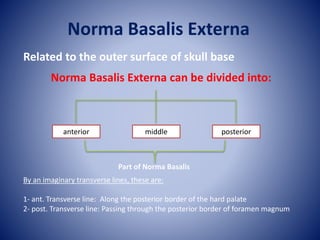
1. Norma Basalis Externa
Related to the outer surface of skull base
Norma Basalis Externa can be divided into:
anterior middle posterior
By an imaginary transverse lines, these are:
1- ant. Transverse line: Along the posterior border of the hard palate
2- post. Transverse line: Passing through the posterior border of foramen magnum
Part of Norma Basalis
2. Anterior part of Norma Basalis Externa
middle part of Norma Basalis Externa
posterior part of Norma Basalis Externa
Ant T. line
post T. line
3. Anterior part of Norma Basalis Externa
Formed by hard (bony) palate which is bounded
within the Alveolar arch carrying the sockets for
the roots of upper teeth
it is divided by the median palatine suture into
right and left halves.
each half is formed by two parts:
1- Ant. ¾ formed by palatine pro. Of maxilla.
2- Post. ¼ formed by the horizontal plate of the
palatine bone.
These two parts unite at the palatomaxillary suture.
palatine
pro.
Of
maxilla
palatine bone
To be continued
Ant. ¾
Post. ¼
4. Particular features
Alveolar arch: carries the sockets for the upper 16 teeth
posterior free border of the hard palate: Is sharp and give attachment to the palatine
aponeurosis of the soft palate.
Post. Nasal spine: is a sharp median projection of the posterior border of the hard
palate, it gives origin to a muscle of the soft palate called musculus uvulae.
Palatine crest: a transverse ridge behind the lateral part of the palatomaxillary suture
opposite to the last molar tooth.
Maxillary tuberosity: lies at the posterior end of the alveolar arch of maxilla it gives
origin to the superfacial head of the med. Pterygoid m.
To be continued
5. Hard palate
Greater palatine foramen
lesser palatine foramina
Stensen and scarpa foramina within incisive fossa
Maxillary tuberosity
Palatine
crest
Posterior nasal spine
6. Foramina in the anterior part of the base:
A- incisive fossa: at the anterior part of the intermaxillary suture behind the
incisors.
It contain small foramina:
2 median (ant& Post.) transmitting Lt&Rt long
sphenopalatine nerve.
2 lateral (Lt&Rt) transmitting the terminal branches
of Lt&Rt greater palatine nerve and vesseles.
B- Greater palatine foramen:
Lies med to the last molar socket, infront of the palatine crest, it is the lower end
of the greater palatine canal. It transmits the greater palatine nerve and vessels
through a groove on the bony palate supplying its mucous membrane.
C- Lesser palatine foramina (usually 2): lie on the pyramidal process of palatine
bone behind the palatine crest. They transmit lesser palatine n & vessels.
8. Middle part of Norma Basalis Externa
Bones forming it:
Anteriorly in the middle vomer
the body of sphenoid
Anteriorly (sphenoid bone): pterygoid process
infratemporal surface of greater wing
Posteriorly: petrous part of temporal bone
tympanic part of temporal bone
mastoid part of temporal bone
Posteriorly in the middle: Basilar part of the occipital bone
2 lateral part of the occipital bone.
To be continued
9. 2 lateral parts of
occipital bone
Vomer
Body of the
sphenoid
Pteryoid process
Lateral
Medial
Petrous part of
temporal bone
Tympanic part of
temporal bone
Mastoid part of
temporal bone
Basilar part of
occipital bone
10. Particular features
Posterior nasal openings (choanae): Separated from each other by the vomer
Post. Nasal spine
choana
Ala of Vomer
vaginal process of med. Pterygoid plate
vomero-vaginal
canal
palato-
vaginal canal
Vomer
To be continued
The vomer:
- median vertical bony plate.
- the ala of the vomer is the upper extended part, articulating with the body
of the sphenoid.
- lateral to the ala of the there is the vaginal process of med. Pterygoid plate
which is separated from the ala of vomer by the vomero-vaginal canal.
11. Pterygoid process of sphenoid: (lateral to choana)
- anteriorly it is separated from maxilla by the pterygomaxillary fissure.
- posteriorly it presents med& lat. Pterygoid plates separated by ptergoid
fossa
A- lateral pterygoid plateL:
- it forms the lateral boundary of the infratemporal fossa.
- its lat. Surface gives origin to lower head of lat. Pterygoid muscle.
- its med. Surface gives origin to deep head of med. Pterygoid muscle.
B- medial pterygoid plate:
- it forms the lateral boundary of the post. Nasal opening.
- its post. Border gives attachment to the pharyngeo-basilar fascia & is
related to the pharyngeo-tympanic tube in the upper part.
the upper end of the posterior border divided into scaphoid fossa lat.
pterygoid tubercle medially.
medial pterygoid plate:
lateral Pterygoid plate:
12. the lower end of the posterior border ----- pterygoid hamulus (hook)
Pterygoid fossa:
V- shape space bet. Med. & lat. Pterygoid plate.
Infra temporal surface of greater wing of sphenoid:
shows
1- spine of sphenoid
2- foramen ovale
3- foramen spinosum
1
2
3
13. Petrous part of temporal bone:
bet. Greater wing of sphenoid and basilar part of occipital bone.
It shows:
1- foramen lacerum
2- a rough quadrate area
3- carotid canal
4- Jujular foramen.
Basilar part of occipital bone:
articulateanteriorly with body of sphenoid
Pharyngeal tubercle: median elevation in the basilar part of occipital bone.
14. (NJF – normal jugular fossa; BJF – blocked jugular fossa; BPO – basilar part of the
occipital bone; PT- petrous part of the temporal; MF – mandibular foramen)
1- foramen lacerum
2- a rough quadrate area
3- carotid canal
4- Jugular foramen.
1
2
3
4
15. the styloid and mastoid parts of temporal bone:
Styloid process: lat. To jugular f. & infront of mastoid process
mastoid process: behind the Styloid process
mastoid notch: medial to mastoid process
occipital groove: medial to notch --- occipital artery
Stylomastoid foramen: bet. Styloid & mastoid processes
styloid process
occipital groove
Stylomastoid foramen
mastoid foramen
Mastoid process
16. Articular surfaces of Norma basalis externa:
a- mandibular fossa: concave depression in the squamous part of temporal
bone- articulate with the head of mandible in the TMJ.
b- articular eminence: an elevation infront of mandibular fossa.
c- occipital condyles: 2 kidney- shaped articular facets situated on each side of
the anterior part of foramen magnum.
* Tympanic plate of temporal bone: behind the articular fossa
mandibular fossa
articular eminence
occipital condyles
17. foramina related to occipital condyles:
1- foramen magnum: largest foramen of the skull- ovale in shape
2- ant. Condylar foramen lies antero-superior to the occipital condyle on
( hypoglossal canal): each side.
3- Condylar fossa: a depression behind the occipital condyle – may
be perforated (post. Condylar foramen).
ant. Condylar foramen 21
foramen magnum
Condylar fossa
21
21
18. Inferior view of the left side of the cranial base. Insertions of the styloid muscles at the styloid
process are shown. The arrow indicates the inferior tympanic canaliculus, and the star indicates
the fossa of the mandibular condyle. CC = carotid canal; DG = digastric groove; FL = foramen
lacerum; FO = foramen ovale; FS = foramen spinosum; JF = jugular foramen; OC = occipital
condyle; SF = stylomastoid foramen.
19. 1. Anterior Palatine Foramen
2. Palatine Process of Maxilla
3. Palatine
4. Greater Palatine Foramen
5. Lesser Palatine Foramen
6. Pterygoid Processes of Sphenoid
7. Zygomatic Process
8. Squamous Part of Temporal Bone
9. Mandibular Fossa
10. Styloid Process
11. Stylomastoid Foramen
12. Mastoid Process
13. Mastoid Foramen
14. Superior Nuchal Line
15. External Occipital Protruberance
16. Median Nuchal Line
17. Inferior Nuchal Line
18. Foramen Magnum
19. Condyloid Canal
20. Occipital Condyle
21. Hypoglossal Canal
22. Jugular Foramen
23. Carotid Canal
24. Foramen Spinosum
25. Foramen Ovale
26. Foramen Lacerum
27. Vomer
28. Transverse Palatine Suture
29. Median Palatine Suture
20. posterior part of Norma Basalis Externa
Shows the following features:
1- external occipital protuberance
2- external occipital crest
3- sup. Nuchal line
4- inf. Nuchal line.