Introduction to Niosomes
•Definition: Niosomes are non-ionic surfactant-based vesicles that are used in
drug delivery. They are similar to liposomes but are made from non-ionic
surfactants rather than phospholipids.
• Structure: Niosomes are composed of surfactants, cholesterol, and possibly
other components, forming a bilayer vesicle structure.
• Applications: Niosomes are used in drug delivery systems, gene therapy,
vaccines, and cosmetic formulations.
3.
Advantages of Niosomes
•Cost-Effective: Non-ionic surfactants are often less expensive than
phospholipids, making niosomes a more cost-effective alternative to
liposomes.
• Stability: Niosomes can offer better stability in terms of temperature and pH
compared to liposomes.
• Controlled Release: They provide controlled release of encapsulated drugs,
improving the therapeutic effect.
• Improved Bioavailability: They enhance the bioavailability of poorly soluble
drugs.
4.
Types of Niosomes
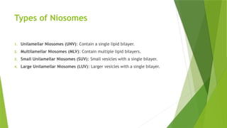
1.Unilamellar Niosomes (UNV): Contain a single lipid bilayer.
2. Multilamellar Niosomes (MLV): Contain multiple lipid bilayers.
3. Small Unilamellar Niosomes (SUV): Small vesicles with a single bilayer.
4. Large Unilamellar Niosomes (LUV): Larger vesicles with a single bilayer.
5.
Composition of Niosomes
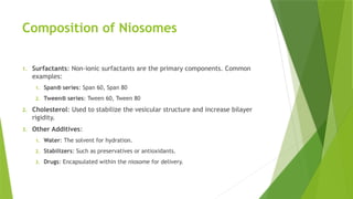
1.Surfactants: Non-ionic surfactants are the primary components. Common
examples:
1. Span® series: Span 60, Span 80
2. Tween® series: Tween 60, Tween 80
2. Cholesterol: Used to stabilize the vesicular structure and increase bilayer
rigidity.
3. Other Additives:
1. Water: The solvent for hydration.
2. Stabilizers: Such as preservatives or antioxidants.
3. Drugs: Encapsulated within the niosome for delivery.
6.
Manufacturing Techniques forNiosomes
1. Ether Injection Method:
1. A solution of surfactants and cholesterol is injected into water under controlled
conditions, forming niosomes.
2. Advantages: Simple and efficient.
2. Thin Film Hydration Method:
1. Surfactants and cholesterol are dissolved in a solvent and evaporated to form a thin
film. The film is then hydrated with aqueous media to form niosomes.
2. Advantages: High encapsulation efficiency.
3. Reverse Phase Evaporation Method:
1. This method involves creating an emulsion between an aqueous phase and an organic
solvent, followed by evaporation.
2. Advantages: Suitable for encapsulating hydrophilic and lipophilic drugs.
7.
1. Proliposome Method:
1.Dry surfactant and cholesterol are hydrated with an aqueous phase under
controlled conditions to form niosomes.
2. Advantages: Increased stability and encapsulation efficiency.
2. Sonication:
1. Sonication uses ultrasonic waves to break down large multilamellar vesicles into
smaller unilamellar ones.
2. Advantages: Good control over vesicle size.
8.
Factors Affecting NiosomeFormulation
1. Surfactant Type:
1. The choice of surfactant influences the size, stability, and drug encapsulation
efficiency.
2. Cholesterol Content:
1. The amount of cholesterol affects the rigidity and stability of the niosome membrane.
3. Drug Properties:
1. Hydrophilic or lipophilic drugs require different formulation strategies for effective
encapsulation.
4. Solvent and Hydration Conditions:
1. The solvent used for film formation and the hydration temperature impact the size
and encapsulation efficiency of niosomes.
9.
Evaluation of Niosomes
1.Physical Appearance:
1. Niosomes should be clear or slightly opalescent suspensions, with no visible
aggregates or particles.
2. Size and Zeta Potential:
1. Dynamic Light Scattering (DLS): Measures the size distribution of niosomes.
2. Zeta Potential: Assesses the surface charge of niosomes, which influences their
stability and interaction with biological membranes.
3. Encapsulation Efficiency:
1. The percentage of drug encapsulated in niosomes is evaluated by separating the
free drug from the encapsulated drug (usually using ultracentrifugation or dialysis).
10.
•Shape and Morphology:
•TransmissionElectron Microscopy (TEM) or Scanning Electron Microscopy
(SEM): Used to observe the shape, structure, and surface morphology of
niosomes.
•Drug Release Studies:
•In vitro Release Testing: Measures how the drug is released from niosomes
over time using techniques like dialysis or Franz diffusion cells.
•Release kinetics are often analyzed to determine if the release follows zero-
order, first-order, or Higuchi models.
•Stability Studies:
•Evaluate niosome stability under various storage conditions (e.g., temperature,
pH, and light exposure). Stability is important for ensuring that the niosomes
maintain their size, drug encapsulation, and drug release characteristics over
time.
11.
In Vivo Evaluation
1.Biodistribution:
1. Using radiolabeled or fluorescently labeled niosomes, researchers can track the
distribution of niosomes in vivo.
2. Pharmacokinetics:
1. Blood samples are taken at various intervals to determine how long the niosomes
stay in circulation and their concentration in different tissues.
3. Toxicity Studies:
1. Animal models are used to assess the safety and potential toxicity of niosomes.
12.
Applications of Niosomes
1.Drug Delivery:
1. Niosomes can encapsulate both hydrophobic and hydrophilic drugs, enhancing bioavailability
and providing controlled release.
2. Example: Delivery of anticancer drugs, antifungal agents, and anti-inflammatory drugs.
2. Gene Delivery:
1. Niosomes can be used as carriers for DNA, RNA, and other genetic materials, improving the
delivery and uptake of genes into target cells.
3. Vaccine Delivery:
1. Niosomes serve as adjuvants or carriers for vaccines, enhancing immune response and stability.
4. Cosmetic and Dermatological Applications:
1. Niosomes are used in skin care products to deliver active ingredients like vitamins,
antioxidants, and anti-aging compounds.
13.
Challenges in NiosomeDevelopment
• Scalability: Large-scale manufacturing of niosomes while maintaining
consistency and quality can be difficult.
• Stability: Niosomes may face challenges in maintaining stability over time,
particularly under varying storage conditions.
• Toxicity: Long-term exposure to non-ionic surfactants may induce toxicity or
irritation in some cases.
• Controlled Release: Achieving precise control over drug release rates in vivo
is challenging.
14.
Recent Advances inNiosome Research
• Targeted Niosomes: Niosomes functionalized with targeting ligands (e.g.,
antibodies, peptides) for specific cell targeting, such as cancer cells.
• Stimuli-Responsive Niosomes: Niosomes that release their payload in
response to specific stimuli (e.g., pH, temperature, enzymes).
• Niosome-Polymer Conjugates: Hybrid systems combining niosomes and
polymers for enhanced stability and targeted delivery.
15.
Conclusion
• Summary:
• Niosomesare versatile and cost-effective drug delivery carriers that can be used to
improve the bioavailability and controlled release of drugs.
• They have potential applications in a wide range of fields, including drug delivery,
gene therapy, vaccines, and cosmetics.
• Future Directions:
• Research is focused on improving the stability, scalability, and targeting abilities of
niosomes, making them an increasingly attractive option for therapeutic
applications.


![Hypothalamus short ppt by Dr. Neha [PT].pptx](https://cdn.slidesharecdn.com/ss_thumbnails/hypothalamusbydr-260124145759-b9f94a93-thumbnail.jpg?width=640&height=640&fit=bounds)


