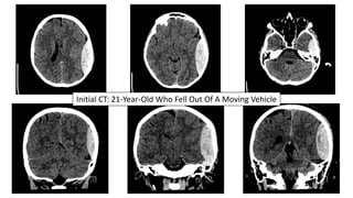
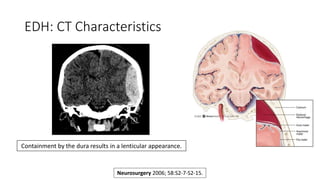
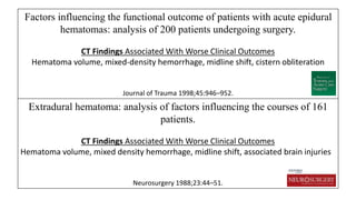
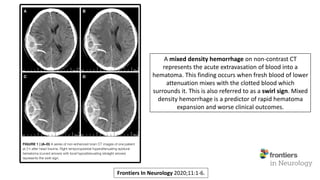
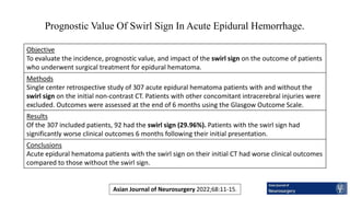
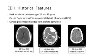
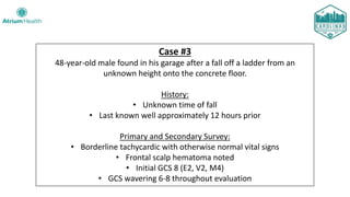
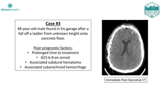
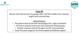
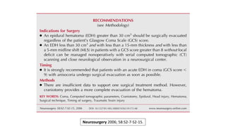
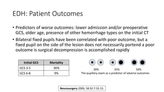
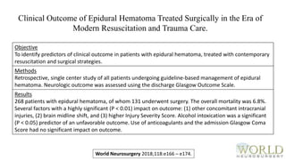
Acute Epidural Hematomas
Madison Watts, MD, Faith Meyers, MD, Steven Perry, MD
Carolinas Medical Center & Levine Children’s Hospital
Department of Emergency Medicine
Charlotte Neurosurgery & Spine Associates
Michael Gibbs, MD, Imaging Mastery Project Lead Editor
Neuroimaging Mastery Project
Presentation #4
Disclosures
 This ongoing series is proudly sponsored by the Emergency Medicine and
Neurosurgery Residency Programs at Carolinas Medical Center.
 The goal is to promote widespread mastery of imaging interpretation.
 There is no personal health information [PHI] within, and all ages have
been changed to protect patient confidentiality.
Meet Our Neuroimaging Editorial Team
Andrew Asimos, MD, FACEP
Medical Director, Atrium Health Stroke Network
Neurosciences Institute
Clinical Professor, Department of Emergency Medicine
Jonathan Clemente, MD, FACR
Chief, Department of Radiology, Carolinas Medical Center
Charlotte Radiology, Neuroradiology Section
Adjunct Clinical Associate Professor, Department of Radiology
Andrew Perron, MD, FACEP
Associate Dean of Graduate Medical Education and DIO
Professor of Emergency Medicine
Department of Graduate Medical Education
Dartmouth Hitchcock Medical Center
Meet Our Neuroimaging Editorial Team
Christa Swisher, MD, FNCS, FACNS
Neurocritical Care/Pulmonary Critical Care Consultants
Department of Medicine, Atrium Health
Clinical Assistant Professor, Department of Neurology
Scott Wait, MD, FAANS
Chief, Pediatric Neurosurgery, Levine Children’s Hospital
Carolina Neurosurgery & Spine Associates
Adjunct Clinical Associate Professor, Department of Neurosurgery
Visit Our Website
www.EMGuidewire.com
For A Complete Archive Of Imaging Presentations And Much More!
Epidural Hematoma (EDH)
Cases From Carolinas Medical Center
Case #1
21-year-old female brought into the ED after she fell out of an open-door
Jeep traveling at approximately 10 miles per hour.
History:
• Unknown loss of consciousness
• Friends say she “wasn’t acting right”
• Vomiting before arriving
• The patient is also clinically intoxicated
Primary and Secondary Survey:
• Hemodynamically stable
• GCS 13 (E3, V4, M6)
• Interacting appropriately and following commands
Initial CT: 21-Year-Old Who Fell Out Of A Moving Vehicle
Case #1
21-year-old female brought into the ED after she fell out of an open-door
Jeep traveling at approximately 10 miles per hour.
Clinical Course:
• Intubated with immediate pre-intubation GCS of 13
• Transferred to our Trauma Center with the neurosurgical team awaiting
• Underwent emergent left craniotomy
• Extubated one day after surgery and found to be neurologically intact
• Discharged home on Post-Operative Day #2
21-Year-Old Who Fell Out Of A Moving Vehicle
Left Temporal EDH CT On Post-Operative Day #2
EDH: Pathophysiology
Classic teaching is that epidural hematomas result from blunt
force trauma to the temporal regional along the distribution
of the middle meningeal artery and its branches.
Neurosurgery 2006; 58:S2-7-S2-15.
EDH: Pathophysiology
• An arterial source of bleeding is
identified in only about 1/3 of
adults with epidural hematoma
• EDHs may also be caused by
injuries to middle meningeal
vein, diploic veins or venous
sinuses. These are especially
likely in children.
Neurosurgery 2006; 58:S2-7-S2-15.
EDH: CT Characteristics
Containment by the dura results in a lenticular appearance.
Neurosurgery 2006; 58:S2-7-S2-15.
EDH: CT Characteristics
Coronal Suture
Middle Meningeal Artery
Fracture of the Squamosal Portion of the Left temporal Bone
Brain Window Bone Window
Factors influencing the functional outcome of patients with acute epidural
hematomas: analysis of 200 patients undergoing surgery.
CT Findings Associated With Worse Clinical Outcomes
Hematoma volume, mixed-density hemorrhage, midline shift, cistern obliteration
Journal of Trauma 1998;45:946–952.
Extradural hematoma: analysis of factors influencing the courses of 161
patients.
CT Findings Associated With Worse Clinical Outcomes
Hematoma volume, mixed density hemorrhage, midline shift, associated brain injuries
Neurosurgery 1988;23:44–51.
Frontiers In Neurology 2020;11:1-6.
A mixed density hemorrhage on non-contrast CT
represents the acute extravasation of blood into a
hematoma. This finding occurs when fresh blood of lower
attenuation mixes with the clotted blood which
surrounds it. This is also referred to as a swirl sign. Mixed
density hemorrhage is a predictor of rapid hematoma
expansion and worse clinical outcomes.
Asian Journal of Neurosurgery 2022;68:11-15.
Prognostic Value Of Swirl Sign In Acute Epidural Hemorrhage.
Objective
To evaluate the incidence, prognostic value, and impact of the swirl sign on the outcome of patients
who underwent surgical treatment for epidural hematoma.
Methods
Single center retrospective study of 307 acute epidural hematoma patients with and without the
swirl sign on the initial non-contrast CT. Patients with other concomitant intracerebral injuries were
excluded. Outcomes were assessed at the end of 6 months using the Glasgow Outcome Scale.
Results
Of the 307 included patients, 92 had the swirl sign (29.96%). Patients with the swirl sign had
significantly worse clinical outcomes 6 months following their initial presentation.
Conclusions
Acute epidural hematoma patients with the swirl sign on their initial CT had worse clinical outcomes
compared to those without the swirl sign.
Case #2
15-year-old male brought into the ED by his parents after he sustained an
un-helmeted fall off of his skateboard.
History:
• Somnolent and confused
• Brief seizure as parents were putting him in the car to come to the ED
• Additional seizure in route with EMS, now combative and post-ictal
Primary and Secondary Survey:
• Tachycardic with normal blood pressure and oxygen saturations
• Scalp hematoma on physical exam
• Initial GCS 12 (E4, V3, M5)... declined to GCS 9 (E2, V3, M4)
• Patient seized again in the ED and was promptly intubated
15-Year-Old Struck His Head Skateboarding
Case #2
15-year-old male brought into the ED by his parents after un-helmeted fall
off skateboard.
Clinical Course:
• Taken to Operating Room with neurosurgery for craniotomy
• Uncomplicated post-operative course
• Discharged on post-operative Day #3 with a prescription for levetiracetam
• Seen by pediatric neurology at 1.5 months, no further seizures
• Plan for discontinuation of antiepileptics if a 6-month EEG is negative
• Unfortunately, the patient was lost to follow up
EDH: Historical Features
• Peak incidence between ages 20 and 30 years
• Classic “lucid interval” in approximately half of patients (47%)
• Clinical presentation ranges from alert to comatose
38-Year-Old
Assaulted With A Bat
40-Year-Old
Fall With Head Strike
66-Year-Old
Pedestrian Struck
Case #3
48-year-old male found in his garage after a fall off a ladder from an
unknown height onto the concrete floor.
History:
• Unknown time of fall
• Last known well approximately 12 hours prior
Primary and Secondary Survey:
• Borderline tachycardic with otherwise normal vital signs
• Frontal scalp hematoma noted
• Initial GCS 8 (E2, V2, M4)
• GCS wavering 6-8 throughout evaluation
Initial CT: 48-Year-Old Fall Off A
Ladder
Several Finding On CT
• Large right epidural hematoma with midline shift
• Left subdural hematoma as a countrecoup injury
• Multiple foci of intraparenchymal hemorrhage
• Trace subarachnoid hemorrhage
*
*
Case #3
48-year-old male found in his garage after a
fall off a ladder from unknown height onto
concrete floor.
Poor prognostic factors:
• Prolonged time to treatment
• GCS 6-8 on arrival
• Associated subdural hematoma
• Associated subarachnoid hemorrhage
Immediate Post-Operative CT
Case #3
48-year-old male found in his garage after a fall off a ladder from unknown
height onto concrete floor.
Clinical Course:
• The patient went to OR with neurosurgery for a right craniotomy
• He had an external ventricular drain placed the following day
• Despite maximal medical therapies his ICPs remained high
• Given the poor prognosis the family opted to withdraw support
Neurosurgery 2006; 58:S2-7-S2-15.
Patient Population
350 patients (182 adults/168 children) with isolated epidural hematomas:
• 207 underwent immediate surgical evacuation
• 143 were observed with serial CT scans and neurologic examinations
Indications For Surgery
Adult patients
Pediatric patients
Patients initially observed
• EDH volume >30 ml
• EDH volume >20 ml
• EDH expansion and/or clinical deterioration within 24 hours
Complication Rates
• 6.7% of those who underwent immediate surgery had rebleeding requiring return to the OR
• 11% of those who were initially observed ultimately required delayed surgical intervention
World Neurosurgery 2023; 177:e686-e692.
Conservative vs. Surgical Management of Post- Traumatic Epidural
Hematoma: A Case and Review of Literature.
Glasgow Outcome Scale
5
Good outcome
(Fully independent
with limited deficits)
1
Death
3
Severe disability
(Need assistance with
tasks of daily living)
4
Moderate disability
(Independent with daily living,
may be able to work with accommodations)
2
Vegetative
state
Conservative vs. Surgical Management of Post- Traumatic Epidural
Hematoma: A Case and Review of Literature.
World Neurosurgery 2023; 177:e686-e692.
A Case of Conservatively Managed EDH
Admissions 6 hours
20 days
5 days
24 hours
American Journal of Case Reports 2015; 16: 811-817.
EDH: Patient Outcomes
• With prompt surgical
evacuation, patients with
epidural hematomas have
better outcomes when
compared with other
significant brain injury types
• 10% mortality in all comers
undergoing surgical
intervention
Neurosurgery 2006; 58:S2-7-S2-15.
Epidural Hematoma
Better Outcomes
Subdural Hematoma
Worse Outcomes
EDH: Patient Outcomes
• Predictors of worse outcomes: lower admission and/or preoperative
GCS, older age, presence of other hemorrhage types on the initial CT
• Bilateral fixed pupils have been correlated with poor outcome, but a
fixed pupil on the side of the lesion does not necessarily portend a poor
outcome is surgical decompression is accomplished rapidly
Initial GCS Mortality
GCS 3-5 36%
GCS 6-8 9%
30% 35% 50%
The pupillary exam as a predictor of adverse outcomes
Neurosurgery 2006; 58:S2-7-S2-15.
Objective
To identify predictors of clinical outcome in patients with epidural hematoma, treated with contemporary
resuscitation and surgical strategies.
Methods
Retrospective, single center study of all patients undergoing guideline-based management of epidural
hematoma. Neurologic outcome was assessed using the discharge Glasgow Outcome Scale.
Results
268 patients with epidural hematoma, of whom 131 underwent surgery. The overall mortality was 6.8%.
Several factors with a highly significant (P < 0.01) impact on outcome: (1) other concomitant intracranial
injuries, (2) brain midline shift, and (3) higher Injury Severity Score. Alcohol intoxication was a significant
(P < 0.05) predictor of an unfavorable outcome. Use of anticoagulants and the admission Glasgow Coma
Score had no significant impact on outcome.
World Neurosurgery 2018;118:e166 – e174.
Clinical Outcome of Epidural Hematoma Treated Surgically in the Era of
Modern Resuscitation and Trauma Care.
Let’s Revisit Our Three CMC Patients And Their Outcomes
Patient #1
21-year-old fell out of a moving Jeep
Best pre-intubation GCS=13, following commands
No high-risk features on the initial head CT
Surgical evacuation shortly after injury
Outcome
Full neurological recovery
Patient #2
15-year-old struck his head while skateboarding
Best pre-intubation GCS=9, several pre-intubation seizures
No high-risk features on the initial CT
Surgical evacuation shortly after injury
Outcome
Full neurological recovery
Patient #3
48-year-old found on the ground after a fall
Best pre-intubation GCS=6
CT with significant shift & associated head injury findings
Surgical evacuation delayed due to a long down-time
Outcome
Intractably ⇧ICP
Death
References:
• Surgical Management of Acute Epidural Hematomas. Neurosurgery 2006; 58:S2-7-S2-15.
• Factors influencing the functional outcome of patients with acute epidural hematomas: analysis
of 200 patients undergoing surgery. Journal of Trauma 1998;45:946–952.
• Extradural hematoma: analysis of factors influencing the courses of 161 patients. Neurosurgery
1988;23:44–51.
• Risk Factors and Prognostic Value of Swirl Sign in Traumatic Acute Epidural Hematoma. Frontiers
In Neurology 2020;11:1-6.
• Prognostic Value of Swirl Sign in Acute Epidural Hemorrhage. Asian Journal of Neurosurgery
2022;68:11-15.
• Management of Epidural Hematomas in Pediatric and Adult Population: A Hospital-Based
Retrospective Study. World Neurosurgery 2023;177:e686-e692.
• Conservative vs. Surgical Management of Post- Traumatic Epidural Hematoma: A Case and
Review of Literature. American Journal of Case Reports 2015;16: 811-817.
• Clinical Outcome of Epidural Hematoma Treated Surgically in the Era of Modern Resuscitation
and Trauma Care. World Neurosurgery 2018;118:e166 – e174.
See You Next Month!

Neuroimaging Mastery Project Presentation #4: Acute Epidural Hematomas

  • 1.
    Acute Epidural Hematomas MadisonWatts, MD, Faith Meyers, MD, Steven Perry, MD Carolinas Medical Center & Levine Children’s Hospital Department of Emergency Medicine Charlotte Neurosurgery & Spine Associates Michael Gibbs, MD, Imaging Mastery Project Lead Editor Neuroimaging Mastery Project Presentation #4
  • 2.
    Disclosures  This ongoingseries is proudly sponsored by the Emergency Medicine and Neurosurgery Residency Programs at Carolinas Medical Center.  The goal is to promote widespread mastery of imaging interpretation.  There is no personal health information [PHI] within, and all ages have been changed to protect patient confidentiality.
  • 3.
    Meet Our NeuroimagingEditorial Team Andrew Asimos, MD, FACEP Medical Director, Atrium Health Stroke Network Neurosciences Institute Clinical Professor, Department of Emergency Medicine Jonathan Clemente, MD, FACR Chief, Department of Radiology, Carolinas Medical Center Charlotte Radiology, Neuroradiology Section Adjunct Clinical Associate Professor, Department of Radiology Andrew Perron, MD, FACEP Associate Dean of Graduate Medical Education and DIO Professor of Emergency Medicine Department of Graduate Medical Education Dartmouth Hitchcock Medical Center
  • 4.
    Meet Our NeuroimagingEditorial Team Christa Swisher, MD, FNCS, FACNS Neurocritical Care/Pulmonary Critical Care Consultants Department of Medicine, Atrium Health Clinical Assistant Professor, Department of Neurology Scott Wait, MD, FAANS Chief, Pediatric Neurosurgery, Levine Children’s Hospital Carolina Neurosurgery & Spine Associates Adjunct Clinical Associate Professor, Department of Neurosurgery
  • 5.
    Visit Our Website www.EMGuidewire.com ForA Complete Archive Of Imaging Presentations And Much More!
  • 6.
    Epidural Hematoma (EDH) CasesFrom Carolinas Medical Center
  • 7.
    Case #1 21-year-old femalebrought into the ED after she fell out of an open-door Jeep traveling at approximately 10 miles per hour. History: • Unknown loss of consciousness • Friends say she “wasn’t acting right” • Vomiting before arriving • The patient is also clinically intoxicated Primary and Secondary Survey: • Hemodynamically stable • GCS 13 (E3, V4, M6) • Interacting appropriately and following commands
  • 8.
    Initial CT: 21-Year-OldWho Fell Out Of A Moving Vehicle
  • 9.
    Case #1 21-year-old femalebrought into the ED after she fell out of an open-door Jeep traveling at approximately 10 miles per hour. Clinical Course: • Intubated with immediate pre-intubation GCS of 13 • Transferred to our Trauma Center with the neurosurgical team awaiting • Underwent emergent left craniotomy • Extubated one day after surgery and found to be neurologically intact • Discharged home on Post-Operative Day #2
  • 10.
    21-Year-Old Who FellOut Of A Moving Vehicle Left Temporal EDH CT On Post-Operative Day #2
  • 11.
    EDH: Pathophysiology Classic teachingis that epidural hematomas result from blunt force trauma to the temporal regional along the distribution of the middle meningeal artery and its branches. Neurosurgery 2006; 58:S2-7-S2-15.
  • 12.
    EDH: Pathophysiology • Anarterial source of bleeding is identified in only about 1/3 of adults with epidural hematoma • EDHs may also be caused by injuries to middle meningeal vein, diploic veins or venous sinuses. These are especially likely in children. Neurosurgery 2006; 58:S2-7-S2-15.
  • 13.
    EDH: CT Characteristics Containmentby the dura results in a lenticular appearance. Neurosurgery 2006; 58:S2-7-S2-15.
  • 14.
    EDH: CT Characteristics CoronalSuture Middle Meningeal Artery Fracture of the Squamosal Portion of the Left temporal Bone Brain Window Bone Window
  • 15.
    Factors influencing thefunctional outcome of patients with acute epidural hematomas: analysis of 200 patients undergoing surgery. CT Findings Associated With Worse Clinical Outcomes Hematoma volume, mixed-density hemorrhage, midline shift, cistern obliteration Journal of Trauma 1998;45:946–952. Extradural hematoma: analysis of factors influencing the courses of 161 patients. CT Findings Associated With Worse Clinical Outcomes Hematoma volume, mixed density hemorrhage, midline shift, associated brain injuries Neurosurgery 1988;23:44–51.
  • 16.
    Frontiers In Neurology2020;11:1-6. A mixed density hemorrhage on non-contrast CT represents the acute extravasation of blood into a hematoma. This finding occurs when fresh blood of lower attenuation mixes with the clotted blood which surrounds it. This is also referred to as a swirl sign. Mixed density hemorrhage is a predictor of rapid hematoma expansion and worse clinical outcomes.
  • 17.
    Asian Journal ofNeurosurgery 2022;68:11-15. Prognostic Value Of Swirl Sign In Acute Epidural Hemorrhage. Objective To evaluate the incidence, prognostic value, and impact of the swirl sign on the outcome of patients who underwent surgical treatment for epidural hematoma. Methods Single center retrospective study of 307 acute epidural hematoma patients with and without the swirl sign on the initial non-contrast CT. Patients with other concomitant intracerebral injuries were excluded. Outcomes were assessed at the end of 6 months using the Glasgow Outcome Scale. Results Of the 307 included patients, 92 had the swirl sign (29.96%). Patients with the swirl sign had significantly worse clinical outcomes 6 months following their initial presentation. Conclusions Acute epidural hematoma patients with the swirl sign on their initial CT had worse clinical outcomes compared to those without the swirl sign.
  • 18.
    Case #2 15-year-old malebrought into the ED by his parents after he sustained an un-helmeted fall off of his skateboard. History: • Somnolent and confused • Brief seizure as parents were putting him in the car to come to the ED • Additional seizure in route with EMS, now combative and post-ictal Primary and Secondary Survey: • Tachycardic with normal blood pressure and oxygen saturations • Scalp hematoma on physical exam • Initial GCS 12 (E4, V3, M5)... declined to GCS 9 (E2, V3, M4) • Patient seized again in the ED and was promptly intubated
  • 19.
    15-Year-Old Struck HisHead Skateboarding
  • 20.
    Case #2 15-year-old malebrought into the ED by his parents after un-helmeted fall off skateboard. Clinical Course: • Taken to Operating Room with neurosurgery for craniotomy • Uncomplicated post-operative course • Discharged on post-operative Day #3 with a prescription for levetiracetam • Seen by pediatric neurology at 1.5 months, no further seizures • Plan for discontinuation of antiepileptics if a 6-month EEG is negative • Unfortunately, the patient was lost to follow up
  • 21.
    EDH: Historical Features •Peak incidence between ages 20 and 30 years • Classic “lucid interval” in approximately half of patients (47%) • Clinical presentation ranges from alert to comatose 38-Year-Old Assaulted With A Bat 40-Year-Old Fall With Head Strike 66-Year-Old Pedestrian Struck
  • 22.
    Case #3 48-year-old malefound in his garage after a fall off a ladder from an unknown height onto the concrete floor. History: • Unknown time of fall • Last known well approximately 12 hours prior Primary and Secondary Survey: • Borderline tachycardic with otherwise normal vital signs • Frontal scalp hematoma noted • Initial GCS 8 (E2, V2, M4) • GCS wavering 6-8 throughout evaluation
  • 23.
    Initial CT: 48-Year-OldFall Off A Ladder Several Finding On CT • Large right epidural hematoma with midline shift • Left subdural hematoma as a countrecoup injury • Multiple foci of intraparenchymal hemorrhage • Trace subarachnoid hemorrhage * *
  • 24.
    Case #3 48-year-old malefound in his garage after a fall off a ladder from unknown height onto concrete floor. Poor prognostic factors: • Prolonged time to treatment • GCS 6-8 on arrival • Associated subdural hematoma • Associated subarachnoid hemorrhage Immediate Post-Operative CT
  • 25.
    Case #3 48-year-old malefound in his garage after a fall off a ladder from unknown height onto concrete floor. Clinical Course: • The patient went to OR with neurosurgery for a right craniotomy • He had an external ventricular drain placed the following day • Despite maximal medical therapies his ICPs remained high • Given the poor prognosis the family opted to withdraw support
  • 26.
  • 27.
    Patient Population 350 patients(182 adults/168 children) with isolated epidural hematomas: • 207 underwent immediate surgical evacuation • 143 were observed with serial CT scans and neurologic examinations Indications For Surgery Adult patients Pediatric patients Patients initially observed • EDH volume >30 ml • EDH volume >20 ml • EDH expansion and/or clinical deterioration within 24 hours Complication Rates • 6.7% of those who underwent immediate surgery had rebleeding requiring return to the OR • 11% of those who were initially observed ultimately required delayed surgical intervention World Neurosurgery 2023; 177:e686-e692. Conservative vs. Surgical Management of Post- Traumatic Epidural Hematoma: A Case and Review of Literature.
  • 28.
    Glasgow Outcome Scale 5 Goodoutcome (Fully independent with limited deficits) 1 Death 3 Severe disability (Need assistance with tasks of daily living) 4 Moderate disability (Independent with daily living, may be able to work with accommodations) 2 Vegetative state Conservative vs. Surgical Management of Post- Traumatic Epidural Hematoma: A Case and Review of Literature. World Neurosurgery 2023; 177:e686-e692.
  • 29.
    A Case ofConservatively Managed EDH Admissions 6 hours 20 days 5 days 24 hours American Journal of Case Reports 2015; 16: 811-817.
  • 30.
    EDH: Patient Outcomes •With prompt surgical evacuation, patients with epidural hematomas have better outcomes when compared with other significant brain injury types • 10% mortality in all comers undergoing surgical intervention Neurosurgery 2006; 58:S2-7-S2-15. Epidural Hematoma Better Outcomes Subdural Hematoma Worse Outcomes
  • 31.
    EDH: Patient Outcomes •Predictors of worse outcomes: lower admission and/or preoperative GCS, older age, presence of other hemorrhage types on the initial CT • Bilateral fixed pupils have been correlated with poor outcome, but a fixed pupil on the side of the lesion does not necessarily portend a poor outcome is surgical decompression is accomplished rapidly Initial GCS Mortality GCS 3-5 36% GCS 6-8 9% 30% 35% 50% The pupillary exam as a predictor of adverse outcomes Neurosurgery 2006; 58:S2-7-S2-15.
  • 32.
    Objective To identify predictorsof clinical outcome in patients with epidural hematoma, treated with contemporary resuscitation and surgical strategies. Methods Retrospective, single center study of all patients undergoing guideline-based management of epidural hematoma. Neurologic outcome was assessed using the discharge Glasgow Outcome Scale. Results 268 patients with epidural hematoma, of whom 131 underwent surgery. The overall mortality was 6.8%. Several factors with a highly significant (P < 0.01) impact on outcome: (1) other concomitant intracranial injuries, (2) brain midline shift, and (3) higher Injury Severity Score. Alcohol intoxication was a significant (P < 0.05) predictor of an unfavorable outcome. Use of anticoagulants and the admission Glasgow Coma Score had no significant impact on outcome. World Neurosurgery 2018;118:e166 – e174. Clinical Outcome of Epidural Hematoma Treated Surgically in the Era of Modern Resuscitation and Trauma Care.
  • 33.
    Let’s Revisit OurThree CMC Patients And Their Outcomes Patient #1 21-year-old fell out of a moving Jeep Best pre-intubation GCS=13, following commands No high-risk features on the initial head CT Surgical evacuation shortly after injury Outcome Full neurological recovery Patient #2 15-year-old struck his head while skateboarding Best pre-intubation GCS=9, several pre-intubation seizures No high-risk features on the initial CT Surgical evacuation shortly after injury Outcome Full neurological recovery Patient #3 48-year-old found on the ground after a fall Best pre-intubation GCS=6 CT with significant shift & associated head injury findings Surgical evacuation delayed due to a long down-time Outcome Intractably ⇧ICP Death
  • 34.
    References: • Surgical Managementof Acute Epidural Hematomas. Neurosurgery 2006; 58:S2-7-S2-15. • Factors influencing the functional outcome of patients with acute epidural hematomas: analysis of 200 patients undergoing surgery. Journal of Trauma 1998;45:946–952. • Extradural hematoma: analysis of factors influencing the courses of 161 patients. Neurosurgery 1988;23:44–51. • Risk Factors and Prognostic Value of Swirl Sign in Traumatic Acute Epidural Hematoma. Frontiers In Neurology 2020;11:1-6. • Prognostic Value of Swirl Sign in Acute Epidural Hemorrhage. Asian Journal of Neurosurgery 2022;68:11-15. • Management of Epidural Hematomas in Pediatric and Adult Population: A Hospital-Based Retrospective Study. World Neurosurgery 2023;177:e686-e692. • Conservative vs. Surgical Management of Post- Traumatic Epidural Hematoma: A Case and Review of Literature. American Journal of Case Reports 2015;16: 811-817. • Clinical Outcome of Epidural Hematoma Treated Surgically in the Era of Modern Resuscitation and Trauma Care. World Neurosurgery 2018;118:e166 – e174.
  • 35.