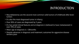
NEUROBLASTOMA
Eretare C Odjugo
OUTLINE
• Introduction
• Developmental anatomy
• Epidemiology
• Aetiology/Genetics
• Pathology
• Clinical presentation
• Diagnosis
• Biochemistry
• Radiology
• Histology
• Staging
OUTLINE
• Grading
• Screening
• Treatment
• Prognosis
• Current and future trends
• Loco-regional issues
• Follow-up
• Conclusion
INTRODUCTION
• Neuroblastomas are the second most common solid tumors of childhood after brain
tumors
• It’s also the most diagnosed tumor in infancy
• Over 90% of cases are diagnosed by 5 years
• It’s has varied clinical features and at diagnosis is believed to have metastasized in
over 70% of cases
• Mean age at diagnosis is ~18months
• Despite advances in diagnosis and treatment, outcomes for aggressive disease
remains poor
DEVELOPMENTAL
ANATOMY
• Arise as derivative of neural crest cells
• These migrate to final organs and
tissues
• The tumor can arise at any site where
these cells are found
• Adrenal medulla 50%
• Paraspinal ganglia and retroperitoneal
25%
• Posterior mediastinum 20%
• Pelvic ganglia <5%
• Cervical <5%
• Metastasis – Liver, bone and bone
marrow, lymph node, skin
LOCO-REGIONAL EPIDEMIOLOGY
• In a 6-month period 15 December 2021 to 14 June 2022 (UBTH)
• Paediatric oncology admissions – 48
• Neuroblastoma – 3 (6.25%)
• Ibadan 13 year study of childhood tumors
• Lymphoma 59%
• Neuroblastomas – 2.6%
• Kano
• Retinoblastoma – 30.6%
• Burkitts lymphoma – 19.9%
• Neuroblastoma – 2.7%
EPIDEMIOLOGY
• Found in 1 in 7500 to 10,000 live births
• 10% of all childhood tumors with 15% of deaths are attributable to it
• 1.2:1 Male to female ratio
• Racial predeliction in Caucasians and Asians.
• Not as common in Africans and people of African descent
AETIOLOGY/GENETICS
• The exact aetiology is unknown in most cases i.e they are spontaneous
• Familial cases account for 1 – 2% and are usually associated with younger age at diagnosis
(median age 9 months) as well as PHOX2B and ALK gene mutations
• BARD2 gene is said to also be a risk factor
• No single environmental factor has been linked to the disease
• MYCN (n-MYC) gene amplification is seen in 20% of tumors and is indicative of advanced
disease and confers a worse prognosis
• Ploidy: Hyperdiploidy when found in children less than 1 year confers a good prognosis
• Loss of heterozygosity at 1p, 11q, and 14q as well as a 17q gain confer a worse prognosis
• However, complete gain of chromosome 17 gives a better outcome
AETIOLOGY/GENETICS
• Neuroblastoma has been observedin infants with
• Beckwith-Wiedemann syndrome,
• Neurofibromatosis (von Recklinghausen disease),
• Hirschsprung disease,
• Central hypoventilation syndrome (Ondine’s curse),
• Fetal alcohol syndrome, and
• In offspring of mothers taking phenytoin (fetal hydantoin syndrome) for seizure disorders.
• In twins, concordance in younger aged twins and divergence in older twins indicates a
familial or spontaneous mutation respectively
PATHOLOGY
• Classic subtypes
• Neuroblastoma
• Ganglion neuroblastoma
• Ganglion neuroma
• Neuroblastoma
• Small round blue uniform cells with
hyperchromatic nuclei and scanty cytoplasm
• Homer-Wright pseudorosette
• Necrosis
• Calcification
CLINICAL PRESENTATION
• Very heterogenous clinical presentation and course
• Spontaneous regression
• Aggressive tumors
• Presentation may be as
• Mass effect of the tumors at site(s)
• Metabolic abnormalities
• Evidence of metastatic disease
• Constitutional symptoms
• Anorexia
• Weight loss
• Malaise
• Fever
CLINICAL PRESENTATION
• Abdominal swelling and pain
• Failure to thrive
• Respiratory distress
• Displacement of the diaphragm by a
large intra-abdominal tumor or liver
• Thoracic inlet partially occluded (cough,
breathlessness)
• Horner’s syndrome
• Neurologic deficits
• Urinary retention
• Severe diarrhoea
• Bone or joint pain
• Protopsis and peri-orbital ecchymoses
• Opsoclonus-Myoclonus syndrome
• Multi-vectorial eye movements
• Twitching of muscle groups
• Ataxia
• Hypokalemia
• Cytopenias
• Blueberry muffin sign
• Paroxysmal hypertension
• Tumor lysis syndrome
DIAGNOSIS
• Laboratory evaluation
• Full blood count
• Renal function test
• Liver function test
• Urinalysis
• Urine assay for catecholamines [Homovanillic acid (HVA) and Vanillylmandellic acid
(VMA)]
• Tumor markers
• Neuron specific enolase
• Chromogranin A
• Ferritin
RADIOLOGY
• Ultrasound
• First line investigation
• Plain radiographs
• Can show calcified tumors in abdomen and posterior mediastinum
• CT scan
• MRI
• Radionuclide scan
• Bone scan
CT SCANS
• Can delineate tumors properly
and are useful when assessing
image derived risk factors
• Calcification easily noted
• Helps in distinguishing neuro-
from nephroblastoma
• Nodal status may be assessed
MRI
• Excellent for visualizing
intra-spinal extensions of
the tumors
• Also good for delineating
vascular arrangements
• Absence of radiation is a
benefit
MIBG SCAN
• Based on the selective uptake
of meta-iodobenzylguanidine
by the neuroblastoma
• Also a great too for monitoring
response to treatment
• Iodine-123 is used for
investigative purposes
BONE SCAN
• Uses Technetium 99
• Useful only in MIBG positive
cases
• Helps detect cortical bone
involvement
BIOPSY
• Tissue diagnosis is essential
• Needle biopsy
• Bone marrow biopsy or aspiration
• Needle biopsies are image guided with several cores collected from different areas
of the tumor
• Immunohistiochemistry and cytogenetics are an important part of diagnosis,
treatment planning and prognostication
• MYCN amplification (~30%)
• DNA ploidy
• 11q deletions and other variants
BONE MARROW ASPIRATION AND
BIOPSY
Spread to bone is observed in up to 40% of cases at the time of diagnosis
A positive finding of cancer cells in biopsy or aspirate with urinary catecholamines is sufficient to make a
diagnosis without the need for biopsying the primary tumor.
2 biopsies and aspirates from opposite sides are recommended by INSS
Biopsies are not done in infants < 6 months old
STAGING
STAGING
GRADING
GRADING
GRADING
SCREENING
• This is done in several developed countries like Japan
• Urine assays for Vanillylmandellic acid (VMA) and Homovanillic acid (HVA) are the
commonest screening tools
• Its role is controversial
• Supporters say the good prognosis for those diagnosed early and the high rate of metastasis
at the time of diagnosis
• Opponents: No survival benefits with screening as found in a large randomized trial.
• Screen detected disease are usually those with a favourable outcome while screening
negative cases that subsequently develop the disease have an unfavourable histology
• Screening was halted in 2004 after studies in Canada and Germany showed no reduction in
deaths due to neuroblastoma, but rather caused an increase in diagnoses that would have
disappeared without treatment, subjecting those infants to unnecessary surgery and
chemotherapy.
TREATMENT
• Multi-modal
• Surgery
• Chemotherapy including immunotherapy
• Radiotherapy
• Multidisciplinary and includes but not limited to
• Surgeon
• Paediatric oncologist
• Nurses
• Radiologic oncologist/radiotherapist
• Psychologists
• Neurologist
SURGERY
• Aims at
• Establishing a diagnosis
• Staging the tumor
• Excision (if localized and resectable)
• Provision of tissue for further
evaluation
• Excision is the mainstay of INSS 1, 2
and 4S patients
• Lymph node sampling is done at
surgery
• Care to be taken to avoid undue
morbidity
• Radical excision is avoided
• Sacrifice of major organs like kidneys
and spleen should be avoided
especially in children < 1 year old
• Approach varies based on location of
tumor
• A liver biopsy is done for 4S cases
CHEMOTHERAPY
• Neoadjuvant or adjuvant
• Multi-agent therapy
• May be low, moderate or high intensity
chemotherapydependent on the stage
and the risk stratification
• Indications
• Respiratory distress
• Spinal cord compression
• Progressive disease
• Recurrence
• Common agents used
• Cisplatin
• Carboplatin
• Etoposide
• Cyclophosphamide
• Vincristine
• Doxorubicin
• Others are
• Thiotepa
• Mephalan
• Topotecan
• busulphan
CHEMOTHERAPY
RADIOTHERAPY
• Neuroblastomas are radio-sensitive
• Radiation exposure and dosage may be kept to a minimum due to good long-term
survival
• Very useful in down-staging a large rapidly enlarging tumor with mass effect
• Also a part of palliative care in symptomatic patients with end-stage disease
• May be used pre-op, intra-op or post-op
• Dosage administered depends on the risk stratification
• Typically delivered as external beam radiation
• Usually avoided in spinal cord lesions due to effect on growth of the spine
LOW RISK (50%)
• Observation
• Asymptomatic 4S disease
• Small adrenal masses in newborns
• Surgery
• Primary mode of treatment
• Sufficient for most cases
• 5 year Survival >95%
• Radiotherapy
• Usually not indicated
• Chemotherapy
• Short cause usually for symptomatic
cord compression
• May be given prior to surgery to
debulk prior to resection
• Adjuvant chemo may be given in cases
of MYCN amplification
INTERMEDIATE RISK (10 – 15%)
• Surgery
• Primary resection where possible
• Chemotherapy
• Multi-agent moderately intensive
chemotherapy.
• 4 cycles for favourable histology, 8 for
unfavourable.
• Neoadjuvant chemo for unresectable
tumor or tumors with intraspinal
extension
• Radiotherapy
• Given when there’s disease progression
despite surgery and chemotherapy
HIGH RISK
• Induction
• Intensive chemotherapy (8 months)
• Shrinks primary tumor and helps with
metastasis
• Local control
• Attained by surgery and radiotherapy
• Surgery is planned for 13 – 18 weeks
post induction
• Radiation of the tumor bed helps
prevent recurrence
• Consolidation
• High dose chemotherapeutic agents
plus melphalan or busalfan (myelo-
ablative therapy)
• Autologous haematopoietic stem cell
rescue + GM CSF therapy
• Maintenance
• Aims to manage minimal residual
disease
• 13 cis-Retinoic acid given every 14 days
for 6 months
• Anti-GD2 antibody and IL-2 give better
outcomes than retinoic acid alone
OUTCOMES
Survival probability of 2196 patients with neuroblastoma treated using the COG and POG guidelines
CURRENT/FUTURE
TRENDS
• PET scans are been evaluated for use vs
CT and MIBG scans
• Pros
• Indicates activity
• Excellent at identifying tissues of
interest
• Great contrasts
• Cons
• Expensive
• High radiation dose
• Not readily available
• Resolution and details still poor
CURRENT/FUTURE TRENDS
• High doses of 131-iodinated MIBG is being trialled in the treatment of
neuroblastoma due to its preferential uptake.
• Good responses
• Challenges with side effects and toxicity
• Targeted anti-ALK therapy (crizitinib, Lorlatinib)
• Targeted autologous T-cells (NK cells)
• Use of anti-angiogenic agents (Bevacizumab)
• Apoptosis inducers (Fenretinide)
FOLLOW-UP
• Post treatment tumors can increase in size by
• Maturation
• Progression
• Histology of new tumor is needed to differentiate both
• Recurrence/relapse
• Low risk
• Intermediate risk
• High risk
• Prognostication
TREATMENT OPTIONS FOR
RECURRENCE
TREATMENT OPTIONS FOR
RECURRENCE
• High risk:
• Candidates for clinical trials
• Palliative care
• Treat as high risk disease
• Palliation
LOCOREGIONAL
• Late presentation
• Financial challenges
• Poor infracstructure
• Religious beliefs
CONCLUSION
• Neuroblastoma despite being the most common extra-cranial solid tumor is a rare
tumor.
• It is a treatable malignancy with a long life expectancy if diagnosed early and
treated appropriately
• Surgery remains the mainstay of treatment with chemo- and radiotherapy being
supportive pillars
• Late presentation results in especially poor outcomes in our environment
• Despite advances in knowledge and treatment, outcomes are still poor in those with
unfavourable histology
REFERENCES
• Coran GA et al (editors); Paediatric surgery; Seventh edition (1)-
441 - 458
• Ochicha, O., Gwarzo, A. K., & Gwarzo, D. (2012). Pediatric
malignancies in Kano, Northern Nigeria. World Journal of Pediatrics,
8(3), 235–239.
• Williams, A. O. (1975). Tumors of childhood in Ibadan, Nigeria.
Cancer, 36(2), 370–378.
• Neuroblastoma – Wikipedia accessed 10/06/22 at 22:35
• Neuroblastoma (image-defined risk factors) | Radiology Reference Article | Radiopaedia.org
• Sci-Hub | Immunotherapeutic Strategies for Neuroblastoma: Present, Past and Future.
Vaccines, 9(1), 43 | 10.3390/vaccines9010043
• International Neuroblastoma Risk Group Task Force (inrgdb.org)
REFERENCES
• Neuroblastoma Cancer | American Cancer Society
• Sci-Hub | Revision of the International Neuroblastoma Pathology Classification.
Cancer, 98(10), 2274–2281 | 10.1002/cncr.11773
NEUROBLASTOMA.pptx

NEUROBLASTOMA.pptx

  • 1.
  • 2.
    OUTLINE • Introduction • Developmentalanatomy • Epidemiology • Aetiology/Genetics • Pathology • Clinical presentation • Diagnosis • Biochemistry • Radiology • Histology • Staging
  • 3.
    OUTLINE • Grading • Screening •Treatment • Prognosis • Current and future trends • Loco-regional issues • Follow-up • Conclusion
  • 4.
    INTRODUCTION • Neuroblastomas arethe second most common solid tumors of childhood after brain tumors • It’s also the most diagnosed tumor in infancy • Over 90% of cases are diagnosed by 5 years • It’s has varied clinical features and at diagnosis is believed to have metastasized in over 70% of cases • Mean age at diagnosis is ~18months • Despite advances in diagnosis and treatment, outcomes for aggressive disease remains poor
  • 5.
    DEVELOPMENTAL ANATOMY • Arise asderivative of neural crest cells • These migrate to final organs and tissues • The tumor can arise at any site where these cells are found • Adrenal medulla 50% • Paraspinal ganglia and retroperitoneal 25% • Posterior mediastinum 20% • Pelvic ganglia <5% • Cervical <5% • Metastasis – Liver, bone and bone marrow, lymph node, skin
  • 6.
    LOCO-REGIONAL EPIDEMIOLOGY • Ina 6-month period 15 December 2021 to 14 June 2022 (UBTH) • Paediatric oncology admissions – 48 • Neuroblastoma – 3 (6.25%) • Ibadan 13 year study of childhood tumors • Lymphoma 59% • Neuroblastomas – 2.6% • Kano • Retinoblastoma – 30.6% • Burkitts lymphoma – 19.9% • Neuroblastoma – 2.7%
  • 7.
    EPIDEMIOLOGY • Found in1 in 7500 to 10,000 live births • 10% of all childhood tumors with 15% of deaths are attributable to it • 1.2:1 Male to female ratio • Racial predeliction in Caucasians and Asians. • Not as common in Africans and people of African descent
  • 8.
    AETIOLOGY/GENETICS • The exactaetiology is unknown in most cases i.e they are spontaneous • Familial cases account for 1 – 2% and are usually associated with younger age at diagnosis (median age 9 months) as well as PHOX2B and ALK gene mutations • BARD2 gene is said to also be a risk factor • No single environmental factor has been linked to the disease • MYCN (n-MYC) gene amplification is seen in 20% of tumors and is indicative of advanced disease and confers a worse prognosis • Ploidy: Hyperdiploidy when found in children less than 1 year confers a good prognosis • Loss of heterozygosity at 1p, 11q, and 14q as well as a 17q gain confer a worse prognosis • However, complete gain of chromosome 17 gives a better outcome
  • 9.
    AETIOLOGY/GENETICS • Neuroblastoma hasbeen observedin infants with • Beckwith-Wiedemann syndrome, • Neurofibromatosis (von Recklinghausen disease), • Hirschsprung disease, • Central hypoventilation syndrome (Ondine’s curse), • Fetal alcohol syndrome, and • In offspring of mothers taking phenytoin (fetal hydantoin syndrome) for seizure disorders. • In twins, concordance in younger aged twins and divergence in older twins indicates a familial or spontaneous mutation respectively
  • 10.
    PATHOLOGY • Classic subtypes •Neuroblastoma • Ganglion neuroblastoma • Ganglion neuroma • Neuroblastoma • Small round blue uniform cells with hyperchromatic nuclei and scanty cytoplasm • Homer-Wright pseudorosette • Necrosis • Calcification
  • 11.
    CLINICAL PRESENTATION • Veryheterogenous clinical presentation and course • Spontaneous regression • Aggressive tumors • Presentation may be as • Mass effect of the tumors at site(s) • Metabolic abnormalities • Evidence of metastatic disease • Constitutional symptoms • Anorexia • Weight loss • Malaise • Fever
  • 12.
    CLINICAL PRESENTATION • Abdominalswelling and pain • Failure to thrive • Respiratory distress • Displacement of the diaphragm by a large intra-abdominal tumor or liver • Thoracic inlet partially occluded (cough, breathlessness) • Horner’s syndrome • Neurologic deficits • Urinary retention • Severe diarrhoea • Bone or joint pain • Protopsis and peri-orbital ecchymoses • Opsoclonus-Myoclonus syndrome • Multi-vectorial eye movements • Twitching of muscle groups • Ataxia • Hypokalemia • Cytopenias • Blueberry muffin sign • Paroxysmal hypertension • Tumor lysis syndrome
  • 13.
    DIAGNOSIS • Laboratory evaluation •Full blood count • Renal function test • Liver function test • Urinalysis • Urine assay for catecholamines [Homovanillic acid (HVA) and Vanillylmandellic acid (VMA)] • Tumor markers • Neuron specific enolase • Chromogranin A • Ferritin
  • 14.
    RADIOLOGY • Ultrasound • Firstline investigation • Plain radiographs • Can show calcified tumors in abdomen and posterior mediastinum • CT scan • MRI • Radionuclide scan • Bone scan
  • 15.
    CT SCANS • Candelineate tumors properly and are useful when assessing image derived risk factors • Calcification easily noted • Helps in distinguishing neuro- from nephroblastoma • Nodal status may be assessed
  • 16.
    MRI • Excellent forvisualizing intra-spinal extensions of the tumors • Also good for delineating vascular arrangements • Absence of radiation is a benefit
  • 17.
    MIBG SCAN • Basedon the selective uptake of meta-iodobenzylguanidine by the neuroblastoma • Also a great too for monitoring response to treatment • Iodine-123 is used for investigative purposes
  • 18.
    BONE SCAN • UsesTechnetium 99 • Useful only in MIBG positive cases • Helps detect cortical bone involvement
  • 19.
    BIOPSY • Tissue diagnosisis essential • Needle biopsy • Bone marrow biopsy or aspiration • Needle biopsies are image guided with several cores collected from different areas of the tumor • Immunohistiochemistry and cytogenetics are an important part of diagnosis, treatment planning and prognostication • MYCN amplification (~30%) • DNA ploidy • 11q deletions and other variants
  • 20.
    BONE MARROW ASPIRATIONAND BIOPSY Spread to bone is observed in up to 40% of cases at the time of diagnosis A positive finding of cancer cells in biopsy or aspirate with urinary catecholamines is sufficient to make a diagnosis without the need for biopsying the primary tumor. 2 biopsies and aspirates from opposite sides are recommended by INSS Biopsies are not done in infants < 6 months old
  • 21.
  • 22.
  • 25.
  • 26.
  • 27.
  • 28.
    SCREENING • This isdone in several developed countries like Japan • Urine assays for Vanillylmandellic acid (VMA) and Homovanillic acid (HVA) are the commonest screening tools • Its role is controversial • Supporters say the good prognosis for those diagnosed early and the high rate of metastasis at the time of diagnosis • Opponents: No survival benefits with screening as found in a large randomized trial. • Screen detected disease are usually those with a favourable outcome while screening negative cases that subsequently develop the disease have an unfavourable histology • Screening was halted in 2004 after studies in Canada and Germany showed no reduction in deaths due to neuroblastoma, but rather caused an increase in diagnoses that would have disappeared without treatment, subjecting those infants to unnecessary surgery and chemotherapy.
  • 29.
    TREATMENT • Multi-modal • Surgery •Chemotherapy including immunotherapy • Radiotherapy • Multidisciplinary and includes but not limited to • Surgeon • Paediatric oncologist • Nurses • Radiologic oncologist/radiotherapist • Psychologists • Neurologist
  • 30.
    SURGERY • Aims at •Establishing a diagnosis • Staging the tumor • Excision (if localized and resectable) • Provision of tissue for further evaluation • Excision is the mainstay of INSS 1, 2 and 4S patients • Lymph node sampling is done at surgery • Care to be taken to avoid undue morbidity • Radical excision is avoided • Sacrifice of major organs like kidneys and spleen should be avoided especially in children < 1 year old • Approach varies based on location of tumor • A liver biopsy is done for 4S cases
  • 31.
    CHEMOTHERAPY • Neoadjuvant oradjuvant • Multi-agent therapy • May be low, moderate or high intensity chemotherapydependent on the stage and the risk stratification • Indications • Respiratory distress • Spinal cord compression • Progressive disease • Recurrence • Common agents used • Cisplatin • Carboplatin • Etoposide • Cyclophosphamide • Vincristine • Doxorubicin • Others are • Thiotepa • Mephalan • Topotecan • busulphan
  • 32.
  • 33.
    RADIOTHERAPY • Neuroblastomas areradio-sensitive • Radiation exposure and dosage may be kept to a minimum due to good long-term survival • Very useful in down-staging a large rapidly enlarging tumor with mass effect • Also a part of palliative care in symptomatic patients with end-stage disease • May be used pre-op, intra-op or post-op • Dosage administered depends on the risk stratification • Typically delivered as external beam radiation • Usually avoided in spinal cord lesions due to effect on growth of the spine
  • 34.
    LOW RISK (50%) •Observation • Asymptomatic 4S disease • Small adrenal masses in newborns • Surgery • Primary mode of treatment • Sufficient for most cases • 5 year Survival >95% • Radiotherapy • Usually not indicated • Chemotherapy • Short cause usually for symptomatic cord compression • May be given prior to surgery to debulk prior to resection • Adjuvant chemo may be given in cases of MYCN amplification
  • 35.
    INTERMEDIATE RISK (10– 15%) • Surgery • Primary resection where possible • Chemotherapy • Multi-agent moderately intensive chemotherapy. • 4 cycles for favourable histology, 8 for unfavourable. • Neoadjuvant chemo for unresectable tumor or tumors with intraspinal extension • Radiotherapy • Given when there’s disease progression despite surgery and chemotherapy
  • 36.
    HIGH RISK • Induction •Intensive chemotherapy (8 months) • Shrinks primary tumor and helps with metastasis • Local control • Attained by surgery and radiotherapy • Surgery is planned for 13 – 18 weeks post induction • Radiation of the tumor bed helps prevent recurrence • Consolidation • High dose chemotherapeutic agents plus melphalan or busalfan (myelo- ablative therapy) • Autologous haematopoietic stem cell rescue + GM CSF therapy • Maintenance • Aims to manage minimal residual disease • 13 cis-Retinoic acid given every 14 days for 6 months • Anti-GD2 antibody and IL-2 give better outcomes than retinoic acid alone
  • 39.
    OUTCOMES Survival probability of2196 patients with neuroblastoma treated using the COG and POG guidelines
  • 40.
    CURRENT/FUTURE TRENDS • PET scansare been evaluated for use vs CT and MIBG scans • Pros • Indicates activity • Excellent at identifying tissues of interest • Great contrasts • Cons • Expensive • High radiation dose • Not readily available • Resolution and details still poor
  • 41.
    CURRENT/FUTURE TRENDS • Highdoses of 131-iodinated MIBG is being trialled in the treatment of neuroblastoma due to its preferential uptake. • Good responses • Challenges with side effects and toxicity • Targeted anti-ALK therapy (crizitinib, Lorlatinib) • Targeted autologous T-cells (NK cells) • Use of anti-angiogenic agents (Bevacizumab) • Apoptosis inducers (Fenretinide)
  • 42.
    FOLLOW-UP • Post treatmenttumors can increase in size by • Maturation • Progression • Histology of new tumor is needed to differentiate both • Recurrence/relapse • Low risk • Intermediate risk • High risk • Prognostication
  • 43.
  • 44.
    TREATMENT OPTIONS FOR RECURRENCE •High risk: • Candidates for clinical trials • Palliative care • Treat as high risk disease • Palliation
  • 45.
    LOCOREGIONAL • Late presentation •Financial challenges • Poor infracstructure • Religious beliefs
  • 46.
    CONCLUSION • Neuroblastoma despitebeing the most common extra-cranial solid tumor is a rare tumor. • It is a treatable malignancy with a long life expectancy if diagnosed early and treated appropriately • Surgery remains the mainstay of treatment with chemo- and radiotherapy being supportive pillars • Late presentation results in especially poor outcomes in our environment • Despite advances in knowledge and treatment, outcomes are still poor in those with unfavourable histology
  • 47.
    REFERENCES • Coran GAet al (editors); Paediatric surgery; Seventh edition (1)- 441 - 458 • Ochicha, O., Gwarzo, A. K., & Gwarzo, D. (2012). Pediatric malignancies in Kano, Northern Nigeria. World Journal of Pediatrics, 8(3), 235–239. • Williams, A. O. (1975). Tumors of childhood in Ibadan, Nigeria. Cancer, 36(2), 370–378. • Neuroblastoma – Wikipedia accessed 10/06/22 at 22:35 • Neuroblastoma (image-defined risk factors) | Radiology Reference Article | Radiopaedia.org • Sci-Hub | Immunotherapeutic Strategies for Neuroblastoma: Present, Past and Future. Vaccines, 9(1), 43 | 10.3390/vaccines9010043 • International Neuroblastoma Risk Group Task Force (inrgdb.org)
  • 48.
    REFERENCES • Neuroblastoma Cancer| American Cancer Society • Sci-Hub | Revision of the International Neuroblastoma Pathology Classification. Cancer, 98(10), 2274–2281 | 10.1002/cncr.11773

Editor's Notes

  • #7 Leukemia 10 (20.8%) Retinoblastoma 9 (18.75%) Nephroblastoma 12 (25%)
  • #9 Familial disease tends to have an autosomal dominant mode of inheritance Hook et al, 1991; Muraji et al 1993(MYCN amplification) ALK (anaplastic lymphoma kinase)
  • #11 Rosette: Eosinophillic neutrophils surrounded by neuroblasts Neuropil: Located at the center of the rosette
  • #12 Esthesioneuroblastoma (olfactory-mass, epistaxis)
  • #14 Ferritin is directly secreted by the tumor. Indicates poor prognosis NSE is a glycolytic enzyme. The version is the gamma subtype. A non specific marker shared with Ewings sarcoma, Wilms tumor, AML, NHL, CgA; Non specific. Secreted by neuroendocrine tumors carcinoid, phaeochromocytoma, neuroblastoma
  • #22 INSS done after surgery Based on resection and disease extent Historically the more popular system
  • #23 Children’s Oncology Group [COG] (North America and Australia), the German Gesellschaft für Pädiatrische Onkologie und Hämatologie [GPOH], the Japanese Advanced Neuroblastoma Study Group [JANB], the Japanese Infantile Neuroblastoma Co-operative Study Group [JINCS], the International Society of Paediatric Oncology European Neuroblastoma Research [SIOPEN] and China with expertise in neuroblastoma established an INRG Task Force. Image defined risk factors are those which make complete resction of a tumor unsafe Multicompartment disease Encasement of major vessels Contiguous involvement of other structures Intraspinal extension >1/3 diameter of the canal Compression of the tracheobronchial airway
  • #31 Complications Atelectasis, tumor rupture, infection, ileus, haemorrhage
  • #32 The choice of agents and the combinations depends on the indication such as induction, consolidation, maintenance or rescue.
  • #33 COG A3961 protocol
  • #34 Not indicated in INSS 1 and 2 Kidneys and ovaries are shielded Given as adjuvant therapy in LR and IR disease 3 – 6Gy total @1.5Gy/# (low risk disease) Up to 21Gy @1.5Gy/# (for residual disease in IR) Up to 1.8Gy/# for HR disease with total of 36Gy for residual tumors
  • #42 The latter are based on the infusion of natural killer (NK) cells, as support of consolidation therapy in addition to standard treatments, or chimeric antigen receptor (CAR) T cells directed against neuroblastoma associated antigens (e.g., disialoganglioside GD2). Finally, future perspectives of adoptive cell therapies represented by γδ T lymphocyes and CAR NK cells are envisaged.
  • #43 Age, INSS stage, MYCN status, time from initial diagnosis to first relapse, LDH level, ploidy, grade of tumor
  • #44 High risk: Candidates for clinical trials Palliative care Treat as high risk disease