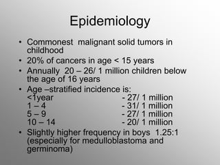
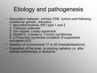
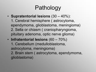
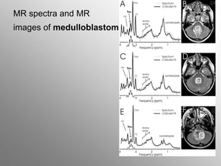
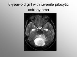
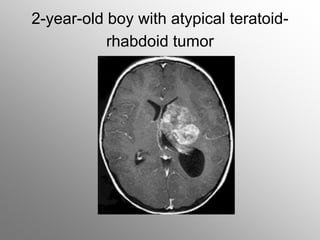
Central nervous system
malignancies
• Commonest malignant solid tumors in
childhood
• 20% of cancers in age < 15 years
• Annually 20 – 26/ 1 million children below
the age of 16 years
• Age –stratified incidence is:
<1year - 27/ 1 million
1 – 4 - 31/ 1 million
5 – 9 - 27/ 1 million
10 – 14 - 20/ 1 million
• Slightly higher frequency in boys 1.25:1
(especially for medulloblastoma and
germinoma)
Epidemiology
Etiology and pathogenesis
• Association between primary CNS tumors and following
conditions/ genetic disorders :
1. Neurofibromatosis (NF) type 1 and 2
2. Tuberous sclerosis
3. Von Hippel- Lindau syndrome
4. Gordlin’s, Cowden’s, Turcot’s syndromes
5. Li-Fraumeni syndrome (mutation of suppressor
oncogene p53)
• Deletion of chromosome 17 or 20 (medulloblastoma)
• Exposition of the brain to ionizing radiation i.e. after
cranial radiotherapy in leukemia
Pathology
• Supratentorial lesions (30 – 40%):
1. Cerebral hemisphere ( astrocytoma,
ependymoma, glioblastoma, meningioma)
2. Sella or chiasm ( craniopharyngioma,
pituitary adenoma, optic nerve glioma)
• Infratentorial lesions (60 – 70%)
1. Cerebellum (medulloblastoma,
astrocytoma, meningioma)
2. Brain stem ( astrocytoma, ependymoma,
glioblastoma)
Classification
• Based on histogenesis and predominance
of cell type
• Degree of malignancy is defined by
grading system e.i. WHO grade based on
cellular morphology, mitotic index,
anaplasia and necrosis:
grades I and II represent benign tumors
grades III and IV - malignant tumors
Clinical presentation
• Depends on :
-age
-anatomical site
-tumor type
• -raised intracranial pressure (ICP)
-localizing neurological deficits
Signs of increased ICP
• Direct tumor infiltration
• Compression of normal structures
• Secondary to obstruction of the
cerebrospinal fluid (CSF)
• Older children:
-inilially behavioural changes and declining school
performance prior to development of the more classical
features of headache, nausea and vomiting ,
headaches start as generalized and intermittent >
increase in both intensity and frequency with time - the
child may awake with headache at night, with the pain
generally being worse in the morning and improving
during the day with an upright posture
-School-age children complain of visual disturbances
• Infants and younger children:
plasticity of the developing skull and inability to
communicate symptoms >
-infant may be irritable, with failure to thrive,
associated with anorexia and vomiting
-regression of developmental milestones
-increase head circumference with widened
sutures and a tense anterior fontanelle
„sun-setting” sign
Symptoms and signs according to
anatomical site of CNS tumors
• Supratentorial (30-40%)
Cerebral hemisphere- hemiparesis, spasticity, seizures (focal or
generalized)
- para/suprasellar – endocrinopathy (growth failure, diabetes
insipidus, pubertal abnormality)
- hypothalamus – diencephalic syndrome (infants), developmental
and behavioural abnormalities
- optic pathway – visual field acuity, color vision deficits, optic
atrophy, nystagmus, head tilt
- pineal - Parinaud’s syndrome, sleep abnormalities
- thalamus, basal ganglia – pain, sensory loss, memory disturbances
- intraventicular
- meningeal
• Infratentorial (60 – 70%)
- posterior fossa – ataxia, nystagmus, dysmetria (presents as
clumsiness or worse handwriting)
- brainstem – multiple cranial nerve palsies, hemiparesis, spasticity,
mood changes
• Spinal (2 – 5%)
- primary intramedullary – pain (local back and root pain), motor and
sensory disturbance
- spinal metastases – scoliosis, sphincter (bowel, bladder)
disturbances, reflex changes
Diagnostic evaluation
• Magnetic resonance and computed tomography –
basic imaging techniques for brain tumors
• Positron emission tomography – help to distinguish
tumors or lesions with a volume greater than 1 cm3
• Conventional radiography of the skull: bone structure,
separating off sutures ( due to ICP), calcification within
the brain
• Special methods (for special indications):
- brain scintigraphy
- angiography
-ultrasonography
-myelography
Additional diagnosis
• Cerebral fluid analysis ( to determine
spread of the tumor to the spinal fluid)
• Electroencephalography
• Stereotactic biopsy
Therapy
• Neurosurgery for maximum tumor removal and low
morbidity depending on the location and extent of the
tumor
-often preoperative relief of intracranial pressure by
ventriculoperitoneal or ventriculoarterial shunt
- preoperative reduction of tumor edema by
corticosteroids
- in patients with seizures - anticonvulsive therapy
• Radiotherapy – extension and volume of
irradiation depend on the biology and
histology of the tumor, age of the child and
combination with chemotherapy and
neurosurgery
- irradiation in children < 3 years of life only
in special cases
• Chemotherapy – depends on tumor type,
location and age -efficacy and penetration
depend on vascularization of the tumor
MR spectra and MR
images of medulloblastoma
8-year-old girl with juvenile pilocytic
astrocytoma
2-year-old boy with atypical teratoid-
rhabdoid tumor
Neuroblastoma
• Malignant, embryonal tumor derived from precursor cells
of sympathetic ganglia and adrenal medulla
• Other types of tumors derived from sympathetic nervous
system:
-ganglioneuroblastoma
-ganglioneuroma
-pheochromocytoma
• Possibility to spontaneous regression and differentiation
to benign tumor in infants less 1 year of age
extremely malignant in older children
Epidemiology
• 8% of all neoplasms in children
• Most frequent malignant neoplasm in
infants
• Mean age at diagnosis 2 – 5 years
Pathology
Two distinct entities:
• Infant:
1. possibility of spontaneous regression
(apoptosis or differentiation into
ganglioneuroblastoma)
2. chemosensitive, chemocurable
• Older
3. chemoresistant malignancies
Molecular cytogenetics
• MYCN oncogene amplification.
MYCN is located on chromosome 2p.
Independent prognostic factor:
in stage III EFS for patients with a single copy is curable 80%;
for those with amplification MYCN – 20%
• DNA ploidy: hyperploidy = good prognosis
• Nerve growth factor receptor: ligands for high –affinity tyrosine
kinase receptors TRKA, TRKB, TRKC:
-TRKA expression is associated with MYCN single copy, low stage
and good prognosis
- TRKA (-) + MYCN amplification= very poor survival
• Structural and numerical abnormalities of chromosome 1
Clinical manifestation
• Occurence in any area with sympathetic nervous
system
Primary location:
-abdomen 65%
-adrenal medulla or sympathetic ganglia 46%
-posterior mediastinum 15%
-pelvic 4%
-head and neck 3%
-others 8%
Common symptoms
• Weight loss
• Fever
• Abdominal disturbances
• Irratability
• Pain of bones and joints
• Child not stand up, not walk
• Pallor
• Lassitude
Symptoms associated
with catecholamine production
• Paroxysmal attacks of sweating, flushing,
pallor
• Headache
• Hypertension
• Palpitation
Paraneoplastic syndromes
• VIP syndrome: untreatable diarrhea,with low
level of potassium
• Opsoclonus- myoclonus
• Anemia, trombocytopenia, leukopenia
( in bone marrow infiltration or massive
hemorrhage)
Local symptoms
• Abdomen:
-intra-abdominal tumor
–paravertebral and presacral -neurological dysfunction
-abdominal distension
• Liver:
-hepatomegaly
• Chest, posterior mediastinum, vertebrae:
-compression of trachea > coughing, dyspnea
-infiltration in vertebral foramina > dumbbell tumor
-compression of nerves >disturbances of gait, muscle
weakness, parasthesia, bladder dysfunction,
constipation
• Eyes:
-periorbital edema, swelling, yellow- brown
ecchymoses
-proptosis and exophthalmos, strabismus,
opsoclonus
-papillary edema, bleeding of the retina, atrophy
of the optic nerve
• Neck:
-cervical lymphadenopathy
-supraclavicular tumor
-Horner syndrome: enophthalmos, miosis, ptosis,
Raccon eyes
• Skin:
- subcutaneous nodules of blue color > reddish > white
owing to vasoconstriction from release of
catecholamines after palpation
- nodules are mainly observed in neonates or infants
with disseminated NBL
• Bone:
-pain
involvement mainly in the skull and long bones
- in X-rays – lytic defects with irregular margins and
periosteal reaction
• Bone marrow:
-trombocytopenia, anemia
Metastases
• Lymphatic and/or hematogenous spread
• Often initially present in children
(40 – 50% children < 1 year and 70%
children > 1 year)
• Metastatic spread mostly in bone marrow,
bone, liver, skin
2-year-old-girl with abdominal neuroblastoma
CT image with large abdominal neuroblastoma
123-MIBG
scintscan
International Staging System for NBL(INSS)
• 1 - localized tumor with complete excision,lymph nodes negative
• 2a - localized tumor without incomplete gross excision, representative,
ipsilateral nonadherent lymph nodes negative for tumor
microscopically
• 2b – ipsilateral nonadherent lymph nodes positive for tumor. Enlarged
contralateral lymph nodes negative microscopically
• 3 – unresectable unilateral tumor infiltrating across the midline,with or
without regional lymph node involvement or localized unilateral tumor
with contralateral regional lymph node involvement or – midline tumor
with bilateral extension by infiltration or by lymph node involvement
• 4 – any primary tumor with dissemination to distant lymph nodes, bone,
bone marrow, liver, skin or other organs (except as defined for stage
4S
• 4s localized primary tumor (as defined for stages 1, 2a, 2b) with
dissemination limited to skin, liver or bone marrow; limited to
infants aged less than 1 year)
Laboratory findings
• Tumor markers:
-catecholamines:
vanillylmandelic acid (VMA), homovanillic acid
(HVA) dopamine in urine/ plasma
adrenaline, noradrenaline
-neuron-specific enolase (glycolitic enzyme of
brain and neuroendocrine tissues) -NSE
• Ferritin
• Lactate dehydrogenease (LDH)
• Bone marrow (aspiration and biopsy)
Locoregional involvement
• Computed tomography scan and/or
• Ultrasound and/or
• magnetic resonance imaging →
localize the mass, provide measurements,
give anatomical information about intra- and
extraperitoneal structures, differentiate cystic
from solid tumors, define the extent of a primary
tumor and its relationship with other structures,
detect small calcification
Evaluation of metastases
• Bone marrow metastases – bone marrow
aspiration and trephine biopsy
• Skeletal metastases - X-ray, Tc-99
scintigraphy,
• mIBG scintigraphy demonstrates primary,
residual tumor masses,diffuse bone marrow
infiltration, skeletal, lymph node and soft tissue
metastases
• FDG-PET scanning
Therapy
Depends on: age, stage, localization and molecular
features at diagnosis
• Surgery
• Chemotherapy
IV stage
• Radiotherapy
• Target radiotherapy (I-131-mIBG)
• Differentiation therapy (retinoids 13-cis and all-trans)
• Immunotherapy: anti-GD2 antibodies
• Auto-BMT
Prognosis
• Depends on:
-age (favorable if less than 18 months of age at
diagnosis), -stage and localization (favorable in primary
NBL of thorax, presacral and cervical)
-involvement of lymph nodes (poor prognosis)
• Low-risk group - 90% long-term survival
• Intermediate and high-risk groups:
-response to initial treatment 60-70% of children with
complete or partial remission
-after consolidation therapy (high-dose chemotherapy+
autologous stem cell support) –EFS after 3 years is 40-
60%
Hepatic tumors
• Malignant:
-hepatoblastoma 43%
-hepatocellular carcinoma 23%
-sarcoma 6%
• Benign:
-vascular (haemangioma,haemangioendothelioma) 13%
-hamartoma 6%
-others 9%
Incidence (malignant)
• -1.2 –5% of all neoplasms in childhood
• Boys: girls 1.4 –2 :1
• High incidence in genetically associated syndromes:
-Beckwith-Wiedemann syndrome, familial adematous
polyposis, trisomy 18, glycogen storage disease,
hereditary tyrosinemia, Li-Fraumeni syndrome
• HBL –mostly in infants, rarely after the age of 3 years
• Hepatocellular Ca – in older children, in adolescents,
after hepatitis B
Clinical symptoms
• Abdominal mass and/ or abdominal distention
• Anorexia, weight loss
• Lethargy
• Jaundice and ascites
• Abdominal pain
• Pallor
• Precocious puberty (hepatoblastoma), virilization (due
to gonadotrophin production by the tumor)
• Generalized osteoporosis (HBL)- tumor cells secrete
osteoclast-activating factor
Laboratory diagnosis
• Serum AFP elevated in 80 - 90% of children with HBL
and in 60-70% with hepatocellular Ca
• B-HCG elevated in both
• Increased bilirubin
• Increased serum aspartate aminotransferase (AST),
alanine transaminase (ALT) because of associated
hepatitis or cirrhosis
• Anemia, thrombocytosis, trombocytopenia (rarely)
• biopsy
Radiological diagnosis
• Ultrasound – liver enlarged, displacement of stomach
and colon, elevated diaphragm on the right side
• CT, MRI
• Liver scintigraphy
• metastases: chest X-ray, lung CT
HBL hepatocellular carcinoma
Treatment
• Presurgical chemotherapy (makes HBL resectable)
• Complete resection (initially > 50% of liver tumors are
not totally resectable)
• Liver transplantation
Prognosis:
• Depends on stage of the tumor
after complete tumor resection and chemotherapy
65-75 % children with HBL
and 40-60% with hepatocellular Ca survive
Germ cell tumors (GCT)
• Develop from embryonal germ cells
• Represent ectodermal, endodermal and mesodermal
lineages
• Approximately 3% of childhood malignancies
• Annually 2.4 – 3.8/ 1 million/per year
• 2/3 GCTs in children occur in extragonadal sites
• The commonest GCT - sacrococcygeal teratoma
• Bimodal age distribution:
one peak in children < 3y ears of age (sacrococcygeal
tumors, yolk sac tumors of testis)
- the later- the later GCT of the ovary, testis, and
intracranial sites
Histological classification of gonadal and
extragonadal tumors
• Germ cell and germinoma/ dysgerminoma and embryonal yolk sac
tumor (pluripotent cells)
a)extraembryonic structures
- yolk sac or endodermal sinus tumor
- choriocarcinoma
b) embryonal ecto-, meso-, endodermal origin tissues represented
-teratoma
c) embryonal carcinoma
• Gonadal germ cells and stroma tumor (Sertoli and Leydig cells)
• Epithelial cells (ovarian origin) and granulosa cell tumor or mixted
form as well as epithelial cell tumors more common in adults
Clinical symptoms
• Testicular GCT :scrotal enlargement,hydrocele
• Ovarian tumors: abdominal pain, acute abdomen,
abdominal mass
• Extragonadal GCT: main sites of involvement:
-sacrococcygeal:
t.1- (47%) predominantly external,
t.2 -both external and intrapelvic
t.3 -external, pelvix and abdominal
t.4 –entitely presacral,
-mediastinal (dyspnea, wheezing, thoracic pain,
superior vena cava syndrome),
-intracranial (visual disturbances, diabetes insipidus,
hypopituitarism, anorexia, precocious puberty)
Ovarian and testicular GCT
Sacrococcygeal tumor in newborn
Diagnostics
Radiological diagnosis:
ultrasound, computed tomography or magnetic
resonance imaging
• Tumor markers:
alpha-fetoprotein (AFP)- globulin produced in the
fetal yolk sac, in embryonal hepatocytes and in
gastrointestinal tract. Increase in malignant GCT,
hepatoblastoma
• Newborns 48,000+/-34,000IU
• Up to 1 month 9,000+/-12,000
• Up to 2 months 320 +/-280
• Up to 4 months 74 +/-56
• Up to 6 months 12+/-10
• Up to 8 months 8+/-5
beta -human chorionic gonadotropin (HCG) –
increase in germinoma/ dysgerminoma,
choriocarcinoma
Therapy
• Depend on:
• stage, location, tumor markers level
• Surgery in stage I
• Advanced stages: chemotherapy, surgery, radiotherapy
Survival
95% - sacrococcygeal teratoma
80% -yolk sac tumor
Wilms’ tumor
Nephroblastoma
• Malignant embryonal tumor of renal tissue consisting of
varying proportion of blastema, stroma and epithelium
• Epidemiology:
6% of all neoplasms in children
80 % of children at diagnosis are less than 5 years old
peak incidence between 2nd and 3rd year of age
- incidence slightly higher in boys
Genetics
Chromosomal association:
• Chromosome 11p13 with Wilms tumor suppressor gene WT1
in 10-30% of nephroblastoma
• Chromosome 11p15 with Wilms tumor suppressor gene WT2
• Chromosome 17q with familial FWT-2
Association with congenital anomalies:
-WAGR syndrome: Wilms tumor, aniridia, genital malformation,
mental retardation)
-Denys+Drash syndrome – pseudohermaphroditism,
glomerulopathy, mutation on chromosome 11p
-Beckwith Wiedemann syndrome- hemihypertrophy,
macroglossia, omphalocele, visceromegaly, associated with WT2
Clinical manifestation
• Abdominal mass- visible and/or palpable - 70%.
Palpation must be done with care – risk of tumor rupture or
dissemination!
• Fever
• Vomiting, anorexia
• Hematuria (micro or macro) 20-25%
• Hypertension in renin-producing tumors
• Pain 44%
• Special symptoms in association with congenital
anomalies
Laboratory diagnosis
• Urine: hematuria
• Chemistry: high serum calcium in children with rhabdoid
nephroblastoma
• Acquired von Willebrand coagulopathy in about 8% of
patients
• Differential diagnosis of NBL: 24-h urine catecholamine
analysis
• Biopsy – only in children with unclear presentation or
diagnosis
The child with Wilms tumor
Radiological diagnosis
• Ultrasound
• Computed tomography
• Magnetic resonance imaging
=anatomy of tumor extend of any spread within abdomen
Metastases:
• Chest radiographs (posteroanterior and lateral) to exclude
pulmonary metastases
• Angiography may be indicated in bilateral nephroblastoma
• Radioisotope scans , skeletal survey in patients with suspected
skeletal metastates
• CNS MRI in clear-cell sarcoma or rhabdoid kidney sarcoma and in
patients with possible brain metastases
Wilms tumor – pulmonary metastatic disease
Wilms tumor – intraoperative view
Prognostic factors
• Histological appearance „ favourable, unfavourable”
• Histology: rhabdoid tumor
• Diffuse anaplasia
• Viable malignant cells after preoperative chemotherapy
• Infiltration of tumor capsule
• Invasion of tumor cells into vessels
• Nonradical surgical resection of tumor
• Lymph nodes involvement
• Tumor rupture
• Metastatic spread
• Large tumor volume
Stage
National Wilms’ Tumor Study Group staging system
I – tumor confined to the kidney and completely resected
II- tumor extend beyond the kidney but is completely resected (none
at margins, no lymph nodes). At least one of the following has
occured: a) penetration of the renal capsule
b) invasion of the renal sinus vessels
c)biopsy of tumor before removal
III- gross or microscopic residual tumor remains postoperatively
including inoperable tumor, tumor at surgical margins, tumor spillage
involving peritoneal surfaces, regional lymph node metastases or
transected tumor thrombus
IV- hematogenous metastases or lymph node metastases outside the
abdomen (lung, liver, bone, brain)
V – bilateral renal Wilms’tumor at onset
Treatment
• Preoperative chemotherapy > surgery> postoperative
chemotherapy with or without radiotherapy
• Primary surgery in infants less than 6 months old and in
adolescents (>15years)
• Prognosis
I stage 3year EFS 90%
II 85%
III 82%
IV 58%

cns,_nbl,_hbl,gonads,_nephro.ppt

  • 1.
  • 2.
    • Commonest malignantsolid tumors in childhood • 20% of cancers in age < 15 years • Annually 20 – 26/ 1 million children below the age of 16 years • Age –stratified incidence is: <1year - 27/ 1 million 1 – 4 - 31/ 1 million 5 – 9 - 27/ 1 million 10 – 14 - 20/ 1 million • Slightly higher frequency in boys 1.25:1 (especially for medulloblastoma and germinoma) Epidemiology
  • 3.
    Etiology and pathogenesis •Association between primary CNS tumors and following conditions/ genetic disorders : 1. Neurofibromatosis (NF) type 1 and 2 2. Tuberous sclerosis 3. Von Hippel- Lindau syndrome 4. Gordlin’s, Cowden’s, Turcot’s syndromes 5. Li-Fraumeni syndrome (mutation of suppressor oncogene p53) • Deletion of chromosome 17 or 20 (medulloblastoma) • Exposition of the brain to ionizing radiation i.e. after cranial radiotherapy in leukemia
  • 4.
    Pathology • Supratentorial lesions(30 – 40%): 1. Cerebral hemisphere ( astrocytoma, ependymoma, glioblastoma, meningioma) 2. Sella or chiasm ( craniopharyngioma, pituitary adenoma, optic nerve glioma) • Infratentorial lesions (60 – 70%) 1. Cerebellum (medulloblastoma, astrocytoma, meningioma) 2. Brain stem ( astrocytoma, ependymoma, glioblastoma)
  • 5.
    Classification • Based onhistogenesis and predominance of cell type • Degree of malignancy is defined by grading system e.i. WHO grade based on cellular morphology, mitotic index, anaplasia and necrosis: grades I and II represent benign tumors grades III and IV - malignant tumors
  • 6.
    Clinical presentation • Dependson : -age -anatomical site -tumor type • -raised intracranial pressure (ICP) -localizing neurological deficits
  • 7.
    Signs of increasedICP • Direct tumor infiltration • Compression of normal structures • Secondary to obstruction of the cerebrospinal fluid (CSF)
  • 8.
    • Older children: -iniliallybehavioural changes and declining school performance prior to development of the more classical features of headache, nausea and vomiting , headaches start as generalized and intermittent > increase in both intensity and frequency with time - the child may awake with headache at night, with the pain generally being worse in the morning and improving during the day with an upright posture -School-age children complain of visual disturbances
  • 9.
    • Infants andyounger children: plasticity of the developing skull and inability to communicate symptoms > -infant may be irritable, with failure to thrive, associated with anorexia and vomiting -regression of developmental milestones -increase head circumference with widened sutures and a tense anterior fontanelle „sun-setting” sign
  • 10.
    Symptoms and signsaccording to anatomical site of CNS tumors • Supratentorial (30-40%) Cerebral hemisphere- hemiparesis, spasticity, seizures (focal or generalized) - para/suprasellar – endocrinopathy (growth failure, diabetes insipidus, pubertal abnormality) - hypothalamus – diencephalic syndrome (infants), developmental and behavioural abnormalities - optic pathway – visual field acuity, color vision deficits, optic atrophy, nystagmus, head tilt - pineal - Parinaud’s syndrome, sleep abnormalities - thalamus, basal ganglia – pain, sensory loss, memory disturbances - intraventicular - meningeal
  • 11.
    • Infratentorial (60– 70%) - posterior fossa – ataxia, nystagmus, dysmetria (presents as clumsiness or worse handwriting) - brainstem – multiple cranial nerve palsies, hemiparesis, spasticity, mood changes • Spinal (2 – 5%) - primary intramedullary – pain (local back and root pain), motor and sensory disturbance - spinal metastases – scoliosis, sphincter (bowel, bladder) disturbances, reflex changes
  • 12.
    Diagnostic evaluation • Magneticresonance and computed tomography – basic imaging techniques for brain tumors • Positron emission tomography – help to distinguish tumors or lesions with a volume greater than 1 cm3 • Conventional radiography of the skull: bone structure, separating off sutures ( due to ICP), calcification within the brain • Special methods (for special indications): - brain scintigraphy - angiography -ultrasonography -myelography
  • 13.
    Additional diagnosis • Cerebralfluid analysis ( to determine spread of the tumor to the spinal fluid) • Electroencephalography • Stereotactic biopsy
  • 14.
    Therapy • Neurosurgery formaximum tumor removal and low morbidity depending on the location and extent of the tumor -often preoperative relief of intracranial pressure by ventriculoperitoneal or ventriculoarterial shunt - preoperative reduction of tumor edema by corticosteroids - in patients with seizures - anticonvulsive therapy
  • 15.
    • Radiotherapy –extension and volume of irradiation depend on the biology and histology of the tumor, age of the child and combination with chemotherapy and neurosurgery - irradiation in children < 3 years of life only in special cases • Chemotherapy – depends on tumor type, location and age -efficacy and penetration depend on vascularization of the tumor
  • 16.
    MR spectra andMR images of medulloblastoma
  • 17.
    8-year-old girl withjuvenile pilocytic astrocytoma
  • 18.
    2-year-old boy withatypical teratoid- rhabdoid tumor
  • 19.
  • 20.
    • Malignant, embryonaltumor derived from precursor cells of sympathetic ganglia and adrenal medulla • Other types of tumors derived from sympathetic nervous system: -ganglioneuroblastoma -ganglioneuroma -pheochromocytoma • Possibility to spontaneous regression and differentiation to benign tumor in infants less 1 year of age extremely malignant in older children
  • 21.
    Epidemiology • 8% ofall neoplasms in children • Most frequent malignant neoplasm in infants • Mean age at diagnosis 2 – 5 years
  • 22.
    Pathology Two distinct entities: •Infant: 1. possibility of spontaneous regression (apoptosis or differentiation into ganglioneuroblastoma) 2. chemosensitive, chemocurable • Older 3. chemoresistant malignancies
  • 23.
    Molecular cytogenetics • MYCNoncogene amplification. MYCN is located on chromosome 2p. Independent prognostic factor: in stage III EFS for patients with a single copy is curable 80%; for those with amplification MYCN – 20% • DNA ploidy: hyperploidy = good prognosis • Nerve growth factor receptor: ligands for high –affinity tyrosine kinase receptors TRKA, TRKB, TRKC: -TRKA expression is associated with MYCN single copy, low stage and good prognosis - TRKA (-) + MYCN amplification= very poor survival • Structural and numerical abnormalities of chromosome 1
  • 24.
    Clinical manifestation • Occurencein any area with sympathetic nervous system Primary location: -abdomen 65% -adrenal medulla or sympathetic ganglia 46% -posterior mediastinum 15% -pelvic 4% -head and neck 3% -others 8%
  • 25.
    Common symptoms • Weightloss • Fever • Abdominal disturbances • Irratability • Pain of bones and joints • Child not stand up, not walk • Pallor • Lassitude
  • 26.
    Symptoms associated with catecholamineproduction • Paroxysmal attacks of sweating, flushing, pallor • Headache • Hypertension • Palpitation
  • 27.
    Paraneoplastic syndromes • VIPsyndrome: untreatable diarrhea,with low level of potassium • Opsoclonus- myoclonus • Anemia, trombocytopenia, leukopenia ( in bone marrow infiltration or massive hemorrhage)
  • 28.
    Local symptoms • Abdomen: -intra-abdominaltumor –paravertebral and presacral -neurological dysfunction -abdominal distension • Liver: -hepatomegaly • Chest, posterior mediastinum, vertebrae: -compression of trachea > coughing, dyspnea -infiltration in vertebral foramina > dumbbell tumor -compression of nerves >disturbances of gait, muscle weakness, parasthesia, bladder dysfunction, constipation
  • 29.
    • Eyes: -periorbital edema,swelling, yellow- brown ecchymoses -proptosis and exophthalmos, strabismus, opsoclonus -papillary edema, bleeding of the retina, atrophy of the optic nerve • Neck: -cervical lymphadenopathy -supraclavicular tumor -Horner syndrome: enophthalmos, miosis, ptosis, Raccon eyes
  • 30.
    • Skin: - subcutaneousnodules of blue color > reddish > white owing to vasoconstriction from release of catecholamines after palpation - nodules are mainly observed in neonates or infants with disseminated NBL • Bone: -pain involvement mainly in the skull and long bones - in X-rays – lytic defects with irregular margins and periosteal reaction • Bone marrow: -trombocytopenia, anemia
  • 31.
    Metastases • Lymphatic and/orhematogenous spread • Often initially present in children (40 – 50% children < 1 year and 70% children > 1 year) • Metastatic spread mostly in bone marrow, bone, liver, skin
  • 32.
  • 33.
    CT image withlarge abdominal neuroblastoma
  • 34.
  • 35.
    International Staging Systemfor NBL(INSS) • 1 - localized tumor with complete excision,lymph nodes negative • 2a - localized tumor without incomplete gross excision, representative, ipsilateral nonadherent lymph nodes negative for tumor microscopically • 2b – ipsilateral nonadherent lymph nodes positive for tumor. Enlarged contralateral lymph nodes negative microscopically • 3 – unresectable unilateral tumor infiltrating across the midline,with or without regional lymph node involvement or localized unilateral tumor with contralateral regional lymph node involvement or – midline tumor with bilateral extension by infiltration or by lymph node involvement • 4 – any primary tumor with dissemination to distant lymph nodes, bone, bone marrow, liver, skin or other organs (except as defined for stage 4S • 4s localized primary tumor (as defined for stages 1, 2a, 2b) with dissemination limited to skin, liver or bone marrow; limited to infants aged less than 1 year)
  • 36.
    Laboratory findings • Tumormarkers: -catecholamines: vanillylmandelic acid (VMA), homovanillic acid (HVA) dopamine in urine/ plasma adrenaline, noradrenaline -neuron-specific enolase (glycolitic enzyme of brain and neuroendocrine tissues) -NSE • Ferritin • Lactate dehydrogenease (LDH) • Bone marrow (aspiration and biopsy)
  • 37.
    Locoregional involvement • Computedtomography scan and/or • Ultrasound and/or • magnetic resonance imaging → localize the mass, provide measurements, give anatomical information about intra- and extraperitoneal structures, differentiate cystic from solid tumors, define the extent of a primary tumor and its relationship with other structures, detect small calcification
  • 38.
    Evaluation of metastases •Bone marrow metastases – bone marrow aspiration and trephine biopsy • Skeletal metastases - X-ray, Tc-99 scintigraphy, • mIBG scintigraphy demonstrates primary, residual tumor masses,diffuse bone marrow infiltration, skeletal, lymph node and soft tissue metastases • FDG-PET scanning
  • 39.
    Therapy Depends on: age,stage, localization and molecular features at diagnosis • Surgery • Chemotherapy IV stage • Radiotherapy • Target radiotherapy (I-131-mIBG) • Differentiation therapy (retinoids 13-cis and all-trans) • Immunotherapy: anti-GD2 antibodies • Auto-BMT
  • 40.
    Prognosis • Depends on: -age(favorable if less than 18 months of age at diagnosis), -stage and localization (favorable in primary NBL of thorax, presacral and cervical) -involvement of lymph nodes (poor prognosis) • Low-risk group - 90% long-term survival • Intermediate and high-risk groups: -response to initial treatment 60-70% of children with complete or partial remission -after consolidation therapy (high-dose chemotherapy+ autologous stem cell support) –EFS after 3 years is 40- 60%
  • 41.
  • 42.
    • Malignant: -hepatoblastoma 43% -hepatocellularcarcinoma 23% -sarcoma 6% • Benign: -vascular (haemangioma,haemangioendothelioma) 13% -hamartoma 6% -others 9%
  • 43.
    Incidence (malignant) • -1.2–5% of all neoplasms in childhood • Boys: girls 1.4 –2 :1 • High incidence in genetically associated syndromes: -Beckwith-Wiedemann syndrome, familial adematous polyposis, trisomy 18, glycogen storage disease, hereditary tyrosinemia, Li-Fraumeni syndrome • HBL –mostly in infants, rarely after the age of 3 years • Hepatocellular Ca – in older children, in adolescents, after hepatitis B
  • 44.
    Clinical symptoms • Abdominalmass and/ or abdominal distention • Anorexia, weight loss • Lethargy • Jaundice and ascites • Abdominal pain • Pallor • Precocious puberty (hepatoblastoma), virilization (due to gonadotrophin production by the tumor) • Generalized osteoporosis (HBL)- tumor cells secrete osteoclast-activating factor
  • 45.
    Laboratory diagnosis • SerumAFP elevated in 80 - 90% of children with HBL and in 60-70% with hepatocellular Ca • B-HCG elevated in both • Increased bilirubin • Increased serum aspartate aminotransferase (AST), alanine transaminase (ALT) because of associated hepatitis or cirrhosis • Anemia, thrombocytosis, trombocytopenia (rarely) • biopsy
  • 46.
    Radiological diagnosis • Ultrasound– liver enlarged, displacement of stomach and colon, elevated diaphragm on the right side • CT, MRI • Liver scintigraphy • metastases: chest X-ray, lung CT HBL hepatocellular carcinoma
  • 47.
    Treatment • Presurgical chemotherapy(makes HBL resectable) • Complete resection (initially > 50% of liver tumors are not totally resectable) • Liver transplantation Prognosis: • Depends on stage of the tumor after complete tumor resection and chemotherapy 65-75 % children with HBL and 40-60% with hepatocellular Ca survive
  • 48.
  • 49.
    • Develop fromembryonal germ cells • Represent ectodermal, endodermal and mesodermal lineages • Approximately 3% of childhood malignancies • Annually 2.4 – 3.8/ 1 million/per year • 2/3 GCTs in children occur in extragonadal sites • The commonest GCT - sacrococcygeal teratoma • Bimodal age distribution: one peak in children < 3y ears of age (sacrococcygeal tumors, yolk sac tumors of testis) - the later- the later GCT of the ovary, testis, and intracranial sites
  • 50.
    Histological classification ofgonadal and extragonadal tumors • Germ cell and germinoma/ dysgerminoma and embryonal yolk sac tumor (pluripotent cells) a)extraembryonic structures - yolk sac or endodermal sinus tumor - choriocarcinoma b) embryonal ecto-, meso-, endodermal origin tissues represented -teratoma c) embryonal carcinoma • Gonadal germ cells and stroma tumor (Sertoli and Leydig cells) • Epithelial cells (ovarian origin) and granulosa cell tumor or mixted form as well as epithelial cell tumors more common in adults
  • 51.
    Clinical symptoms • TesticularGCT :scrotal enlargement,hydrocele • Ovarian tumors: abdominal pain, acute abdomen, abdominal mass • Extragonadal GCT: main sites of involvement: -sacrococcygeal: t.1- (47%) predominantly external, t.2 -both external and intrapelvic t.3 -external, pelvix and abdominal t.4 –entitely presacral, -mediastinal (dyspnea, wheezing, thoracic pain, superior vena cava syndrome), -intracranial (visual disturbances, diabetes insipidus, hypopituitarism, anorexia, precocious puberty)
  • 52.
  • 53.
  • 54.
    Diagnostics Radiological diagnosis: ultrasound, computedtomography or magnetic resonance imaging • Tumor markers: alpha-fetoprotein (AFP)- globulin produced in the fetal yolk sac, in embryonal hepatocytes and in gastrointestinal tract. Increase in malignant GCT, hepatoblastoma • Newborns 48,000+/-34,000IU • Up to 1 month 9,000+/-12,000 • Up to 2 months 320 +/-280 • Up to 4 months 74 +/-56 • Up to 6 months 12+/-10 • Up to 8 months 8+/-5 beta -human chorionic gonadotropin (HCG) – increase in germinoma/ dysgerminoma, choriocarcinoma
  • 55.
    Therapy • Depend on: •stage, location, tumor markers level • Surgery in stage I • Advanced stages: chemotherapy, surgery, radiotherapy Survival 95% - sacrococcygeal teratoma 80% -yolk sac tumor
  • 56.
  • 57.
    • Malignant embryonaltumor of renal tissue consisting of varying proportion of blastema, stroma and epithelium • Epidemiology: 6% of all neoplasms in children 80 % of children at diagnosis are less than 5 years old peak incidence between 2nd and 3rd year of age - incidence slightly higher in boys
  • 58.
    Genetics Chromosomal association: • Chromosome11p13 with Wilms tumor suppressor gene WT1 in 10-30% of nephroblastoma • Chromosome 11p15 with Wilms tumor suppressor gene WT2 • Chromosome 17q with familial FWT-2 Association with congenital anomalies: -WAGR syndrome: Wilms tumor, aniridia, genital malformation, mental retardation) -Denys+Drash syndrome – pseudohermaphroditism, glomerulopathy, mutation on chromosome 11p -Beckwith Wiedemann syndrome- hemihypertrophy, macroglossia, omphalocele, visceromegaly, associated with WT2
  • 59.
    Clinical manifestation • Abdominalmass- visible and/or palpable - 70%. Palpation must be done with care – risk of tumor rupture or dissemination! • Fever • Vomiting, anorexia • Hematuria (micro or macro) 20-25% • Hypertension in renin-producing tumors • Pain 44% • Special symptoms in association with congenital anomalies
  • 60.
    Laboratory diagnosis • Urine:hematuria • Chemistry: high serum calcium in children with rhabdoid nephroblastoma • Acquired von Willebrand coagulopathy in about 8% of patients • Differential diagnosis of NBL: 24-h urine catecholamine analysis • Biopsy – only in children with unclear presentation or diagnosis
  • 61.
    The child withWilms tumor
  • 62.
    Radiological diagnosis • Ultrasound •Computed tomography • Magnetic resonance imaging =anatomy of tumor extend of any spread within abdomen Metastases: • Chest radiographs (posteroanterior and lateral) to exclude pulmonary metastases • Angiography may be indicated in bilateral nephroblastoma • Radioisotope scans , skeletal survey in patients with suspected skeletal metastates • CNS MRI in clear-cell sarcoma or rhabdoid kidney sarcoma and in patients with possible brain metastases
  • 63.
    Wilms tumor –pulmonary metastatic disease
  • 64.
    Wilms tumor –intraoperative view
  • 65.
    Prognostic factors • Histologicalappearance „ favourable, unfavourable” • Histology: rhabdoid tumor • Diffuse anaplasia • Viable malignant cells after preoperative chemotherapy • Infiltration of tumor capsule • Invasion of tumor cells into vessels • Nonradical surgical resection of tumor • Lymph nodes involvement • Tumor rupture • Metastatic spread • Large tumor volume
  • 66.
    Stage National Wilms’ TumorStudy Group staging system I – tumor confined to the kidney and completely resected II- tumor extend beyond the kidney but is completely resected (none at margins, no lymph nodes). At least one of the following has occured: a) penetration of the renal capsule b) invasion of the renal sinus vessels c)biopsy of tumor before removal III- gross or microscopic residual tumor remains postoperatively including inoperable tumor, tumor at surgical margins, tumor spillage involving peritoneal surfaces, regional lymph node metastases or transected tumor thrombus IV- hematogenous metastases or lymph node metastases outside the abdomen (lung, liver, bone, brain) V – bilateral renal Wilms’tumor at onset
  • 67.
    Treatment • Preoperative chemotherapy> surgery> postoperative chemotherapy with or without radiotherapy • Primary surgery in infants less than 6 months old and in adolescents (>15years) • Prognosis I stage 3year EFS 90% II 85% III 82% IV 58%