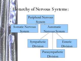
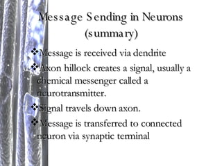
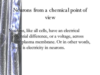
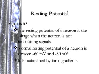
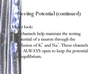
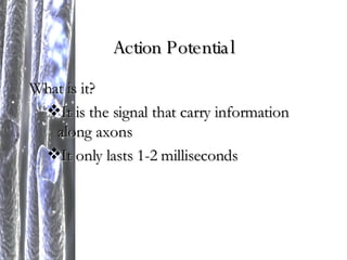
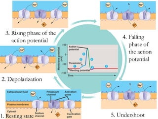
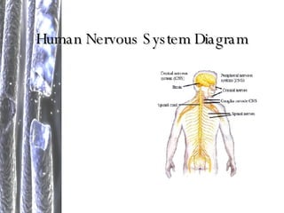
The Nervous System (Chapter 48) Samuel Black, Glasha Marcon, Csilla T óth, Kina Winoto
Background Information Neuron = a nerve cell, makes up nerves Axon = a nerve fiber THEREFORE, a nerve is made of many axons and neurons
Organization of Nervous Systems Nerves, which make up nervous systems, are organized in the following way…
Hierarchy of Nervous Systems: Peripheral Nervous System Somatic Nervous System Automatic Nervous System Sympathetic Division Parasympathetic Division Enteric Division
Central Nervous System (CNS) What is it? Simplest version = a small brain and longitudinal nerve cords BASICALLY, a brain and a mode of transporting “messages” to the brain (i.e. a spinal cord) Ganglia = segmentally arranged clusters of neurons (found in complex CNSs)
Peripheral Nervous System (PNS) What is it? Nerves that connect the CNS with the rest of an organism’s body Examples: sensory receptors, spinal nerves, cranial nerves
Somatic Nervous System What is it? It consists of peripheral nerve fibers that deliver sensory information to the CNS. It also consists of motor nerve fibers that extend to skeletal muscle.
Autonomic Nervous System (ANS) What is it? The ANS regulates the body’s internal environment by controlling smooth and cardiac muscles and vital organs. Examples: lungs, heart, intestines
Sympathetic Division  (part of the ANS) What is it? This division is activated during the “flight-or-fight” response as the heart beats faster, the liver converts glycogen to glucose, and the lungs adapt to support increased gas exchange. Examples/organs involved: heart, liver, lungs
Parasympathetic Division (part of the ANS) What is it? This division promotes calming and a return to the “rest and digest” mode as the heart slows down, the liver starts creating more glycogen, and digestion begins. Examples/organs involved: heart, liver, stomach
Enteric Division  (part of the ANS) What is it? It consists of networks of neurons in the digestive tracts, pancreas, and gallbladder. It controls these organs’ secretions. Examples/organs involved: intestines, pancreas, and gallbladder
Information Processing:   Typical Nerve Pathway Sensory input Integration (brain analyzes) Motor output
Reflexes 1 2 3 4 5 6
Neurons Dendrites Axon hillock Axon Myelin sheath Synaptic terminal
Message Sending in Neurons (summary) Message is received via dendrite Axon hillock creates a signal, usually a chemical messenger called a neurotransmitter. Signal travels down axon. Message is transferred to connected neuron via synaptic terminal
Neurons from a chemical point of view Neurons, like all cells, have an electrical potential difference, or a voltage, across their plasma membrane. Or in other words, there is electricity in neurons.
Resting Potential What is it? The resting potential of a neuron is the voltage when the neuron is not transmitting signals Normal resting potential of a neuron is between -60 mV and -80 mV It is maintained by ionic gradients.
Resting Potential (continued) A closer look: Ion channels help maintain the resting potential of a neuron through the diffusion of K +  and Na + . These channels are ALWAYS open to keep the potential at equilibrium.
Gated Ion Channels These channels open and close in response to stimuli. There are 3 types: Stretch-gated ion channels Ligand-gated ion channels Voltage-gated ion channels
What is it? It is the signal that carry information along axons It only lasts 1-2 milliseconds Action Potential
Production of Action Potentials This process involves the opening and closing of many gates and is best represented with the following diagram…
1. Resting state 2. Depolarization 3. Rising phase of the action potential 4. Falling phase of the action potential 5. Undershoot
Action Potential in Axons 1 2 3
Action Potential in Axons (summary) In the axon hillock, an action potential is created and spreads Na + , which triggers the depolarization of the neighboring regions in the axon.  From this depolarization, the action potential is started again, which yet again triggers depolarization in neighboring regions.  This process is repeated down the length of the axon.
Neurotransmitters Acetylcholine Most common neurotransmitter in invertebrates and vertebrates Biogenic amines Derived from amino acids, involved in indirect synaptic transmission Amino Acids Gamma aminobutyric acid, glycine, glutamate, and aspartate  Gases NO and CO act as local regulators
Impulse Propagation Each action potential is regenerated along the entire length of the axon through depolarization which triggers a new action potential Action potentials normally move in only one direction The speed at which an action potential propagates along an axon relates to the diameter of the axon (faster)  and myelinated neurons which only depolarize at Ranvier nodes
Neuron Communication Chemical Neurotransmitters A presynaptic neuron synthesizes a neurotransmitter and packages it in synaptic vesicles When an impulse reaches the terminal end it depolarizes the terminal membrane, opening voltage-gated calcium channels in the membrane.  The increase in Ca 2+  causes the release of neurotransmitters by exocytosis
The Synapse The following occurs at the synapse: After the Ca 2+  influx occurs the synaptic vesicles fuse with the presynaptic membrane The vesicles release neurotransmitters into the synaptic cleft The neurotransmitters bind to the open receptor of the ligand-gated ion channel Both Na +  and K +  then diffuse through the channels
Graded Potentials Occurrences at synaptic inputs on cell bodies Excitatory postsynaptic potentials (EPSPs) When the membrane depolarizes in the presence of Na +  and K +  the membrane potential reaches a point between E Na  and E K Brinings the membrane potential toward the threshold Inhibitory postsynaptic potentials (IPSPs) A different neurotransmitter only binds to K +  selective channels The postsynaptic membrane hyperpolarizes, which moves the membrane further from the threshold
Nervous System Variations Variations in nervous system occur throughout the animal kingdom Simplest nervous systems were radial around a gastrovascular cavity  More complex systems contain nerve nets and nerves More evolved: Cephalization: centralization of nerves in brain with ganglia extensions
Human Nervous System Diagram
The Human Brain The Brainstem Functions in homeostasis, coordination of movement, and conduction of information to higher brain centers Medulla oblongata Controls breathing, heart and blood vessel activity, swallowing, vomiting, and digestion Pons Works in conjunction with medulla: regulates breathing centers in medulla Midbrain Receipt and integration of sensory information, relays information to specific regions of forebrain, hearing (inferior colliculi) and vision (superior colliculi) Reticular formation (reticular activating system) Diffuse neuron network, which regulates sleep and arousal
The Cerebellum Important for coordination and error checking during motor, perceptual, and cognitive functions Involved with Learning Learned motor skills Coordinates movement and balance Hand-eye coordination The Diencephalon Develops into three adult regions Epithalamus Pineal gland Choroid plexus Capillaries Thalamus Main input center for motor information Information is sorted and sent to the appropriate region of the brain Receives information from the cerebrum and parts of the brain that regulate emotion and arousal Hypothalamus Homeostatic regulation Thermostat, sexual and mating behaviors, fight-or-flight, and pleasure
Circadian Rhythms Biological Clock (Suprachiasmatic nuclei) Hormone release Huger Heightened sensitivity
The Cerebrum Supports olfactory reception as well as audiotry and visual processing Divided into right and left cerebral hemispheres The left hemisphere controls and monitors the right side of the body The right hemisphere controls and monitors the left side of the body Outer covering of grey matter called cerebral cortex Most complex part of the brain Sensory information is analyzed Motor commands are issued Language is generated Internal white matter, and neurons called basal nuclei (deep within) Basal Nuclei Centers for planning and movement sequences The  corpus callosum  enables communication between  the right and left cerebral cortices
Lateralization of Cortical Function Right and left hemispheres become more adapt at certain skills Left Hemisphere Language, math, logic, and processing sequences Right Hemisphere Pattern recognition, face recognition, spatial relations, nonverbal thinking, emotional processing, and multi-tasking
Brain Attributes Limbic system Amygdala, hippocampus,  and olfactory bulb Deals with emotions Memory and Learning Short-term memory Long-term memory Long-term potentiation
Nervous System Diseases and Disorders Schizophrenia Depression Alzheimer’s Disease Parkinson’s Disease
Extra Credit Insect Nervous System Two main divisions Brain Ventral nerve cord Head capsule contains six pairs of ganglia, the first three pairs are fused into the brain The last three pairs are fused into the subesophageal ganglion  The number of ganglia differs depending on the insect species: cockroaches have six ganglia in their abdomen

Nervoussystempresentation 1193558738696238 3

  • 1.
    The Nervous System(Chapter 48) Samuel Black, Glasha Marcon, Csilla T óth, Kina Winoto
  • 2.
    Background Information Neuron= a nerve cell, makes up nerves Axon = a nerve fiber THEREFORE, a nerve is made of many axons and neurons
  • 3.
    Organization of NervousSystems Nerves, which make up nervous systems, are organized in the following way…
  • 4.
    Hierarchy of NervousSystems: Peripheral Nervous System Somatic Nervous System Automatic Nervous System Sympathetic Division Parasympathetic Division Enteric Division
  • 5.
    Central Nervous System(CNS) What is it? Simplest version = a small brain and longitudinal nerve cords BASICALLY, a brain and a mode of transporting “messages” to the brain (i.e. a spinal cord) Ganglia = segmentally arranged clusters of neurons (found in complex CNSs)
  • 6.
    Peripheral Nervous System(PNS) What is it? Nerves that connect the CNS with the rest of an organism’s body Examples: sensory receptors, spinal nerves, cranial nerves
  • 7.
    Somatic Nervous SystemWhat is it? It consists of peripheral nerve fibers that deliver sensory information to the CNS. It also consists of motor nerve fibers that extend to skeletal muscle.
  • 8.
    Autonomic Nervous System(ANS) What is it? The ANS regulates the body’s internal environment by controlling smooth and cardiac muscles and vital organs. Examples: lungs, heart, intestines
  • 9.
    Sympathetic Division (part of the ANS) What is it? This division is activated during the “flight-or-fight” response as the heart beats faster, the liver converts glycogen to glucose, and the lungs adapt to support increased gas exchange. Examples/organs involved: heart, liver, lungs
  • 10.
    Parasympathetic Division (partof the ANS) What is it? This division promotes calming and a return to the “rest and digest” mode as the heart slows down, the liver starts creating more glycogen, and digestion begins. Examples/organs involved: heart, liver, stomach
  • 11.
    Enteric Division (part of the ANS) What is it? It consists of networks of neurons in the digestive tracts, pancreas, and gallbladder. It controls these organs’ secretions. Examples/organs involved: intestines, pancreas, and gallbladder
  • 12.
    Information Processing: Typical Nerve Pathway Sensory input Integration (brain analyzes) Motor output
  • 13.
    Reflexes 1 23 4 5 6
  • 14.
    Neurons Dendrites Axonhillock Axon Myelin sheath Synaptic terminal
  • 15.
    Message Sending inNeurons (summary) Message is received via dendrite Axon hillock creates a signal, usually a chemical messenger called a neurotransmitter. Signal travels down axon. Message is transferred to connected neuron via synaptic terminal
  • 16.
    Neurons from achemical point of view Neurons, like all cells, have an electrical potential difference, or a voltage, across their plasma membrane. Or in other words, there is electricity in neurons.
  • 17.
    Resting Potential Whatis it? The resting potential of a neuron is the voltage when the neuron is not transmitting signals Normal resting potential of a neuron is between -60 mV and -80 mV It is maintained by ionic gradients.
  • 18.
    Resting Potential (continued)A closer look: Ion channels help maintain the resting potential of a neuron through the diffusion of K + and Na + . These channels are ALWAYS open to keep the potential at equilibrium.
  • 19.
    Gated Ion ChannelsThese channels open and close in response to stimuli. There are 3 types: Stretch-gated ion channels Ligand-gated ion channels Voltage-gated ion channels
  • 20.
    What is it?It is the signal that carry information along axons It only lasts 1-2 milliseconds Action Potential
  • 21.
    Production of ActionPotentials This process involves the opening and closing of many gates and is best represented with the following diagram…
  • 22.
    1. Resting state2. Depolarization 3. Rising phase of the action potential 4. Falling phase of the action potential 5. Undershoot
  • 23.
  • 24.
    Action Potential inAxons (summary) In the axon hillock, an action potential is created and spreads Na + , which triggers the depolarization of the neighboring regions in the axon. From this depolarization, the action potential is started again, which yet again triggers depolarization in neighboring regions. This process is repeated down the length of the axon.
  • 25.
    Neurotransmitters Acetylcholine Mostcommon neurotransmitter in invertebrates and vertebrates Biogenic amines Derived from amino acids, involved in indirect synaptic transmission Amino Acids Gamma aminobutyric acid, glycine, glutamate, and aspartate Gases NO and CO act as local regulators
  • 26.
    Impulse Propagation Eachaction potential is regenerated along the entire length of the axon through depolarization which triggers a new action potential Action potentials normally move in only one direction The speed at which an action potential propagates along an axon relates to the diameter of the axon (faster) and myelinated neurons which only depolarize at Ranvier nodes
  • 27.
    Neuron Communication ChemicalNeurotransmitters A presynaptic neuron synthesizes a neurotransmitter and packages it in synaptic vesicles When an impulse reaches the terminal end it depolarizes the terminal membrane, opening voltage-gated calcium channels in the membrane. The increase in Ca 2+ causes the release of neurotransmitters by exocytosis
  • 28.
    The Synapse Thefollowing occurs at the synapse: After the Ca 2+ influx occurs the synaptic vesicles fuse with the presynaptic membrane The vesicles release neurotransmitters into the synaptic cleft The neurotransmitters bind to the open receptor of the ligand-gated ion channel Both Na + and K + then diffuse through the channels
  • 29.
    Graded Potentials Occurrencesat synaptic inputs on cell bodies Excitatory postsynaptic potentials (EPSPs) When the membrane depolarizes in the presence of Na + and K + the membrane potential reaches a point between E Na and E K Brinings the membrane potential toward the threshold Inhibitory postsynaptic potentials (IPSPs) A different neurotransmitter only binds to K + selective channels The postsynaptic membrane hyperpolarizes, which moves the membrane further from the threshold
  • 30.
    Nervous System VariationsVariations in nervous system occur throughout the animal kingdom Simplest nervous systems were radial around a gastrovascular cavity More complex systems contain nerve nets and nerves More evolved: Cephalization: centralization of nerves in brain with ganglia extensions
  • 31.
  • 32.
    The Human BrainThe Brainstem Functions in homeostasis, coordination of movement, and conduction of information to higher brain centers Medulla oblongata Controls breathing, heart and blood vessel activity, swallowing, vomiting, and digestion Pons Works in conjunction with medulla: regulates breathing centers in medulla Midbrain Receipt and integration of sensory information, relays information to specific regions of forebrain, hearing (inferior colliculi) and vision (superior colliculi) Reticular formation (reticular activating system) Diffuse neuron network, which regulates sleep and arousal
  • 33.
    The Cerebellum Importantfor coordination and error checking during motor, perceptual, and cognitive functions Involved with Learning Learned motor skills Coordinates movement and balance Hand-eye coordination The Diencephalon Develops into three adult regions Epithalamus Pineal gland Choroid plexus Capillaries Thalamus Main input center for motor information Information is sorted and sent to the appropriate region of the brain Receives information from the cerebrum and parts of the brain that regulate emotion and arousal Hypothalamus Homeostatic regulation Thermostat, sexual and mating behaviors, fight-or-flight, and pleasure
  • 34.
    Circadian Rhythms BiologicalClock (Suprachiasmatic nuclei) Hormone release Huger Heightened sensitivity
  • 35.
    The Cerebrum Supportsolfactory reception as well as audiotry and visual processing Divided into right and left cerebral hemispheres The left hemisphere controls and monitors the right side of the body The right hemisphere controls and monitors the left side of the body Outer covering of grey matter called cerebral cortex Most complex part of the brain Sensory information is analyzed Motor commands are issued Language is generated Internal white matter, and neurons called basal nuclei (deep within) Basal Nuclei Centers for planning and movement sequences The corpus callosum enables communication between the right and left cerebral cortices
  • 36.
    Lateralization of CorticalFunction Right and left hemispheres become more adapt at certain skills Left Hemisphere Language, math, logic, and processing sequences Right Hemisphere Pattern recognition, face recognition, spatial relations, nonverbal thinking, emotional processing, and multi-tasking
  • 37.
    Brain Attributes Limbicsystem Amygdala, hippocampus, and olfactory bulb Deals with emotions Memory and Learning Short-term memory Long-term memory Long-term potentiation
  • 38.
    Nervous System Diseasesand Disorders Schizophrenia Depression Alzheimer’s Disease Parkinson’s Disease
  • 39.
    Extra Credit InsectNervous System Two main divisions Brain Ventral nerve cord Head capsule contains six pairs of ganglia, the first three pairs are fused into the brain The last three pairs are fused into the subesophageal ganglion The number of ganglia differs depending on the insect species: cockroaches have six ganglia in their abdomen