Basic Concepts
Nerve conduction
Normalvalues
Repetitive stim
Normal anatomy
Median nerve
Ulnar nerve
Peroneal nerve
Radial nerve
Other compressions
Anomalous innervation
Axonal vs. demyel.
Root and plexus
Cranial nerves
A Self-Study Curriculum in Nerve
Conduction Studies for Technologists
Content developed by Zachary N. London, MD, Gary W. Gallagher, MD, and Matthew J.
Ebright, MD as part of a A Self-Study Curriculum in Electromyography and Nerve
Conduction Studies for Residents and Fellows. Content has been tailored for the
technologist’s role.
2.
Basic Concepts
Nerve conduction
Normalvalues
Repetitive stim
Normal anatomy
Median nerve
Ulnar nerve
Peroneal nerve
Radial nerve
Other compressions
Anomalous innervation
Axonal vs. demyel.
Root and plexus
Cranial nerves
Basic Concepts
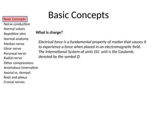
What is charge?
Electrical force is a fundamental property of matter that causes it
to experience a force when placed in an electromagnetic field.
The International System of units (SI) unit is the Coulomb,
denoted by the symbol Ǫ.
3.
Basic Concepts
Nerve conduction
Normalvalues
Repetitive stim
Normal anatomy
Median nerve
Ulnar nerve
Peroneal nerve
Radial nerve
Other compressions
Anomalous innervation
Axonal vs. demyel.
Root and plexus
Cranial nerves
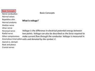
What is voltage?
Voltage is the difference in electrical potential energy between
two points. Voltage can also be described as the force required to
make current flow through the conductor. Voltage is measured in
volts and denoted by the symbol, E.
Basic Concepts
4.
Basic Concepts
Nerve conduction
Normalvalues
Repetitive stim
Normal anatomy
Median nerve
Ulnar nerve
Peroneal nerve
Radial nerve
Other compressions
Anomalous innervation
Axonal vs. demyel.
Root and plexus
Cranial nerves
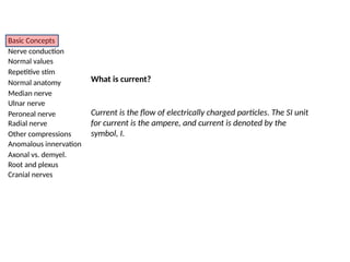
What is current?
Current is the flow of electrically charged particles. The SI unit
for current is the ampere, and current is denoted by the
symbol, I.
5.
Basic Concepts
Nerve conduction
Normalvalues
Repetitive stim
Normal anatomy
Median nerve
Ulnar nerve
Peroneal nerve
Radial nerve
Other compressions
Anomalous innervation
Axonal vs. demyel.
Root and plexus
Cranial nerves
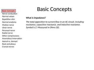
What is impedance?
The total opposition to current flow in an AC circuit, including
resistance, capacitive reactance, and inductive reactance.
Symbol is Z. Measured in Ohms (Ω).
Basic Concepts
6.
Basic Concepts
Nerve conduction
Normalvalues
Repetitive stim
Normal anatomy
Median nerve
Ulnar nerve
Peroneal nerve
Radial nerve
Other compressions
Anomalous innervation
Axonal vs. demyel.
Root and plexus
Cranial nerves
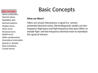
What are filters?
Filters are circuits that process a signal (i.e. remove
unwanted electrical noise). Electrodiagnostic studies use low-
frequency (high-pass) and high-frequency (low-pass) filters to
exclude high- and low-frequency electrical noise to reproduce
the signal of interest.
Basic Concepts
7.
Basic Concepts
Nerve conduction
Normalvalues
Repetitive stim
Normal anatomy
Median nerve
Ulnar nerve
Peroneal nerve
Radial nerve
Other compressions
Anomalous innervation
Axonal vs. demyel.
Root and plexus
Cranial nerves
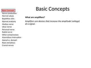
What are amplifiers?
Amplifiers are devices that increase the amplitude (voltage)
of a signal.
Basic Concepts
8.
Basic Concepts
Nerve conduction
Normalvalues
Repetitive stim
Normal anatomy
Median nerve
Ulnar nerve
Peroneal nerve
Radial nerve
Other compressions
Anomalous innervation
Axonal vs. demyel.
Root and plexus
Cranial nerves
Nerve Conduction Studies
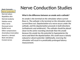
What is the difference between an anode and a cathode?
An anode is the terminal on the stimulator where current
flows in. The cathode is the terminal on the stimulator where
current flows out. Depolarization of a nerve occurs under the
cathode, and the depolarization proceeds in both directions
(orthodromic and antidromic). The cathode should be placed
closer to the active recording electrode than the anode
because the anode has the potential to hyperpolarize the
nerve and block the depolarization; this could cause a falsely
reduced or absent potential. Additionally, reversing the
stimulator will result in a predictably prolonged latency
measurement.
9.
Basic Concepts
Nerve conduction
Normalvalues
Repetitive stim
Normal anatomy
Median nerve
Ulnar nerve
Peroneal nerve
Radial nerve
Other compressions
Anomalous innervation
Axonal vs. demyel.
Root and plexus
Cranial nerves
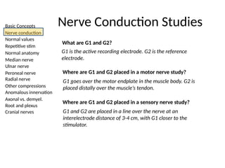
What are G1 and G2?
G1 is the active recording electrode. G2 is the reference
electrode.
Where are G1 and G2 placed in a motor nerve study?
G1 goes over the motor endplate in the muscle body. G2 is
placed distally over the muscle’s tendon.
Where are G1 and G2 placed in a sensory nerve study?
G1 and G2 are placed in a line over the nerve at an
interelectrode distance of 3-4 cm, with G1 closer to the
stimulator.
Nerve Conduction Studies
10.
Basic Concepts
Nerve conduction
Normalvalues
Repetitive stim
Normal anatomy
Median nerve
Ulnar nerve
Peroneal nerve
Radial nerve
Other compressions
Anomalous innervation
Axonal vs. demyel.
Root and plexus
Cranial nerves
Nerve Conduction Studies
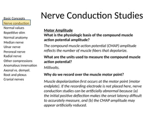
Motor Amplitude
What is the physiologic basis of the compound muscle
action potential amplitude?
The compound muscle action potential (CMAP) amplitude
reflects the number of muscle fibers that depolarize.
What are the units used to measure the compound muscle
action potential?
Millivolts.
Why do we record over the muscle motor point?
Muscle depolarization first occurs at the motor point (motor
endplate). If the recording electrode is not placed here, nerve
conduction studies can be artificially abnormal because (a)
the initial positive deflection makes the onset latency difficult
to accurately measure, and (b) the CMAP amplitude may
appear artificially reduced.
11.
Basic Concepts
Nerve conduction
Normalvalues
Repetitive stim
Normal anatomy
Median nerve
Ulnar nerve
Peroneal nerve
Radial nerve
Other compressions
Anomalous innervation
Axonal vs. demyel.
Root and plexus
Cranial nerves
Nerve Conduction Studies
Sensory Amplitude
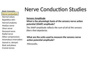
What is the physiologic basis of the sensory nerve action
potential (SNAP) amplitude?
The SNAP amplitude reflects the sum of all of the sensory
fibers that depolarize.
What are the units used to measure the sensory nerve
action potential amplitude?
Microvolts.
12.
Basic Concepts
Nerve conduction
Normalvalues
Repetitive stim
Normal anatomy
Median nerve
Ulnar nerve
Peroneal nerve
Radial nerve
Other compressions
Anomalous innervation
Axonal vs. demyel.
Root and plexus
Cranial nerves
Nerve Conduction Studies
Motor latency
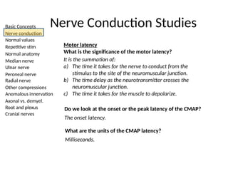
What is the significance of the motor latency?
It is the summation of:
a) The time it takes for the nerve to conduct from the
stimulus to the site of the neuromuscular junction.
b) The time delay as the neurotransmitter crosses the
neuromuscular junction.
c) The time it takes for the muscle to depolarize.
Do we look at the onset or the peak latency of the CMAP?
The onset latency.
What are the units of the CMAP latency?
Milliseconds.
13.
Basic Concepts
Nerve conduction
Normalvalues
Repetitive stim
Normal anatomy
Median nerve
Ulnar nerve
Peroneal nerve
Radial nerve
Other compressions
Anomalous innervation
Axonal vs. demyel.
Root and plexus
Cranial nerves
Nerve Conduction Studies
Sensory latency
What is the significance of the onset and peak sensory
latencies?
The onset latency measures the time from stimulation to
initial deflection of the SNAP. It represents the fastest and
largest nerve fibers. The peak latency is measured at the
midpoint of the first negative peak of the SNAP.
Do we look at onset or peak latency for the SNAP? Why?
The peak latency. It is more reliable and less subject to
artifact than the onset latency.
What are the units of the SNAP latency?
Milliseconds.
14.
Basic Concepts
Nerve conduction
Normalvalues
Repetitive stim
Normal anatomy
Median nerve
Ulnar nerve
Peroneal nerve
Radial nerve
Other compressions
Anomalous innervation
Axonal vs. demyel.
Root and plexus
Cranial nerves
Nerve Conduction Studies
Conduction Velocity
What is the physiologic significance of a slow conduction
velocity?
Conduction velocity measures the speed of the fastest and
largest conducting axons. Slowing is most commonly
associated with demyelination, but can also be seen
secondary to loss of these particular fastest and largest
axons.
What are the units of conduction velocity?
Meters/second.
15.
Basic Concepts
Nerve conduction
Normalvalues
Repetitive stim
Normal anatomy
Median nerve
Ulnar nerve
Peroneal nerve
Radial nerve
Other compressions
Anomalous innervation
Axonal vs. demyel.
Root and plexus
Cranial nerves
Nerve Conduction Studies
Conduction Velocity
How do you calculate conduction velocity in a motor nerve?
1) Stimulate at two different sites of the motor nerve.
2) Measure the distance between the two stimulation sites.
3) Divide the distance by the difference between the onset
latencies.
Conduction velocity = distance / (proximal latency – distal latency)
16.
Basic Concepts
Nerve conduction
Normalvalues
Repetitive stim
Normal anatomy
Median nerve
Ulnar nerve
Peroneal nerve
Radial nerve
Other compressions
Anomalous innervation
Axonal vs. demyel.
Root and plexus
Cranial nerves
Nerve Conduction Studies
Conduction Velocity
Why do we stimulate at two different sites along the nerve
for a motor conduction study, but not a sensory conduction
velocity?
Since you are recording CMAP over a muscle, the time from
stimulation to response includes the time to cross the
neuromuscular junction and depolarize the muscle. However,
you can calculate the conduction velocity between the two
sites by subtracting out the time and distance involved
between the distal site and the muscle.
In sensory studies, the neuromuscular junction and muscle
are not involved, so the latency only reflects the time it takes
for the nerve to depolarize. Thus, you can simply measure
distance/time.
17.
Basic Concepts
Nerve conduction
Normalvalues
Repetitive stim
Normal anatomy
Median nerve
Ulnar nerve
Peroneal nerve
Radial nerve
Other compressions
Anomalous innervation
Axonal vs. demyel.
Root and plexus
Cranial nerves
Nerve Conduction Studies
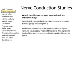
What is the difference between an orthodromic and
antidromic study?
Orthodromic: stimulation in the direction a nerve normally
travels, (going “with the grain”).
Antidromic: stimulation in the opposite direction signals
normally travel, (going “against the grain”). This is proximal
to distal in a sensory nerve and distal to proximal in a motor
nerve.
18.
Basic Concepts
Nerve conduction
Normalvalues
Repetitive stim
Normal anatomy
Median nerve
Ulnar nerve
Peroneal nerve
Radial nerve
Other compressions
Anomalous innervation
Axonal vs. demyel.
Root and plexus
Cranial nerves
Nerve Conduction Studies
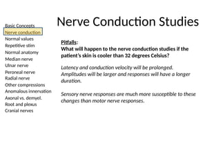
Pitfalls:
What will happen to the nerve conduction studies if the
patient’s skin is cooler than 32 degrees Celsius?
Latency and conduction velocity will be prolonged.
Amplitudes will be larger and responses will have a longer
duration.
Sensory nerve responses are much more susceptible to these
changes than motor nerve responses.
19.
Basic Concepts
Nerve conduction
Normalvalues
Repetitive stim
Normal anatomy
Median nerve
Ulnar nerve
Peroneal nerve
Radial nerve
Other compressions
Anomalous innervation
Axonal vs. demyel.
Root and plexus
Cranial nerves
Nerve Conduction Studies
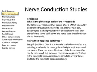
F-response
What is the physiologic basis of the F-response?
The late motor response that occurs after a CMAP. Caused by
antidromic travel up the nerve to the anterior horn cell,
backfiring of a small population of anterior horn cells, and
orthodromic travel back down the nerve past the stimulation
site to the muscle.
How is the F-response performed?
Setup is just like a CMAP, but turn the cathode around so it is
pointing proximally. Increase gain to 200 μV to pick up small
responses. There are several features of the F-response that
can be measured, but the most commonly measured feature
is the minimal F-response latency. Stimulate several times
and take the minimal F-response latency.
20.
Basic Concepts
Nerve conduction
Normalvalues
Repetitive stim
Normal anatomy
Median nerve
Ulnar nerve
Peroneal nerve
Radial nerve
Other compressions
Anomalous innervation
Axonal vs. demyel.
Root and plexus
Cranial nerves
Nerve Conduction Studies
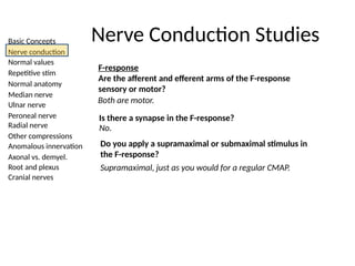
F-response
Are the afferent and efferent arms of the F-response
sensory or motor?
Both are motor.
Is there a synapse in the F-response?
No.
Do you apply a supramaximal or submaximal stimulus in
the F-response?
Supramaximal, just as you would for a regular CMAP.
21.
Basic Concepts
Nerve conduction
Normalvalues
Repetitive stim
Normal anatomy
Median nerve
Ulnar nerve
Peroneal nerve
Radial nerve
Other compressions
Anomalous innervation
Axonal vs. demyel.
Root and plexus
Cranial nerves
Nerve Conduction Studies
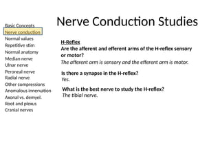
H-Reflex
Are the afferent and efferent arms of the H-reflex sensory
or motor?
The afferent arm is sensory and the efferent arm is motor.
Is there a synapse in the H-reflex?
Yes.
What is the best nerve to study the H-reflex?
The tibial nerve.
22.
Basic Concepts
Nerve conduction
Normalvalues
Repetitive stim
Normal anatomy
Median nerve
Ulnar nerve
Peroneal nerve
Radial nerve
Other compressions
Anomalous innervation
Axonal vs. demyel.
Root and plexus
Cranial nerves
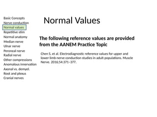
Normal Values
The following reference values are provided
from the AANEM Practice Topic
Chen S, et al. Electrodiagnostic reference values for upper and
lower limb nerve conduction studies in adult populations. Muscle
Nerve. 2016;54:371–377.
23.
Basic Concepts
Nerve conduction
Normalvalues
Repetitive stim
Normal anatomy
Median nerve
Ulnar nerve
Peroneal nerve
Radial nerve
Other compressions
Anomalous innervation
Axonal vs. demyel.
Root and plexus
Cranial nerves
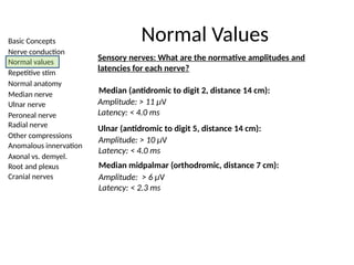
Normal Values
Sensory nerves: What are the normative amplitudes and
latencies for each nerve?
Ulnar (antidromic to digit 5, distance 14 cm):
Amplitude: > 11 µV
Latency: < 4.0 ms
Amplitude: > 10 µV
Latency: < 4.0 ms
Median midpalmar (orthodromic, distance 7 cm):
Amplitude: > 6 µV
Latency: < 2.3 ms
Median (antidromic to digit 2, distance 14 cm):
24.
Basic Concepts
Nerve conduction
Normalvalues
Repetitive stim
Normal anatomy
Median nerve
Ulnar nerve
Peroneal nerve
Radial nerve
Other compressions
Anomalous innervation
Axonal vs. demyel.
Root and plexus
Cranial nerves
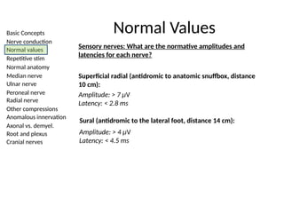
Normal Values
Sensory nerves: What are the normative amplitudes and
latencies for each nerve?
Superficial radial (antidromic to anatomic snuffbox, distance
10 cm):
Amplitude: > 7 µV
Latency: < 2.8 ms
Sural (antidromic to the lateral foot, distance 14 cm):
Amplitude: > 4 µV
Latency: < 4.5 ms
25.
Basic Concepts
Nerve conduction
Normalvalues
Repetitive stim
Normal anatomy
Median nerve
Ulnar nerve
Peroneal nerve
Radial nerve
Other compressions
Anomalous innervation
Axonal vs. demyel.
Root and plexus
Cranial nerves
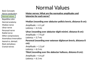
Normal Values
Motor nerves: What are the normative amplitudes and
latencies for each nerve?
Median (recording over abductor pollicis brevis, distance 8 cm):
Amplitude: > 7.9 µV
Latency: < 3.7 ms
Amplitude: > 4.1 µV
Latency: < 4.5 ms
Peroneal (recording over extensor digitorum brevis, distance 8
cm):
Amplitude: > 1.3 µV
Latency: < 6.5 ms
Tibial (recording over the abductor halluces, distance 8 cm):
Amplitude: > 4.4 µV
Latency: < 6.1 ms
Ulnar (recording over abductor digiti minimi. distance 8 cm):
26.
Basic Concepts
Nerve conduction
Normalvalues
Repetitive stim
Normal anatomy
Median nerve
Ulnar nerve
Peroneal nerve
Radial nerve
Other compressions
Anomalous innervation
Axonal vs. demyel.
Root and plexus
Cranial nerves
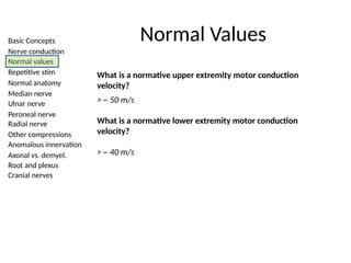
Normal Values
What is a normative upper extremity motor conduction
velocity?
> ~ 50 m/s
What is a normative lower extremity motor conduction
velocity?
> ~ 40 m/s
27.
Basic Concepts
Nerve conduction
Normalvalues
Repetitive stim
Normal anatomy
Median nerve
Ulnar nerve
Peroneal nerve
Radial nerve
Other compressions
Anomalous innervation
Axonal vs. demyel.
Root and plexus
Cranial nerves
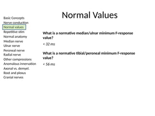
Normal Values
What is a normative median/ulnar minimum F-response
value?
< 32 ms
What is a normative tibial/peroneal minimum F-response
value?
< 56 ms
28.
Basic Concepts
Nerve conduction
Normalvalues
Repetitive stim
Normal anatomy
Median nerve
Ulnar nerve
Peroneal nerve
Radial nerve
Other compressions
Anomalous innervation
Axonal vs. demyel.
Root and plexus
Cranial nerves
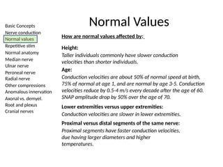
Normal Values
How are normal values affected by:
Height:
Taller individuals commonly have slower conduction
velocities than shorter individuals.
Age:
Conduction velocities are about 50% of normal speed at birth,
75% of normal at age 1, and are normal by age 3-5. Conduction
velocities reduce by 0.5-4 m/s every decade after the age of 60.
SNAP amplitude drop by 50% over the age of 70.
Lower extremities versus upper extremities:
Conduction velocities are slower in lower extremities.
Proximal versus distal segments of the same nerve:
Proximal segments have faster conduction velocities,
due having larger diameters and higher
temperatures.
29.
Basic Concepts
Nerve conduction
Normalvalues
Repetitive stim
Normal anatomy
Median nerve
Ulnar nerve
Peroneal nerve
Radial nerve
Other compressions
Anomalous innervation
Axonal vs. demyel.
Root and plexus
Cranial nerves
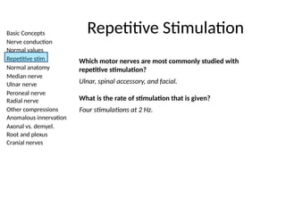
Repetitive Stimulation
Which motor nerves are most commonly studied with
repetitive stimulation?
Ulnar, spinal accessory, and facial.
What is the rate of stimulation that is given?
Four stimulations at 2 Hz.
30.
Basic Concepts
Nerve conduction
Normalvalues
Repetitive stim
Normal anatomy
Median nerve
Ulnar nerve
Peroneal nerve
Radial nerve
Other compressions
Anomalous innervation
Axonal vs. demyel.
Root and plexus
Cranial nerves
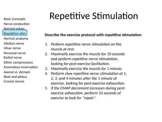
Repetitive Stimulation
Describe the exercise protocol with repetitive stimulation:
1. Perform repetitive nerve stimulation on the
muscle at rest.
2. Maximally exercise the muscle for 10 seconds
and perform repetitive nerve stimulation,
looking for post-exercise facilitation.
3. Maximally exercise the muscle for 1 minute.
4. Perform slow repetitive nerve stimulation at 1,
2, 3, and 4 minutes after the 1 minute of
exercise, looking for post-exercise exhaustion.
5. If the CMAP decrement increases during post-
exercise exhaustion, perform 10 seconds of
exercise to look for “repair”.
31.
Basic Concepts
Nerve conduction
Normalvalues
Repetitive stim
Normal anatomy
Median nerve
Ulnar nerve
Peroneal nerve
Radial nerve
Other compressions
Anomalous innervation
Axonal vs. demyel.
Root and plexus
Cranial nerves
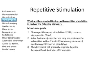
Repetitive Stimulation
What are the expected findings with repetitive stimulation
in each of the following disorders:
Myasthenia gravis:
1. Slow repetitive nerve stimulation (2-3 Hz) causes a
decrement in CMAP.
2. After 1 minute of exercise, you may see post-exercise
exhaustion, with a transiently worsening decrement
on slow repetitive nerve stimulation.
3. The decrement will gradually return to baseline
between 3 and 5 minutes after exercise.
32.
Basic Concepts
Nerve conduction
Normalvalues
Repetitive stim
Normal anatomy
Median nerve
Ulnar nerve
Peroneal nerve
Radial nerve
Other compressions
Anomalous innervation
Axonal vs. demyel.
Root and plexus
Cranial nerves
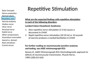
Repetitive Stimulation
What are the expected findings with repetitive stimulation
in each of the following disorders:
Lambert Eaton Myasthenic Syndrome:
1. Slow repetitive nerve stimulation (2-3 Hz) causes a
decrement in CMAP.
2. Rapid repetitive nerve stimulation (30-50 Hz) or 10 seconds
of exercise produces a marked facilitation in CMAP.
For further reading on neuromuscular junction anatomy
and testing, see AAEE minimonograph #33:
Keesey JC. AAEE Minimonograph #33: Electrodiagnostic approach to
defects of neuromuscular transmission. Muscle Nerve.
1989;12(8):613-626
33.
Basic Concepts
Nerve conduction
Normalvalues
Repetitive stim
Normal anatomy
Median nerve
Ulnar nerve
Peroneal nerve
Radial nerve
Other compressions
Anomalous innervation
Axonal vs. demyel.
Root and plexus
Cranial nerves
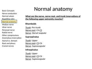
Normal anatomy
What are the nerve, nerve root, and trunk innervations of
the following upper extremity muscles?
Rhomboids
Trunk: Pre-trunk
Nerve root: C4-C5
Nerve: Dorsal scapular
Supraspinatus
Trunk: Upper
Nerve root: C5-C6
Nerve: Suprascapular
Infraspinatus
Trunk: Upper
Nerve root: C5-C6
Nerve: Suprascapular
34.
Basic Concepts
Nerve conduction
Normalvalues
Repetitive stim
Normal anatomy
Median nerve
Ulnar nerve
Peroneal nerve
Radial nerve
Other compressions
Anomalous innervation
Axonal vs. demyel.
Root and plexus
Cranial nerves
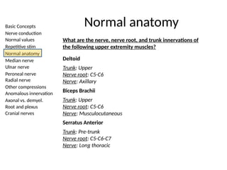
Normal anatomy
What are the nerve, nerve root, and trunk innervations of
the following upper extremity muscles?
Deltoid
Trunk: Upper
Nerve root: C5-C6
Nerve: Axillary
Biceps Brachii
Trunk: Upper
Nerve root: C5-C6
Nerve: Musculocutaneous
Serratus Anterior
Trunk: Pre-trunk
Nerve root: C5-C6-C7
Nerve: Long thoracic
35.
Basic Concepts
Nerve conduction
Normalvalues
Repetitive stim
Normal anatomy
Median nerve
Ulnar nerve
Peroneal nerve
Radial nerve
Other compressions
Anomalous innervation
Axonal vs. demyel.
Root and plexus
Cranial nerves
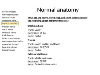
Normal anatomy
What are the nerve, nerve root, and trunk innervations of
the following upper extremity muscles?
Brachioradialis
Trunk: Upper
Nerve root: C5-C6
Nerve: Radial
Triceps
Trunk: Upper, middle, and lower
Nerve root: C6-C7-C8
Nerve: Radial
Extensor digitorum
Trunk: Middle and lower
Nerve root: C7-C8
Nerve: Posterior interosseous
36.
Basic Concepts
Nerve conduction
Normalvalues
Repetitive stim
Normal anatomy
Median nerve
Ulnar nerve
Peroneal nerve
Radial nerve
Other compressions
Anomalous innervation
Axonal vs. demyel.
Root and plexus
Cranial nerves
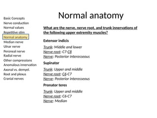
Normal anatomy
What are the nerve, nerve root, and trunk innervations of
the following upper extremity muscles?
Extensor indicis
Trunk: Middle and lower
Nerve root: C7-C8
Nerve: Posterior interosseous
Supinator
Trunk: Upper and middle
Nerve root: C6-C7
Nerve: Posterior interosseous
Pronator teres
Trunk: Upper and middle
Nerve root: C6-C7
Nerve: Median
37.
Basic Concepts
Nerve conduction
Normalvalues
Repetitive stim
Normal anatomy
Median nerve
Ulnar nerve
Peroneal nerve
Radial nerve
Other compressions
Anomalous innervation
Axonal vs. demyel.
Root and plexus
Cranial nerves
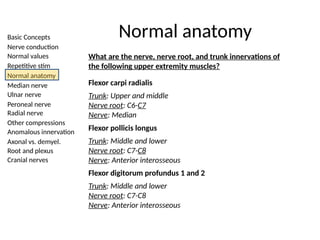
Normal anatomy
What are the nerve, nerve root, and trunk innervations of
the following upper extremity muscles?
Flexor carpi radialis
Trunk: Upper and middle
Nerve root: C6-C7
Nerve: Median
Flexor pollicis longus
Trunk: Middle and lower
Nerve root: C7-C8
Nerve: Anterior interosseous
Flexor digitorum profundus 1 and 2
Trunk: Middle and lower
Nerve root: C7-C8
Nerve: Anterior interosseous
38.
Basic Concepts
Nerve conduction
Normalvalues
Repetitive stim
Normal anatomy
Median nerve
Ulnar nerve
Peroneal nerve
Radial nerve
Other compressions
Anomalous innervation
Axonal vs. demyel.
Root and plexus
Cranial nerves
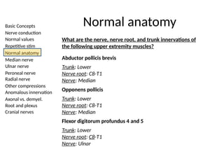
Normal anatomy
What are the nerve, nerve root, and trunk innervations of
the following upper extremity muscles?
Abductor pollicis brevis
Trunk: Lower
Nerve root: C8-T1
Nerve: Median
Opponens pollicis
Trunk: Lower
Nerve root: C8-T1
Nerve: Median
Flexor digitorum profundus 4 and 5
Trunk: Lower
Nerve root: C8-T1
Nerve: Ulnar
39.
Basic Concepts
Nerve conduction
Normalvalues
Repetitive stim
Normal anatomy
Median nerve
Ulnar nerve
Peroneal nerve
Radial nerve
Other compressions
Anomalous innervation
Axonal vs. demyel.
Root and plexus
Cranial nerves
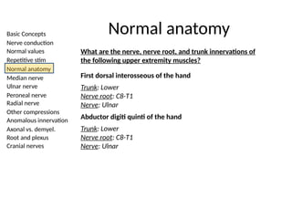
Normal anatomy
What are the nerve, nerve root, and trunk innervations of
the following upper extremity muscles?
First dorsal interosseous of the hand
Trunk: Lower
Nerve root: C8-T1
Nerve: Ulnar
Abductor digiti quinti of the hand
Trunk: Lower
Nerve root: C8-T1
Nerve: Ulnar
40.
Basic Concepts
Nerve conduction
Normalvalues
Repetitive stim
Normal anatomy
Median nerve
Ulnar nerve
Peroneal nerve
Radial nerve
Other compressions
Anomalous innervation
Axonal vs. demyel.
Root and plexus
Cranial nerves
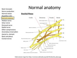
Normal anatomy
Brachial Plexus
Public domain image from https://commons.wikimedia.org/wiki/File:Brachial_plexus.jpg
41.
Basic Concepts
Nerve conduction
Normalvalues
Repetitive stim
Normal anatomy
Median nerve
Ulnar nerve
Peroneal nerve
Radial nerve
Other compressions
Anomalous innervation
Axonal vs. demyel.
Root and plexus
Cranial nerves
Normal anatomy
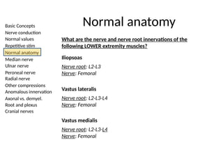
What are the nerve and nerve root innervations of the
following LOWER extremity muscles?
Iliopsoas
Nerve root: L2-L3
Nerve: Femoral
Vastus lateralis
Nerve root: L2-L3-L4
Nerve: Femoral
Vastus medialis
Nerve root: L2-L3-L4
Nerve: Femoral
42.
Basic Concepts
Nerve conduction
Normalvalues
Repetitive stim
Normal anatomy
Median nerve
Ulnar nerve
Peroneal nerve
Radial nerve
Other compressions
Anomalous innervation
Axonal vs. demyel.
Root and plexus
Cranial nerves
Normal anatomy
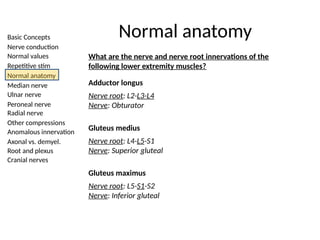
What are the nerve and nerve root innervations of the
following lower extremity muscles?
Adductor longus
Nerve root: L2-L3-L4
Nerve: Obturator
Gluteus medius
Nerve root: L4-L5-S1
Nerve: Superior gluteal
Gluteus maximus
Nerve root: L5-S1-S2
Nerve: Inferior gluteal
43.
Basic Concepts
Nerve conduction
Normalvalues
Repetitive stim
Normal anatomy
Median nerve
Ulnar nerve
Peroneal nerve
Radial nerve
Other compressions
Anomalous innervation
Axonal vs. demyel.
Root and plexus
Cranial nerves
Normal anatomy
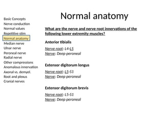
What are the nerve and nerve root innervations of the
following lower extremity muscles?
Anterior tibialis
Nerve root: L4-L5
Nerve: Deep peroneal
Extensor digitorum longus
Nerve root: L5-S1
Nerve: Deep peroneal
Extensor digitorum brevis
Nerve root: L5-S1
Nerve: Deep peroneal
44.
Basic Concepts
Nerve conduction
Normalvalues
Repetitive stim
Normal anatomy
Median nerve
Ulnar nerve
Peroneal nerve
Radial nerve
Other compressions
Anomalous innervation
Axonal vs. demyel.
Root and plexus
Cranial nerves
Normal anatomy
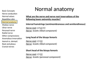
What are the nerve and nerve root innervations of the
following lower extremity muscles?
Internal hamstrings (semimembranosus and semitendinosus)
Nerve root: L4-L5-S1
Nerve: Sciatic (tibial component)
Long head of the biceps femoris
Nerve root: L5-S1
Nerve: Sciatic (tibial component)
Short head of the biceps femoris
Nerve root: L5-S1
Nerve: Sciatic (peroneal component)
45.
Basic Concepts
Nerve conduction
Normalvalues
Repetitive stim
Normal anatomy
Median nerve
Ulnar nerve
Peroneal nerve
Radial nerve
Other compressions
Anomalous innervation
Axonal vs. demyel.
Root and plexus
Cranial nerves
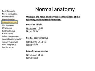
Normal anatomy
What are the nerve and nerve root innervations of the
following lower extremity muscles?
Posterior tibialis
Nerve root: L5-S1
Nerve: Tibial
Medial gastrocnemius
Nerve root: L5-S1-S2
Nerve: Tibial
Lateral gastrocnemius
Nerve root: S1-S2
Nerve: Tibial
46.
Basic Concepts
Nerve conduction
Normalvalues
Repetitive stim
Normal anatomy
Median nerve
Ulnar nerve
Peroneal nerve
Radial nerve
Other compressions
Anomalous innervation
Axonal vs. demyel.
Root and plexus
Cranial nerves
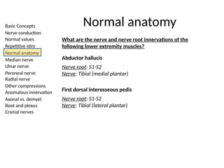
Normal anatomy
What are the nerve and nerve root innervations of the
following lower extremity muscles?
Abductor hallucis
Nerve root: S1-S2
Nerve: Tibial (medial plantar)
First dorsal interosseous pedis
Nerve root: S1-S2
Nerve: Tibial (lateral plantar)
47.
Basic Concepts
Nerve conduction
Normalvalues
Repetitive stim
Normal anatomy
Median nerve
Ulnar nerve
Peroneal nerve
Radial nerve
Other compressions
Anomalous innervation
Axonal vs. demyel.
Root and plexus
Cranial nerves
Uncommon Compression Neuropathies
Advanced Topics
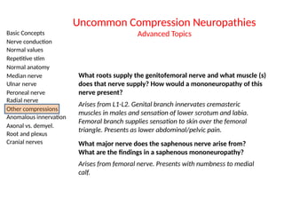
What roots supply the genitofemoral nerve and what muscle (s)
does that nerve supply? How would a mononeuropathy of this
nerve present?
Arises from L1-L2. Genital branch innervates cremasteric
muscles in males and sensation of lower scrotum and labia.
Femoral branch supplies sensation to skin over the femoral
triangle. Presents as lower abdominal/pelvic pain.
What major nerve does the saphenous nerve arise from?
What are the findings in a saphenous mononeuropathy?
Arises from femoral nerve. Presents with numbness to medial
calf.
48.
Basic Concepts
Nerve conduction
Normalvalues
Repetitive stim
Normal anatomy
Median nerve
Ulnar nerve
Peroneal nerve
Radial nerve
Other compressions
Anomalous innervation
Axonal vs. demyel.
Root and plexus
Cranial nerves
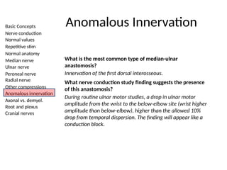
Anomalous Innervation
What is the most common type of median-ulnar
anastomosis?
Innervation of the first dorsal interosseous.
What nerve conduction study finding suggests the presence
of this anastomosis?
During routine ulnar motor studies, a drop in ulnar motor
amplitude from the wrist to the below-elbow site (wrist higher
amplitude than below-elbow), higher than the allowed 10%
drop from temporal dispersion. The finding will appear like a
conduction block.
49.
Basic Concepts
Nerve conduction
Normalvalues
Repetitive stim
Normal anatomy
Median nerve
Ulnar nerve
Peroneal nerve
Radial nerve
Other compressions
Anomalous innervation
Axonal vs. demyel.
Root and plexus
Cranial nerves
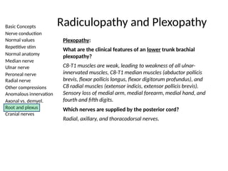
Radiculopathy and Plexopathy
Plexopathy:
What are the clinical features of an lower trunk brachial
plexopathy?
C8-T1 muscles are weak, leading to weakness of all ulnar-
innervated muscles, C8-T1 median muscles (abductor pollicis
brevis, flexor pollicis longus, flexor digitorum profundus), and
C8 radial muscles (extensor indicis, extensor pollicis brevis).
Sensory loss of medial arm, medial forearm, medial hand, and
fourth and fifth digits.
Which nerves are supplied by the posterior cord?
Radial, axillary, and thoracodorsal nerves.
50.
Basic Concepts
Nerve conduction
Normalvalues
Repetitive stim
Normal anatomy
Median nerve
Ulnar nerve
Peroneal nerve
Radial nerve
Other compressions
Anomalous innervation
Axonal vs. demyel.
Root and plexus
Cranial nerves
Radiculopathy and Plexopathy
Plexopathy:
Which nerves are supplied by the lateral cord?
Musculocutaneous nerve (including lateral antebrachial
cutaneous) and the C6-C7 portion of median nerve.
Which nerves are supplied by the medial cord?
Ulnar nerve and the C8-T1 portion of median nerve. (Identical
to lower trunk plexopathy except for normal C8 radial
innervated muscles are not affected).
Are paraspinal muscles affected in plexopathy?
No, though rarely there can be a root avulsion that
accompanies brachial plexus injury.
51.
Basic Concepts
Nerve conduction
Normalvalues
Repetitive stim
Normal anatomy
Median nerve
Ulnar nerve
Peroneal nerve
Radial nerve
Other compressions
Anomalous innervation
Axonal vs. demyel.
Root and plexus
Cranial nerves
Nerve Conduction Studies
Advanced Topics
What are the filter and gain settings for sensory nerve
conduction studies?
Low frequency filter: 10-20 Hz
High frequency filter: 2 kHz
Gain: 20 microvolts/division
What are the filter and gain settings for motor nerve
conduction studies?
Low frequency filter: 10 Hz
High frequency filter: 10 kHz
Gain: 2-5 millivolts/division
52.
Basic Concepts
Nerve conduction
Normalvalues
Repetitive stim
Normal anatomy
Median nerve
Ulnar nerve
Peroneal nerve
Radial nerve
Other compressions
Anomalous innervation
Axonal vs. demyel.
Root and plexus
Cranial nerves
Nerve Conduction Studies
Advanced Topics
What is the signal-to-noise-ratio?
Signal-to-noise ratio is the ratio of the desired signal power
to the background noise signal power. The most common
background noise is 60-Hz noise from electrical devices in the
surrounding environment.
53.
Basic Concepts
Nerve conduction
Normalvalues
Repetitive stim
Normal anatomy
Median nerve
Ulnar nerve
Peroneal nerve
Radial nerve
Other compressions
Anomalous innervation
Axonal vs. demyel.
Root and plexus
Cranial nerves
Nerve Conduction Studies
Advanced Topics
What can be done to improve the response?
Since the signals recorded during nerve conduction
studies and EMG are based on the differences between
the active and reference electrodes, making sure that
the two electrodes have the same impedance will
decrease the background noise. This can be done by
making sure the electrodes are the same type, have
intact wires and good connections, the underlying skin
is clean and intact, a conducting jelly is used between
the skin and electrodes, the electrodes are secured to
the skin with tape, a ground is in place between the
stimulator and recording electrodes, and coaxial cables
are used.
54.
Basic Concepts
Nerve conduction
Normalvalues
Repetitive stim
Normal anatomy
Median nerve
Ulnar nerve
Peroneal nerve
Radial nerve
Other compressions
Anomalous innervation
Axonal vs. demyel.
Root and plexus
Cranial nerves
Nerve Conduction Studies
Advanced Topics
What types of disorders cause a reduction of the CMAP
amplitude and how can these be distinguished
electrodiagnostically?
1. Motor neuron disease
2. Radiculopathy
3. Plexopathy
4. Neuropathy
5. Some myopathies
6. Lambert Eaton myasthenic syndrome
7. Conduction block from demyelination
These can be distinguished by looking for associated
electrodiagnostic findings such as pattern of
weakness/denervation, presence of sensory
involvement, exercise testing for neuromuscular
junction disorder, and/or needle EMG testing to
differentiate neurogenic from myogenic changes.
55.
Basic Concepts
Nerve conduction
Normalvalues
Repetitive stim
Normal anatomy
Median nerve
Ulnar nerve
Peroneal nerve
Radial nerve
Other compressions
Anomalous innervation
Axonal vs. demyel.
Root and plexus
Cranial nerves
Nerve Conduction Studies
Advanced Topics
How can you tell if you are not over the motor point of the
muscle? What errors might this produce?
There will be an initial positive deflection in the CMAP.
This can cause difficulty determining an accurate onset
latency. It can also artificially reduce the amplitude.
56.
Basic Concepts
Nerve conduction
Normalvalues
Repetitive stim
Normal anatomy
Median nerve
Ulnar nerve
Peroneal nerve
Radial nerve
Other compressions
Anomalous innervation
Axonal vs. demyel.
Root and plexus
Cranial nerves
Nerve Conduction Studies
Advanced Topics
What is the significance of supramaximal stimulation, and if
not obtained, what errors occur?
Supramaximal stimulation ensures that all nerve fibers have
been depolarized. If not achieved, latencies may be artificially
prolonged and amplitudes artificially lower.
What does 60 Hz interference look like and what can be
done to eliminate it?
60 Hz noise looks like a sinusoidal 60 Hz wave. This
interference can be reduced by making sure the recording
and reference electrodes are electrically neutral. This includes
cleansing the skin, applying conductive jelly to the electrodes,
and ensuring the electrodes are securely fixed to the skin.
57.
Basic Concepts
Nerve conduction
Normalvalues
Repetitive stim
Normal anatomy
Median nerve
Ulnar nerve
Peroneal nerve
Radial nerve
Other compressions
Anomalous innervation
Axonal vs. demyel.
Root and plexus
Cranial nerves
Nerve Conduction Studies
Advanced Topics
What disease states are correlated with a prolonged
F-response?
Demyelinating polyradiculoneuropathies (AIDP/CIDP), C8/T1
or L5/S1 radiculopathies.
What disease states are correlated with a prolonged
H-reflex?
Polyneuropathies, proximal sciatic and tibial
mononeuropathies, lumbosacral plexopathies, and S1
radiculopathies.
58.
Basic Concepts
Nerve conduction
Normalvalues
Repetitive stim
Normal anatomy
Median nerve
Ulnar nerve
Peroneal nerve
Radial nerve
Other compressions
Anomalous innervation
Axonal vs. demyel.
Root and plexus
Cranial nerves
Normal Values
Advanced Topics
What is a normative tibial/soleus H-reflex value?
34 ms, with a side to side difference of up to 1.5 ms.
59.
Basic Concepts
Nerve conduction
Normalvalues
Repetitive stim
Normal anatomy
Median nerve
Ulnar nerve
Peroneal nerve
Radial nerve
Other compressions
Anomalous innervation
Axonal vs. demyel.
Root and plexus
Cranial nerves
Normal Values
Advanced Topics
You have completed the advanced topics for
this module. Please choose a new module
from the menu on the left.
60.
Basic Concepts
Nerve conduction
Normalvalues
Repetitive stim
Normal anatomy
Median nerve
Ulnar nerve
Peroneal nerve
Radial nerve
Other compressions
Anomalous innervation
Axonal vs. demyel.
Root and plexus
Cranial nerves
Neuromuscular Junction Physiology:
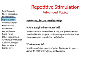
How is acetylcholine synthesized?
Acetylcholine is synthesized in the pre-synaptic nerve
terminal by the enzyme choline acetyltransferase from
the compounds acetyl-CoA and choline.
Repetitive Stimulation
Advanced Topics
What are quanta?
Vesicles containing acetylcholine. Each quanta stores
about 10,000 molecules of acetylcholine.
61.
Basic Concepts
Nerve conduction
Normalvalues
Repetitive stim
Normal anatomy
Median nerve
Ulnar nerve
Peroneal nerve
Radial nerve
Other compressions
Anomalous innervation
Axonal vs. demyel.
Root and plexus
Cranial nerves
Neuromuscular Junction Physiology:
Repetitive Stimulation
Advanced Topics
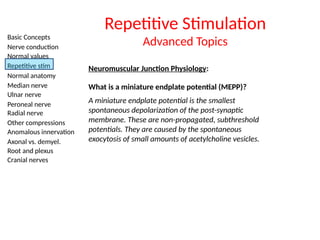
What is a miniature endplate potential (MEPP)?
A miniature endplate potential is the smallest
spontaneous depolarization of the post-synaptic
membrane. These are non-propagated, subthreshold
potentials. They are caused by the spontaneous
exocytosis of small amounts of acetylcholine vesicles.
62.
Basic Concepts
Nerve conduction
Normalvalues
Repetitive stim
Normal anatomy
Median nerve
Ulnar nerve
Peroneal nerve
Radial nerve
Other compressions
Anomalous innervation
Axonal vs. demyel.
Root and plexus
Cranial nerves
Neuromuscular Junction Physiology:
Repetitive Stimulation
Advanced Topics
What is an end plate potential (EPP)?
End plate potentials are the depolarizations of the
skeletal muscle fibers due to binding of acetylcholine
to the post-synaptic membrane of the neuromuscular
junction.
What is a muscle fiber action potential (MFAP)?
The depolarization of the muscle fiber to threshold.
63.
Basic Concepts
Nerve conduction
Normalvalues
Repetitive stim
Normal anatomy
Median nerve
Ulnar nerve
Peroneal nerve
Radial nerve
Other compressions
Anomalous innervation
Axonal vs. demyel.
Root and plexus
Cranial nerves
Repetitive Stimulation
Advanced Topics
Define the primary, secondary, and tertiary stores of
acetylcholine.
1. Primary stores of acetylcholine sit just beneath the pre-
synaptic membrane and are the first quanta released.
2. Secondary stores of acetylcholine consist of nearby
acetylcholine quanta that re-supply the primary stores
quickly.
3. Tertiary stores of acetylcholine exist in the axon and cell
body and are located far from the neuromuscular
junction, functioning as reserves.
64.
Basic Concepts
Nerve conduction
Normalvalues
Repetitive stim
Normal anatomy
Median nerve
Ulnar nerve
Peroneal nerve
Radial nerve
Other compressions
Anomalous innervation
Axonal vs. demyel.
Root and plexus
Cranial nerves
Repetitive Stimulation
Advanced Topics
Describe what happens to the primary, secondary, and
tertiary stores of acetylcholine with slow repetitive
stimulation in a normal subject.
During slow repetitive nerve stimulation, the primary stores
are slowly depleted, with progressively less release of
acetylcholine quanta with each stimulation. This leads to a
progressive decrease in amplitude of the end plate potential.
However, the amplitude remains above the necessary
threshold to illicit a muscle fiber action potential. Within a
few seconds, the secondary store of acetylcholine restores
the depleted quanta, leading to a rise in the amplitude of the
end plate potential.
65.
Basic Concepts
Nerve conduction
Normalvalues
Repetitive stim
Normal anatomy
Median nerve
Ulnar nerve
Peroneal nerve
Radial nerve
Other compressions
Anomalous innervation
Axonal vs. demyel.
Root and plexus
Cranial nerves
Repetitive Stimulation
Advanced Topics
Describe what happens to the primary, secondary, and
tertiary stores of acetylcholine with fast repetitive
stimulation in a normal subject.
During fast repetitive nerve stimulation, the depletion of
primary stores of acetylcholine is fixed by both restoration
from the secondary stores as well as a progressive influx of
calcium into the pre-synaptic membrane. Given the speed of
stimulation, the influx of calcium is faster than its use,
leading to an accumulation of calcium and progressive
increase of quanta. This causes a higher end plate potential
amplitude, which does not change outcome given the muscle
fiber action potential being generated in an all-or-none
manner.
66.
Basic Concepts
Nerve conduction
Normalvalues
Repetitive stim
Normal anatomy
Median nerve
Ulnar nerve
Peroneal nerve
Radial nerve
Other compressions
Anomalous innervation
Axonal vs. demyel.
Root and plexus
Cranial nerves
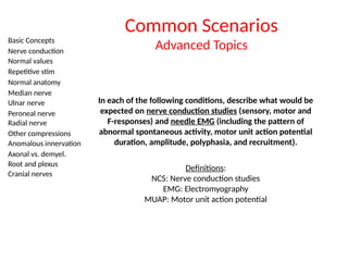
Common Scenarios
Advanced Topics
In each of the following conditions, describe what would be
expected on nerve conduction studies (sensory, motor and
F-responses) and needle EMG (including the pattern of
abnormal spontaneous activity, motor unit action potential
duration, amplitude, polyphasia, and recruitment).
Definitions:
NCS: Nerve conduction studies
EMG: Electromyography
MUAP: Motor unit action potential
67.
Basic Concepts
Nerve conduction
Normalvalues
Repetitive stim
Normal anatomy
Median nerve
Ulnar nerve
Peroneal nerve
Radial nerve
Other compressions
Anomalous innervation
Axonal vs. demyel.
Root and plexus
Cranial nerves
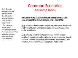
Common Scenarios
Advanced Topics
Neuromuscular junction lesions (excluding abnormalities
seen on repetitive stimulation and single fiber EMG):
NCS: Normal, other than presynaptic disorders have decreased
motor amplitudes, while postsynaptic disorders have normal
motor amplitudes.
EMG: Usually no abnormal spontaneous activity (except
botulism). Usually normal recruitment and morphology, though
if severe, can look like myopathy with early recruitment, short
duration, low amplitude, and polyphasic motor units.
68.
Basic Concepts
Nerve conduction
Normalvalues
Repetitive stim
Normal anatomy
Median nerve
Ulnar nerve
Peroneal nerve
Radial nerve
Other compressions
Anomalous innervation
Axonal vs. demyel.
Root and plexus
Cranial nerves
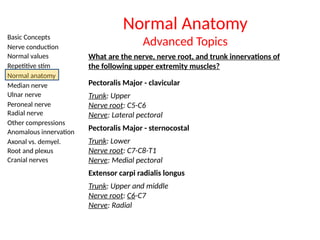
Normal Anatomy
Advanced Topics
What are the nerve, nerve root, and trunk innervations of
the following upper extremity muscles?
Pectoralis Major - clavicular
Trunk: Upper
Nerve root: C5-C6
Nerve: Lateral pectoral
Pectoralis Major - sternocostal
Trunk: Lower
Nerve root: C7-C8-T1
Nerve: Medial pectoral
Extensor carpi radialis longus
Trunk: Upper and middle
Nerve root: C6-C7
Nerve: Radial
69.
Basic Concepts
Nerve conduction
Normalvalues
Repetitive stim
Normal anatomy
Median nerve
Ulnar nerve
Peroneal nerve
Radial nerve
Other compressions
Anomalous innervation
Axonal vs. demyel.
Root and plexus
Cranial nerves
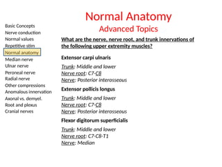
Normal Anatomy
Advanced Topics
What are the nerve, nerve root, and trunk innervations of
the following upper extremity muscles?
Extensor carpi ulnaris
Trunk: Middle and lower
Nerve root: C7-C8
Nerve: Posterior interosseous
Extensor pollicis longus
Trunk: Middle and lower
Nerve root: C7-C8
Nerve: Posterior interosseous
Flexor digitorum superficialis
Trunk: Middle and lower
Nerve root: C7-C8-T1
Nerve: Median
70.
Basic Concepts
Nerve conduction
Normalvalues
Repetitive stim
Normal anatomy
Median nerve
Ulnar nerve
Peroneal nerve
Radial nerve
Other compressions
Anomalous innervation
Axonal vs. demyel.
Root and plexus
Cranial nerves
Normal Anatomy
Advanced Topics
What are the nerve, nerve root, and trunk innervations of
the following upper extremity muscles?
Flexor carpi ulnaris
Trunk: Lower
Nerve root: C8-T1
Nerve: Ulnar
71.
Basic Concepts
Nerve conduction
Normalvalues
Repetitive stim
Normal anatomy
Median nerve
Ulnar nerve
Peroneal nerve
Radial nerve
Other compressions
Anomalous innervation
Axonal vs. demyel.
Root and plexus
Cranial nerves
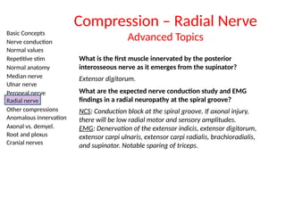
What is the first muscle innervated by the posterior
interosseous nerve as it emerges from the supinator?
Extensor digitorum.
What are the expected nerve conduction study and EMG
findings in a radial neuropathy at the spiral groove?
NCS: Conduction block at the spiral groove. If axonal injury,
there will be low radial motor and sensory amplitudes.
EMG: Denervation of the extensor indicis, extensor digitorum,
extensor carpi ulnaris, extensor carpi radialis, brachioradialis,
and supinator. Notable sparing of triceps.
Compression – Radial Nerve
Advanced Topics
72.
Basic Concepts
Nerve conduction
Normalvalues
Repetitive stim
Normal anatomy
Median nerve
Ulnar nerve
Peroneal nerve
Radial nerve
Other compressions
Anomalous innervation
Axonal vs. demyel.
Root and plexus
Cranial nerves
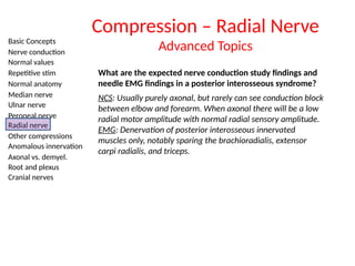
What are the expected nerve conduction study findings and
needle EMG findings in a posterior interosseous syndrome?
NCS: Usually purely axonal, but rarely can see conduction block
between elbow and forearm. When axonal there will be a low
radial motor amplitude with normal radial sensory amplitude.
EMG: Denervation of posterior interosseous innervated
muscles only, notably sparing the brachioradialis, extensor
carpi radialis, and triceps.
Compression – Radial Nerve
Advanced Topics
73.
Basic Concepts
Nerve conduction
Normalvalues
Repetitive stim
Normal anatomy
Median nerve
Ulnar nerve
Peroneal nerve
Radial nerve
Other compressions
Anomalous innervation
Axonal vs. demyel.
Root and plexus
Cranial nerves
END OF PRESENTATION