JEELANI SAIMA HABEEB
LecturerCON BGSBU
INTRODUCTION
Majority (80%) of fetal deaths occur in the antepartum period. The important causes of death are
– (1) chronic maternal hypoxia (IUGR). (2) Maternal complications, e.g. diabetes, hypertension,
infection. (3) Fetal congenital malformations and (4) unexplained cause. There is a progressive
decline in maternal deaths all over the world. Currently more interest is focused to evaluate the
fetal health. The primary objective of antenatal fetal assessment is to avoid fetal death. As such
simultaneously with good maternal care during pregnancy & labor, the fetal health in uteri
should be supervised with equal vigilance .The fetal evaluation measures are used to the fetus is
suspected of being at risk.
Aims of fetal monitoring:
To ensure satisfactory growth & wellbeing of the fetus
throughout pregnancy.
To screen out the high risk factors that affects the growth of fetus .
To prevent the prenatal morbidity and mortality.
Common indications for fetal monitoring:
Pregnancy with obstetric complications: IUGR, multiple pregnancies, polyhydramnios or
oligohydramnios, rhesus alloimmunisation.
Pregnancy with medical complications: Diabetes mellitus, hypertension, epilepsy, renal
or cardiac diseases, infections (tuberculosis), SLE
Others: Advanced maternal age (>35yrs), previous still birth or recurrent abortion,
previous birth of baby with structural or chromosomal abnormalities.
Routine antenatal testing.
Clinical evaluation of fetal wellbeing: At every antenatal visit, the following clinical
parameters are taken into account for assessment of satisfactory progress of gestation.
1. Maternal weight gain: During second half of pregnancy, the average weight gain is 1 kg a
fortnight. Any excess weight gain may be due to excess fluid retension and could be sign
of pre- eclampsia.
2. Blood pressure: Initial recording of blood pressure prior to 12 weeks helps to differentiate
a pre- existing hypertension from a pregnancy induced hypertension.
2.
3. Assessment ofthe size of the uterus and height of the fundus: In early weeks, the size of
uterus is of great value in confirming the calculated duration of gestation. The height of
fundus should be documented at each visit.
4. Clinical assessment excess liquor: Should be recorded, as well as scanty liquor in the last
trimester. Evidence scanty liquor may indicate placental insufficiency and the need for
undertaking other placental function test.
5. Documentation of the girth of the abdomen in the last trimester of pregnancy. This is
measured at the lower border of the umbilicus. Normally, the girth increases steadily upto
term. If the girth gradually diminishes beyond term or earlier, it arouses suspicion of
placental insufficiency.
Biochemical measures (early pregnancy): Antenatal assessment of fetal well being in early
pregnancy is primarily designed to detect fetal congenital abnormalities.
Maternal serum alpha fetoprotein (MSAFP):
AFP is an oncofetal protein, produced by the yolk sac & fetal liver.
Highest levels of AFP in fetal serum & amniotic fluid are reached around 13 weeks &
thereafter it decreases.
MSAFP level is elevated in a number of conditions: wrong gestational age, neural tube
defects(NTDS), IUFD, renal anomalies etc. low levels are found in trisomies (Down’s
syndrome)
Test is done between 15-20 weeks. Elevated MSAFP detects 85% of all neural tube
defects.
Nurse will use a small needle to withdraw blood from a vein, & a laboratory specialist
will analyze the sample.
Triple test:
It is a combined test which includes MSAFP, HCG & UE3 (unconjugated estriol).
In an affected pregnancy, level of MSAFP & UE, tend to be low while that of HCG is
high.
It is performed at 15-18 weeks. The result is considered to be screen positive if the risk
ratio is 1:250 or greater.
Acetyl choline esterase:
Amniotic fluid AChE level is elevated in most cases of open neural tube defects .It has
got better diagnostic value than AFP.
Inhibin A is a dimeric glycoprotein. It is produced by the corpus luteum & the placenta.
Serum level of inhibin A is raised in women carrying a fetus with Down’s syndrome.
3.
Amniocentesis:
“Obtaining a sampleof amniotic fluid surrounding the fetus during pregnancy.” It is an invasive
procedure performed under ultrasonographic guidance. The fetal cells obtained in this procedure
are subjected for cytogenic analysis.
Indications:
Diagnostic (at 11- 20 weeks):
• Chromosomal analysis (Down syndrome),Spina bifida
• Inherited diseases (muscular dystrophy)
• Fetal lung maturation (L/S ratio
Therapeutic( at any time):
• Reduce maternal stress in polyhydramnios
• Mainly in twin-twin transfusion or if abnormality associated
Precautions:
Prophylactic administration of 100mg of anti-D immunoglobulin in Rh-negative non
immunized mother.
Hazards:
Maternal complications: infection, hemorrhage, premature rupture of membranes
Fetal hazards are fetal loss (0.06-0.5%), trauma, fetomaternal hemorrhage, and
oligohydramnios due to leakage of amniotic fluid & may lead to fetal lung hypoplasia.
Chorionic villus sampling:
It is performed for prenatal diagnosis of genetic disorders. It is carried out transcervically
between 10-12 weeks and transabdominally from 10 weeks to term.Sampling is done to the
cyto-trophoblasts. Indications are fetal karyotyping and genetic testing.
Complications:
Fetal loss (1-2%)
Vaginal bleeding
Limb reduction defects are high when CVS was performed at less than 10 weeks of
gestation. Anti – D immunoglobulin 50 micro gm IM should be administered following
the procedure to a Rh negative women.
Cordocentesis:
A 22 gauze spinal needle 13cm in length is inserted through the maternal abdominal and uterine
4.
wall under ultrasonicguidance. The needle tip is progressed carefully & it punctures the
umbilical vein approximately 1-2cm from the placental inertion. Generally, 0.5 to 2 ml of fetal
blood is collected. It is performed under local anesthesia usually from 18 weeks gestation.
Risks: - The invasive procedure may lead to abortion, preterm labor & intrauterine fetal death.
BIOPHYSIAL (early pregnancy): Ultrasonographic examinations of the fetus in early
pregnancy can detect fetal anomalies.
Crown- Rump Length smaller than gestational age is associated with the risk of
chromosomal anomalies.
Nuchal translucency at 10 – 14 weeks is associated with many chromosomal
abnormalities( Trisomy).
Absence of nasal bone on USG at 10-12 weeks is associated with fetal Downs
syndrome.
FETAL MONITORING (late pregnancy):
Objectives:
Prevention of fetal death.
Avoidance of unnecessary interventions.
Methods: biochemical and biophysical.
Biochemical: mainly done for assessment of pulmonary maturity.
Biophysical tests:
Fetal movement counting
Sadovsky technique( 4 movements felt in 1 hour)
• For one hour after meal the woman should lie down and concentrate on fetal movement.
• 4 movements should be felt in one hour. If not, she should count for another hour.
• If after 2 hours four movements are not felt, she should have fetal monitoring
Cardiff technique :Done in the morning, Patient should calculate how long it takes to
have 10 fetal movements.10 movements should be appreciated in 12 hours
Non stress test:
Done using the cardiotocometry with the patient in left lateral position. Record for 20
minutes. The base line is 120-160 beats/minute
5.
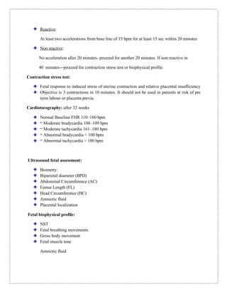
Reactive:
At least twoaccelerations from base line of 15 bpm for at least 15 sec within 20 minutes
Non reactive:
No acceleration after 20 minutes- proceed for another 20 minutes. If non reactive in
40 minutes---proceed for contraction stress test or biophysical profile.
Contraction stress test:
Fetal response to induced stress of uterine contraction and relative placental insufficiency
Objective is 3 contractions in 10 minutes. It should not be used in patients at risk of pre
term labour or placenta previa.
Cardiotocography: after 32 weeks
Normal Baseline FHR 110–160 bpm
– Moderate bradycardia 100–109 bpm
– Moderate tachycardia 161–180 bpm
– Abnormal bradycardia < 100 bpm
– Abnormal tachycardia > 180 bpm
Ultrasound fetal assessment:
Biometry:
Biparietal diameter (BPD)
Abdominal Circumference (AC)
Femur Length (FL)
Head Circumference (HC)
Amniotic fluid
Placental localization
Fetal biophysical profile:
NST
Fetal breathing movements
Gross body movement
Fetal muscle tone
Amniotic fluid