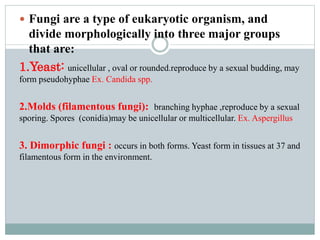
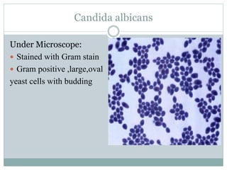
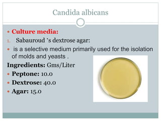
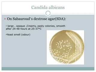
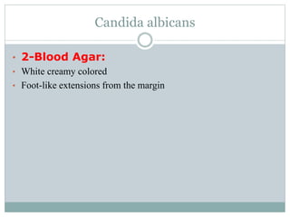
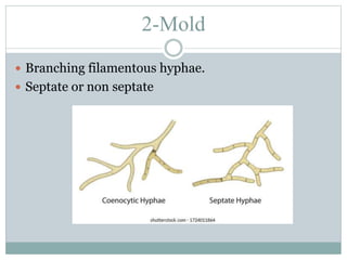
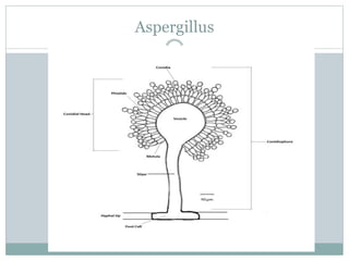
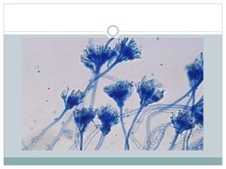
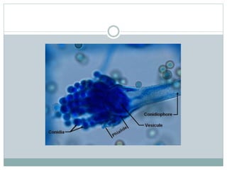
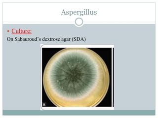
CHRISTINE EFAT TAWFEEK
Mycology Lab
 Fungi are a type of eukaryotic organism, and
divide morphologically into three major groups
that are:
1.Yeast: unicellular , oval or rounded.reproduce by a sexual budding, may
form pseudohyphae Ex. Candida spp.
2.Molds (filamentous fungi): branching hyphae ,reproduce by a sexual
sporing. Spores (conidia)may be unicellular or multicellular. Ex. Aspergillus
3. Dimorphic fungi : occurs in both forms. Yeast form in tissues at 37 and
filamentous form in the environment.
Yeasts
 Yeast is a single-cell organism, which needs food,
warmth, and moisture to thrive.
 Candida albicans is the commonest yeast causing
disease to humans ( UTI, moniliasis, onychomycosis,
vulvovaginitis, systemic mycosis)
Candida albicans
Under Microscope:
 Stained with Gram stain
 Gram positive ,large,oval
yeast cells with budding
Candida albicans
 Culture media:
1. Sabauroud ’s dextrose agar:
 is a selective medium primarily used for the isolation
of molds and yeasts .
Ingredients: Gms/Liter
 Peptone: 10.0
 Dextrose: 40.0
 Agar: 15.0
Candida albicans
 On Sabauroud’s dextrose agar(SDA):
• large , opaque ,Creamy, pasty colonies, smooth
after 24-48 hours at 25-37°C
•Yeast smell (odour)
Candida albicans
• 2-Blood Agar:
• White creamy colored
• Foot-like extensions from the margin
2-Mold
 Branching filamentous hyphae.
 Septate or non septate
Aspergillus
Aspergillus
 Aspergillus species are medically relevant pathogens.
 For humans, a range of diseases such as infection to the external
ear, skin lesions, and ulcers.
Aspergillus
Diagnosis:
 Under microscope:
Stained with Lactophenol cotton blue.
Principle of Lactophenol cotton blue Staining
 Lactophenol cotton blue is used in medical mycology for the examination of fungal
cell structure, by preparing a wet mount.It is used for the preparation of
microscopic slides of fungi for examination. Fungal elements are stained
stained intensely blue
 It is composed of the following components :
 1-methyl blue(is a histological stain which stains collagen blue in tissue sections
sections & stains the chitin in the fungal cell walls)

 2-lactophenol (is a mixer of four components such as phenol, lactic acid, and
and glycerol in water. Where phenol helps to kill any microorganisms, lactic acid
preserves fungal structures & acts as a mountant.)
 3-glycerol inhibits the cellulolytic activity of the fungus
Aspergillus
1.Conidiophore: terminating in apical vesicle
and in the opposite end, a basal foot cell inserted
into the supporting hyphae.
2.Vesicle:covered partially or completely
by flask-shaped phiallides (uniseriate)
or an intervening cells called metula
3.Phiallides: columnar heads, produce
chains of round,Rough conidia (spores) about(2-5micrometer in diameter).
4-Conidia: Non motile
5- Hyphae: Septate
Vesicle+phiallides+metula(if present)+conidia
= conidial head
Aspergillus
 Culture:
On Sabauroud’s dextrose agar (SDA)

Mycology Lab (1).pptx

  • 1.
  • 2.
     Fungi area type of eukaryotic organism, and divide morphologically into three major groups that are: 1.Yeast: unicellular , oval or rounded.reproduce by a sexual budding, may form pseudohyphae Ex. Candida spp. 2.Molds (filamentous fungi): branching hyphae ,reproduce by a sexual sporing. Spores (conidia)may be unicellular or multicellular. Ex. Aspergillus 3. Dimorphic fungi : occurs in both forms. Yeast form in tissues at 37 and filamentous form in the environment.
  • 3.
    Yeasts  Yeast isa single-cell organism, which needs food, warmth, and moisture to thrive.  Candida albicans is the commonest yeast causing disease to humans ( UTI, moniliasis, onychomycosis, vulvovaginitis, systemic mycosis)
  • 4.
    Candida albicans Under Microscope: Stained with Gram stain  Gram positive ,large,oval yeast cells with budding
  • 6.
    Candida albicans  Culturemedia: 1. Sabauroud ’s dextrose agar:  is a selective medium primarily used for the isolation of molds and yeasts . Ingredients: Gms/Liter  Peptone: 10.0  Dextrose: 40.0  Agar: 15.0
  • 7.
    Candida albicans  OnSabauroud’s dextrose agar(SDA): • large , opaque ,Creamy, pasty colonies, smooth after 24-48 hours at 25-37°C •Yeast smell (odour)
  • 8.
    Candida albicans • 2-BloodAgar: • White creamy colored • Foot-like extensions from the margin
  • 10.
    2-Mold  Branching filamentoushyphae.  Septate or non septate
  • 11.
  • 12.
    Aspergillus  Aspergillus speciesare medically relevant pathogens.  For humans, a range of diseases such as infection to the external ear, skin lesions, and ulcers.
  • 13.
  • 14.
    Principle of Lactophenolcotton blue Staining  Lactophenol cotton blue is used in medical mycology for the examination of fungal cell structure, by preparing a wet mount.It is used for the preparation of microscopic slides of fungi for examination. Fungal elements are stained stained intensely blue  It is composed of the following components :  1-methyl blue(is a histological stain which stains collagen blue in tissue sections sections & stains the chitin in the fungal cell walls)   2-lactophenol (is a mixer of four components such as phenol, lactic acid, and and glycerol in water. Where phenol helps to kill any microorganisms, lactic acid preserves fungal structures & acts as a mountant.)  3-glycerol inhibits the cellulolytic activity of the fungus
  • 16.
    Aspergillus 1.Conidiophore: terminating inapical vesicle and in the opposite end, a basal foot cell inserted into the supporting hyphae. 2.Vesicle:covered partially or completely by flask-shaped phiallides (uniseriate) or an intervening cells called metula 3.Phiallides: columnar heads, produce chains of round,Rough conidia (spores) about(2-5micrometer in diameter). 4-Conidia: Non motile 5- Hyphae: Septate Vesicle+phiallides+metula(if present)+conidia = conidial head
  • 18.