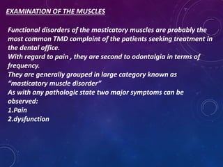
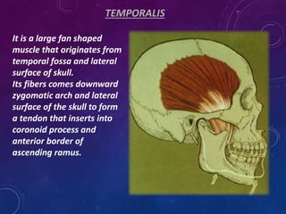
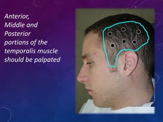
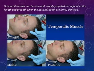
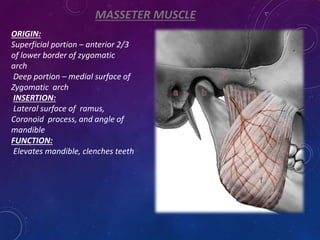
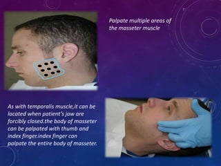
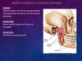
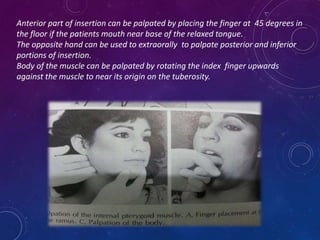
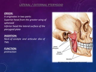
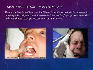
This document discusses the muscles of mastication and their examination. It describes the temporalis, masseter, and medial and lateral pterygoid muscles. These muscles can cause pain and dysfunction when disorders are present. The temporalis and masseter muscles elevate the mandible and can be palpated when the teeth are clenched. The medial and lateral pterygoid muscles are more difficult to palpate due to their deep locations. Examining the muscles involves palpating the different areas to check for tenderness which could indicate a muscle disorder.
A Critique of the Proposed National Education Policy Reform
Examination of Masticatory Muscles (less than 40 chars
1. MUSCLES OF MASTICATION AND
THEIR EXAMINATION
UZMA JAN
BDS 3RD YEAR
ROLL NO 39
ORAL DIAGNOSIS &MEDICINE DEPARTMENT
2. EXAMINATION OF THE MUSCLES
Functional disorders of the masticatory muscles are probably the
most common TMD complaint of the patients seeking treatment in
the dental office.
With regard to pain , they are second to odontalgia in terms of
frequency.
They are generally grouped in large category known as
“masticatory muscle disorder”
As with any pathologic state two major symptoms can be
observed:
1.Pain
2.dysfunction
3. PAIN
Certainly the most common complaint in patients with
masticatory muscle disorder is pain , which may range from slight
tenderness to extreme discomfort.
Pain felt in muscle tissue is called myalgia.
It may arise from increased level of muscle use.
Symptoms are usually associated with a feeling of muscle fatigue
and tightness.
4. DYSFUNCTION
Usually it is seen as decrease in range of mandibular movement.
When muscle tissues have been compromised by overuse , any
contraction or stretching increases the pain.
Therefore to maintain comfort , patient restricts movement within
a range that doesnot increase the pain level.
Clinically this is seen as inability to open mouth widely.
5. TEMPORALIS
It is a large fan shaped
muscle that originates from
temporal fossa and lateral
surface of skull.
Its fibers comes downward
zygomatic arch and lateral
surface of the skull to form
a tendon that inserts into
coronoid process and
anterior border of
ascending ramus.
6. It can be divided into three distinct areas:
Anterior portion : consists of fibers that are direcrted vertically
Middle portion : contains fibers that run obliquely across lateral
aspect of the skull
Posterior portion : that are aligned almost horizontally
When temporal muscle contracts , it elevates mandible.
8. Temporalis muscle can be seen and readily palpated throughout entire
length and breadth when the patient’s teeth are firmly clenched.
9. MASSETER MUSCLE
ORIGIN:
Superficial portion – anterior 2/3
of lower border of zygomatic
arch
Deep portion – medial surface of
Zygomatic arch
INSERTION:
Lateral surface of ramus,
Coronoid process, and angle of
mandible
FUNCTION:
Elevates mandible, clenches teeth
10. Palpate multiple areas of
the masseter muscle
As with temporalis muscle,it can be
located when patient’s jaw are
forcibly closed.the body of masseter
can be palpated with thumb and
index finger.index finger can
palpate the entire body of masseter.
11. MEDIAL PTERYGOID / INTERNAL PTERYGOID
ORIGIN:
Medial surface of lateral pterygoid plate
and tuberosity of maxilla and can not be
palpated
INSERTION:
lower medial surface of ramus of
mandible
FUNCTION:
Elevation and protraction
12. Anterior part of insertion can be palpated by placing the finger at 45 degrees in
the floor if the patients mouth near base of the relaxed tongue.
The opposite hand can be used to extraorally to palpate posterior and inferior
portions of insertion.
Body of the muscle can be palpated by rotating the index finger upwards
against the muscle to near its origin on the tuberosity.
13. LATERAL / EXTERNAL PTERYGOID
ORIGIN:
It originates in two parts:
Superior head from the greater wing of
sphenoid
Inferior head the lateral surface of the
pterygoid plate
INSERTION:
Neck of condyle and articular disc of
TMJ.
FUNCTION:
protraction
14. PALPATION OF LATERAL PTERYGOID MUSCLE
The muscle is palpated by using the little or index finger and placing it lateral to
maxillary tuberosity and medial to coronoid process.The finger presses upwards
and inwards and a painful response can be determined .
15. References-
B D Chaurasia.Human anatomy:Regional and applied dissection amd clinical,5th
edition
Drake L R, Vogl W, Mitchell A W M. Gray’s anatomy for student.InternationalEdition.
Sinnatamby C S. Last’s anatomy regional and applied. 11th edition.
Lippert, L.S. (2011). Clinical Kinesiology and Anatomy, 5th ed. Philadelphia, PA: F.A.
Davis.
Blaschke DD, Solberg WK, Sanders B . Arthrography of the temporomandibular
joint : review of current status . J Am Dent Assoc 1980 ; 100:388 .
Kahan LB . Temporomandibular joint dysfunction : an occasional manifestation of
serious psychopathology . J Oral Surg 1981 ; 39:742 .
Meyer RA. Osteochondroma of coronoid process of mandible . J Oral Surg 1972 ;30 :297
Meyer RA . Clicking sounds owing to temporomandibular joint injury.JAMA 1982 ;248