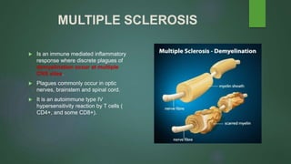
MULTIPLE SCLEROSIS
SAMUEL YEBOAH ASIAMAH
DIFFERENTIAL DIAGNOSIS
 OPTICAL NEURITIS
 Inflammation of the optic nerve.
 PRESENTATION
 Temporary vision loss in one eye lasting about 7-10 days.
 Periocular pain – pain around the eye.
 Dyschromatosia – inability to see colours correctly.
Presentation is similar to MS such that 85% of MS patient with pain and loss
of vision.
Optical neuritis is not disseminated in space nor time. ie no other symptoms
can be found in the body apart from the eye and symptoms do not relapse
and remit.
DIFFERENTIAL DIAGNOSIS
SCHILDER’S DISEASE
Degenerative demyelination of the CNS. Cause is unknown and usually affect
ages 7-10
SYMPTOMS
Fever and headache
Aphasia(difficulty speaking)
Psychosis
Optic neuritis
Deafness and vertigo
Schilder disease is a rare form of MS. The difference is that it starts from
childhood and especially after an infection.
THE NEURONE
ACTION POTENTIAL
NERVE ACTION POTENTIAL
 Action potential is the fast depolarisation and repolarisation of the membrane of an
excitable tissue.
 As the action potential must move along the axon of the neurone with the same shape
and size.
 Mechanical, chemical and electrical stimulus is what generate the action potential.
CONDUCTION VELOCITY
 Conduction velocity: speed at which an action potential is moves along the axon of a
neurone.
CABLE PROPERTIES
 Membrane capacitance (Cm): ability of the membrane to store charges. Reduced
membrane capacitance reduces the time constant.
 time constant: how quickly depolarisation spread along the axon of a neurone. (τ) = Rm
x Cm.
 Length constant: how far depolarisation spread.
λ ∝ √Rm /Ri.
Length constant increase with increasing diameter because internal resistance is reduced.
CONDUCTION VELOCITY
Mechanisms that increase conductance velocity includes;
 Increasing membrane diameter: this reduces internal resistance and
increases conductance velocity.
 Myelination: sphingosine myelin sheath produced by oligodendrocytes or
schwann cells, reduces membrane capacitance and increases membrane
resistance. Action potential is the forced to flow along the axon interior of
low resistance.
SALTATORY CONDUCTION
 At the nodes of Ranvier, which is
about 2-3µm, depolarisation and
repolarisation occur.
 Myelin acts as an insulator
preventing depolarisation.
 The action potential “Jumps” from
node to node called saltatory
conduction.
 Unmyelinated neurons action
potential travel at 0.25m/sec.
 Myelinated neurons action potential
travel at 100m/sec.
MYELINATED AND UNMYELINATED
NEURONE
UNMYELINATED AND MYELINATED
NEURONE
MULTIPLE SCLEROSIS
 Is an immune mediated inflammatory
response where discrete plagues of
demyelination occur at multiple
CNS sites.
 Plagues commonly occur in optic
nerves, brainstem and spinal cord.
 It is an autoimmune type IV
hypersensitivity reaction by T cells (
CD4+, and some CD8+).
EPDEMIOLOGY OF MS
 Prevalence in the US is about 58-95 per 100,000 population.
 World wide 2.1million are affected.
 Age of symptoms onset is 20years and above.
CAUSES OF MS
 Autoimmune disease: type IV hypersensitivity reaction.
 Viruses:
Epstein-Barr virus, mumps, measeles, Herpes simlex serotype 6 activate T cells via
immunological mimicry
 Vitamin D deficiency: results in poor regulation T lymphocytes immune response.
Risk Factors of MS
 Genetics : presence of HLA DRB1 on chromosome 6.
 Identical twins
 Gender: female : male, 3:1
 Smoking
 People living around the north and south pole
PATHOPHYSIOLOGY OF MULTIPLE
SCLEROSIS
PATHOPHYSIOLOGY OF MS
 T helper cells Tн1 and Tн17 T cells cross the blood brain barrier (BBB) to react against
myelin antigens and secrete cytokines.
 Tн1 cells secrete IFN-ϒ, which activates macrophages, and Tн17 cells promotes the
recruitment of leucocytes.
 B cells also secrete antibodies against myelin antigens.
 All these contributes to demyelination which slows conduction velocity of action potential.
 Partial Remyelination may occur by oligodendrocytes or Schwann cells –Remittance.
SYMPTOMS OF MS
 Unilateral optical neuritis(pain on eye movement and rapid reduced central
vision) due to optic nerve involvement making it difficult to perceive visual
signals.
demyelination of brainstem produces
 Ataxia(loss of coordination)
Cranial nerve signs like;
 Scanning speech
 Difficulty swallowing
 Nystagmus – involuntary eye movement (dancing eyes)
SYMPTOMS
Spinal cord lesions give rise to motor and sensory impairment to transmit
signals as seen in;
 Muscle spasticity and weakness.
 Paresthesia, tingling and numbness of trunk and limbs.
 Difficulty in voluntary bladder control
MRI OF MULTIPLE SCLEROSIS
DIAGNOSIS OF MS
 MAINLY CLINICAL DIAGNOSIS McDonald criteria
 Magnetic Resonance Imaging (MRI)
 Lumbar puncture: CSF is evaluated for ↑IgG and oligoclonal IgG bands.
 Evoked potential: measures time it takes the brain to respond to sensory
stimulation.
TYPES OF MS
o Relapsing-remitting MS
o Secondary progressive MS
o Primary progressive MS
o Progressive-relapsing MS
TREATMENT
 Steroids: shorten acute relapses, suppress migration of polymorphonuclear leukocytes
which cause phagocytosis of myelin to slow action potential.
eg Methyl prednisolone
 Monoclonal antibodies
Alemtuzumab acts against T.cells responsible for the autoimmune demyelination in MS.
 Interferon beta 1a:antiproliferative immune protein;
Slows demylination by altering immune response to myelin surface antigen.
TREATMENT

Multiple Sclerosis

  • 1.
  • 2.
    DIFFERENTIAL DIAGNOSIS  OPTICALNEURITIS  Inflammation of the optic nerve.  PRESENTATION  Temporary vision loss in one eye lasting about 7-10 days.  Periocular pain – pain around the eye.  Dyschromatosia – inability to see colours correctly. Presentation is similar to MS such that 85% of MS patient with pain and loss of vision. Optical neuritis is not disseminated in space nor time. ie no other symptoms can be found in the body apart from the eye and symptoms do not relapse and remit.
  • 3.
    DIFFERENTIAL DIAGNOSIS SCHILDER’S DISEASE Degenerativedemyelination of the CNS. Cause is unknown and usually affect ages 7-10 SYMPTOMS Fever and headache Aphasia(difficulty speaking) Psychosis Optic neuritis Deafness and vertigo Schilder disease is a rare form of MS. The difference is that it starts from childhood and especially after an infection.
  • 4.
  • 5.
  • 6.
    NERVE ACTION POTENTIAL Action potential is the fast depolarisation and repolarisation of the membrane of an excitable tissue.  As the action potential must move along the axon of the neurone with the same shape and size.  Mechanical, chemical and electrical stimulus is what generate the action potential.
  • 7.
    CONDUCTION VELOCITY  Conductionvelocity: speed at which an action potential is moves along the axon of a neurone. CABLE PROPERTIES  Membrane capacitance (Cm): ability of the membrane to store charges. Reduced membrane capacitance reduces the time constant.  time constant: how quickly depolarisation spread along the axon of a neurone. (τ) = Rm x Cm.  Length constant: how far depolarisation spread. λ ∝ √Rm /Ri. Length constant increase with increasing diameter because internal resistance is reduced.
  • 8.
    CONDUCTION VELOCITY Mechanisms thatincrease conductance velocity includes;  Increasing membrane diameter: this reduces internal resistance and increases conductance velocity.  Myelination: sphingosine myelin sheath produced by oligodendrocytes or schwann cells, reduces membrane capacitance and increases membrane resistance. Action potential is the forced to flow along the axon interior of low resistance.
  • 9.
    SALTATORY CONDUCTION  Atthe nodes of Ranvier, which is about 2-3µm, depolarisation and repolarisation occur.  Myelin acts as an insulator preventing depolarisation.  The action potential “Jumps” from node to node called saltatory conduction.  Unmyelinated neurons action potential travel at 0.25m/sec.  Myelinated neurons action potential travel at 100m/sec.
  • 10.
  • 11.
  • 12.
    MULTIPLE SCLEROSIS  Isan immune mediated inflammatory response where discrete plagues of demyelination occur at multiple CNS sites.  Plagues commonly occur in optic nerves, brainstem and spinal cord.  It is an autoimmune type IV hypersensitivity reaction by T cells ( CD4+, and some CD8+).
  • 13.
    EPDEMIOLOGY OF MS Prevalence in the US is about 58-95 per 100,000 population.  World wide 2.1million are affected.  Age of symptoms onset is 20years and above.
  • 14.
    CAUSES OF MS Autoimmune disease: type IV hypersensitivity reaction.  Viruses: Epstein-Barr virus, mumps, measeles, Herpes simlex serotype 6 activate T cells via immunological mimicry  Vitamin D deficiency: results in poor regulation T lymphocytes immune response.
  • 15.
    Risk Factors ofMS  Genetics : presence of HLA DRB1 on chromosome 6.  Identical twins  Gender: female : male, 3:1  Smoking  People living around the north and south pole
  • 16.
  • 17.
    PATHOPHYSIOLOGY OF MS T helper cells Tн1 and Tн17 T cells cross the blood brain barrier (BBB) to react against myelin antigens and secrete cytokines.  Tн1 cells secrete IFN-ϒ, which activates macrophages, and Tн17 cells promotes the recruitment of leucocytes.  B cells also secrete antibodies against myelin antigens.  All these contributes to demyelination which slows conduction velocity of action potential.  Partial Remyelination may occur by oligodendrocytes or Schwann cells –Remittance.
  • 18.
    SYMPTOMS OF MS Unilateral optical neuritis(pain on eye movement and rapid reduced central vision) due to optic nerve involvement making it difficult to perceive visual signals. demyelination of brainstem produces  Ataxia(loss of coordination) Cranial nerve signs like;  Scanning speech  Difficulty swallowing  Nystagmus – involuntary eye movement (dancing eyes)
  • 19.
    SYMPTOMS Spinal cord lesionsgive rise to motor and sensory impairment to transmit signals as seen in;  Muscle spasticity and weakness.  Paresthesia, tingling and numbness of trunk and limbs.  Difficulty in voluntary bladder control
  • 20.
    MRI OF MULTIPLESCLEROSIS
  • 21.
    DIAGNOSIS OF MS MAINLY CLINICAL DIAGNOSIS McDonald criteria  Magnetic Resonance Imaging (MRI)  Lumbar puncture: CSF is evaluated for ↑IgG and oligoclonal IgG bands.  Evoked potential: measures time it takes the brain to respond to sensory stimulation.
  • 22.
    TYPES OF MS oRelapsing-remitting MS o Secondary progressive MS o Primary progressive MS o Progressive-relapsing MS
  • 23.
    TREATMENT  Steroids: shortenacute relapses, suppress migration of polymorphonuclear leukocytes which cause phagocytosis of myelin to slow action potential. eg Methyl prednisolone  Monoclonal antibodies Alemtuzumab acts against T.cells responsible for the autoimmune demyelination in MS.  Interferon beta 1a:antiproliferative immune protein; Slows demylination by altering immune response to myelin surface antigen.
  • 24.