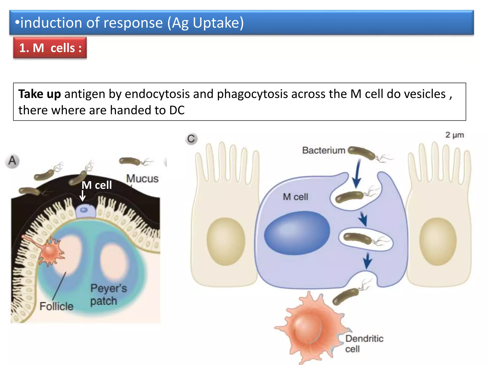
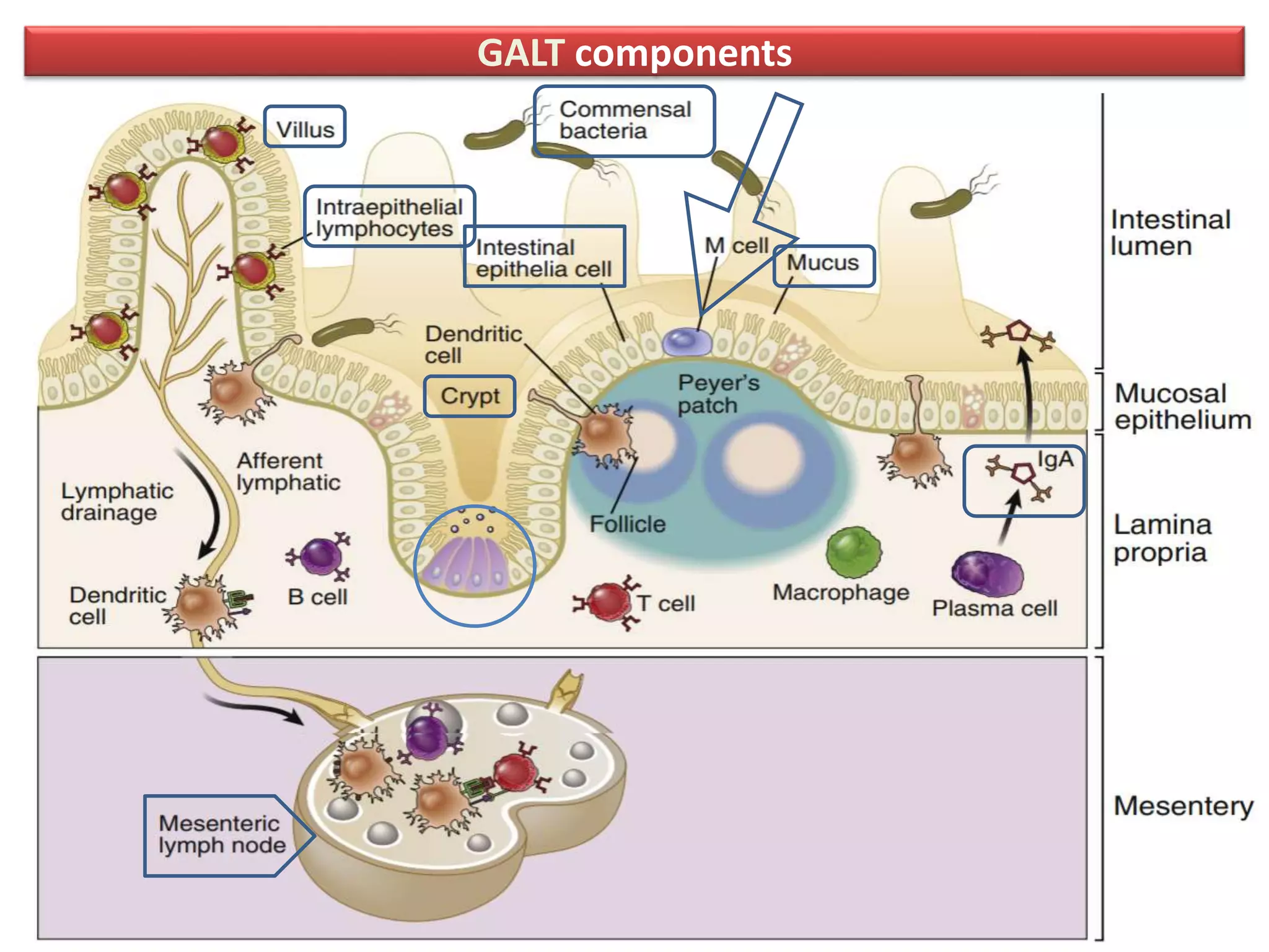
Mucosal immunity
( Regional immunity )
How mucosal immunity is very specialized
than systemic immune system?
By two Challenges (functions )
• Active immune response to pathogenic organisms.
• Regulatory functions : that serve to prevent
responses to nonpathogenic microbes (commensal
bacteria, microbiota ) and foreign substances (ex.
Food molecules )
regional immune system
GALT
Cells and functions
• General immunity Function?
•  prevents microbial invasion, underlying
connective tissue
•  mediate immune responses to organisms
that do invade through the epithelium
•  prevent impair barrier by inflammatory
response
GALT components
Peyer’s patches,
• Found mainly in the distal ileum,
• Peyer’s patches have the structure of lymphoid
follicles,
• with germinal centers containing B lymphocytes,
follicular helper T cells, follicular dendritic cells,
and macrophages.
• Its unencapsulated but organized secondary
lymphoid tissues just under the epithelial barrier.
1. Innate immune protection
Mechanisms :
1. Secrete mucus, Glycocalyx.
2. produce α-defensins, , β-defensins
3. produce C-type lectins
4. Activate Innate lymphoid cells
5. produce proteins that form tight junctions
which block the movement of microbes
between the cells into the lamina propria
• 1. Mucins ;
1. a single layer,
2. its function prevents microbes from contacting with small
intestine epithelial cells
• Most in intestinal layer is composed of MUC2,
• Glycolipids with mucins form  Glycocalyx
• 2. Glycocalyx;
1. glycolipids is coated the apical surface of gastrointestinal
epithelial cells
2. It Function prevent microbial contact
1. Mucus
• Increases in mucins production
• By
1. cytokines (IL-1, IL-4, IL-6, IL-9, IL-13, tumor
necrosis factor [TNF], and type I interferons)
2. neutrophil products (such as elastase),
3. and microbial adhesive proteins.
2. α-defensins
• Defensins are peptides produced by intestinal epithelial
cells that exert lethal toxic effects on microbes
• causing loss of integrity of their outer phospholipid
membranes of microbes
• including human defensin 5 (HD5) and HD6
• produced by Paneth cells response to IL-1 or invasive
bacteria.
• neutrophil granules are rich in α defensins
• In the colon, β-defensins are epithelial cells in the large
intestinal crypts,
• which likely contribute to their antimicrobial functions in
the setting of infections of the bowel wall.
Paneth cells and epithelial cells C-type lectins = (REGIII)
• Paneth cells and other epithelial cells of the
intestine also secrete C-type lectins called
regenerating islet-derived proteins (REGIII),
• Function which block bacterial colonization
of the epithelial surface.
secrete
3. C-type lectins = (REGIII)
TLRs
• Toll-like receptors (TLRs)
• NOD like receptors (NLRs)
• Its recognize PAMPs molecules of Ag
• On or in intestinal epithelial cells and lymphoid cells (as DCs)
• Ligation with Ag can:
1. increase strength of tight junctions
2. Give signal to increases intestinal epithelial motility and
proliferation.
3. Give signal to secretion of defensins, REGIII lectins, and IgA,
• but also limit inflammatory responses to commensal
bacteria.
TLRs
• TLR2 ( peptidoglycan or lipopeptides)
• TLR4 (LPS)
• TLR5 (flagellin)
• TLR6 (lipopeptides)
• TLR7 (ssRNA)
• TLR9 (CpG DNA)
TLRs
Out lecture
2. Adaptive immune response
• 1. sampling Ag by APCs (DCs, Macrophages..)
or across through M-cells
• 2. Differ if it pathogenic or nonpathogenic Ag
by TLRs
Regulation
(tolerance )
4. Class switching Produce IgA
5. Pass IgA to lumen
6. Lymphocyte Homing to blood
3. Presenting to B, naïve T cells
M cell
•induction of response (Ag Uptake)
Take up antigen by endocytosis and phagocytosis across the M cell do vesicles ,
there where are handed to DC
1. M cells :
2. Dendritic cells
• Capture antigen from lumen
• 1. Presentation of antigen to
mesenteric lymph nodes
• 2. or present Ag to T-cells
• An essential link between
innate and adaptive immunity
A. extend dendritic processes, so as Macrophages
B. sample antigens that derived from lumina through the epithelial barrier.
Sampling Ag
Some dendritic cells
•induction of response (Ag Uptake)
2.A pathogen Dc  Effector T cell
• Dendritic cells
• 1. Antigen-sampling DCs extend dendritic processes
between intestinal epithelial cells into the lumen to
sample antigens
• 2. and then migrate to mesenteric lymph nodes,
• 3. where they initiate activation and differentiation
of pro inflammatory effector T cells.
• 4 . These DCs express the CD11b+ integrin chain and
the CX3CR1 chemokine receptor
2. B nonpathogenic Ag Dc  Treg
• Other Dendritic cells
• 1. present in the lamina propria, which
express the integrin CD103,
• 2. present antigen to naïve T cells and induce
there differentiation of regulatory T cells, in
part by secreting TG F-B and retinoic acid (RA)
• The regulatory function of these DCs
depends on factors secreted by intestinal
epithelial cells
FIGURE 13-4 DCs in the intestinal mucosa. 2012
Regulation of Immunity in the Gastrointestinal
Tract by Regulatory T Cells and Cytokines
• Both retinoic acid and TGF-β promote FoxP3
expression and inhibit the generation of Th1 and
Th2 cells
• Treg produce immunosuppressive cytokine IL-10.
• Several cytokines, including TGF-β, IL-10, and IL-2,
play crucial roles in maintaining homeostasis
No immune responses to commensal bacteria
Tolerance
Lack danger signal
(no inflammation)
Oral Tolerance
• Oral Tolerance : a mechanism for prevention
harmful immune responses to harmless antigen
such as food
• Induction of anergy of Ag-specific T cells
• Clonal deletion of antigen-specific T cells
• Producing immunosuppressive cytokines ( IL-4,
IL-10, TGF-B)
4. In T-dependent IgA class switching
• dendritic cells capture bacterial antigens
• delivered by M cells and migrate to the interfollicular
zone, where they present antigen to naive CD4+ T cells.
• The activated T cells differentiate into helper T cells
• B cell class switching to IgA is stimulated through T cell
CD40L binding to B cell CD40, together with the action
of TGF-β.
In T-dependent IgA class switching
• T-independent IgA class switching involves
dendritic cell activation of IgM+ B cells, including B-
1 cells.
• TLR ligand–activated dendritic cells secrete
cytokines that induce IgA class switching, including
BAFF, APRIL, and TGF-β.
• It is Isotypes (A1 and A2) are tissue-specific
• J-chain( joining chain)
• Secretory component (five domains)
Secretory IgA
Critical features of Secretory IgA
•They are resistance against common intestinal proteases
.
• can not interact with compliment or cells in and cause
inflammation Secretory Component
(five domains)
J chain
organization of mucosal immune system
Transported IgA across epithelial cell
• IgA produced by plasma cells in lamina
propria
• Produced as dimer covalently held together
by J chain
• Transported by poly-Ig receptor
• Complex is endocytosed into epithelial cell
and transported to luminal surface
• Poly-ig receptor is cleaved Secretory
component
Transport of the sIgA across the mucosal
epithelium into lumen
T cell Mediated immunity
• CD8+ Predominant
• Lamina propria T cells are mostly CD4+
• 10 % are γδ T cells
• Th17 : its presence dependent on colonization of
certain bacteria
(play a special role in maintaining mucosal epithelial
barrier function because of the actions of the two
signature cytokines they produce, IL-17 and IL-22)
• Th2 : mucus secretion and smooth muscle contraction
• Treg : promoted by CD103+ DC to
• Transforming growth factor beta = TGF-β
• Thymic stromal lymphopoietin (TSLP) is a
protein belonging to the cytokine family. It is
known to play an important role in the
maturation of T cell populations

mucosal immunity.pptx

  • 1.
  • 2.
    How mucosal immunityis very specialized than systemic immune system? By two Challenges (functions ) • Active immune response to pathogenic organisms. • Regulatory functions : that serve to prevent responses to nonpathogenic microbes (commensal bacteria, microbiota ) and foreign substances (ex. Food molecules )
  • 3.
  • 5.
    GALT Cells and functions •General immunity Function? •  prevents microbial invasion, underlying connective tissue •  mediate immune responses to organisms that do invade through the epithelium •  prevent impair barrier by inflammatory response
  • 6.
  • 7.
    Peyer’s patches, • Foundmainly in the distal ileum, • Peyer’s patches have the structure of lymphoid follicles, • with germinal centers containing B lymphocytes, follicular helper T cells, follicular dendritic cells, and macrophages. • Its unencapsulated but organized secondary lymphoid tissues just under the epithelial barrier.
  • 8.
    1. Innate immuneprotection Mechanisms : 1. Secrete mucus, Glycocalyx. 2. produce α-defensins, , β-defensins 3. produce C-type lectins 4. Activate Innate lymphoid cells 5. produce proteins that form tight junctions which block the movement of microbes between the cells into the lamina propria
  • 9.
    • 1. Mucins; 1. a single layer, 2. its function prevents microbes from contacting with small intestine epithelial cells • Most in intestinal layer is composed of MUC2, • Glycolipids with mucins form  Glycocalyx • 2. Glycocalyx; 1. glycolipids is coated the apical surface of gastrointestinal epithelial cells 2. It Function prevent microbial contact 1. Mucus
  • 10.
    • Increases inmucins production • By 1. cytokines (IL-1, IL-4, IL-6, IL-9, IL-13, tumor necrosis factor [TNF], and type I interferons) 2. neutrophil products (such as elastase), 3. and microbial adhesive proteins.
  • 11.
    2. α-defensins • Defensinsare peptides produced by intestinal epithelial cells that exert lethal toxic effects on microbes • causing loss of integrity of their outer phospholipid membranes of microbes • including human defensin 5 (HD5) and HD6 • produced by Paneth cells response to IL-1 or invasive bacteria. • neutrophil granules are rich in α defensins • In the colon, β-defensins are epithelial cells in the large intestinal crypts, • which likely contribute to their antimicrobial functions in the setting of infections of the bowel wall.
  • 12.
    Paneth cells andepithelial cells C-type lectins = (REGIII) • Paneth cells and other epithelial cells of the intestine also secrete C-type lectins called regenerating islet-derived proteins (REGIII), • Function which block bacterial colonization of the epithelial surface. secrete 3. C-type lectins = (REGIII)
  • 13.
    TLRs • Toll-like receptors(TLRs) • NOD like receptors (NLRs) • Its recognize PAMPs molecules of Ag • On or in intestinal epithelial cells and lymphoid cells (as DCs) • Ligation with Ag can: 1. increase strength of tight junctions 2. Give signal to increases intestinal epithelial motility and proliferation. 3. Give signal to secretion of defensins, REGIII lectins, and IgA, • but also limit inflammatory responses to commensal bacteria.
  • 14.
    TLRs • TLR2 (peptidoglycan or lipopeptides) • TLR4 (LPS) • TLR5 (flagellin) • TLR6 (lipopeptides) • TLR7 (ssRNA) • TLR9 (CpG DNA)
  • 15.
  • 17.
    2. Adaptive immuneresponse • 1. sampling Ag by APCs (DCs, Macrophages..) or across through M-cells • 2. Differ if it pathogenic or nonpathogenic Ag by TLRs Regulation (tolerance ) 4. Class switching Produce IgA 5. Pass IgA to lumen 6. Lymphocyte Homing to blood 3. Presenting to B, naïve T cells
  • 18.
    M cell •induction ofresponse (Ag Uptake) Take up antigen by endocytosis and phagocytosis across the M cell do vesicles , there where are handed to DC 1. M cells :
  • 19.
    2. Dendritic cells •Capture antigen from lumen • 1. Presentation of antigen to mesenteric lymph nodes • 2. or present Ag to T-cells • An essential link between innate and adaptive immunity A. extend dendritic processes, so as Macrophages B. sample antigens that derived from lumina through the epithelial barrier. Sampling Ag Some dendritic cells •induction of response (Ag Uptake)
  • 20.
    2.A pathogen Dc Effector T cell • Dendritic cells • 1. Antigen-sampling DCs extend dendritic processes between intestinal epithelial cells into the lumen to sample antigens • 2. and then migrate to mesenteric lymph nodes, • 3. where they initiate activation and differentiation of pro inflammatory effector T cells. • 4 . These DCs express the CD11b+ integrin chain and the CX3CR1 chemokine receptor
  • 21.
    2. B nonpathogenicAg Dc  Treg • Other Dendritic cells • 1. present in the lamina propria, which express the integrin CD103, • 2. present antigen to naïve T cells and induce there differentiation of regulatory T cells, in part by secreting TG F-B and retinoic acid (RA) • The regulatory function of these DCs depends on factors secreted by intestinal epithelial cells
  • 22.
    FIGURE 13-4 DCsin the intestinal mucosa. 2012
  • 24.
    Regulation of Immunityin the Gastrointestinal Tract by Regulatory T Cells and Cytokines • Both retinoic acid and TGF-β promote FoxP3 expression and inhibit the generation of Th1 and Th2 cells • Treg produce immunosuppressive cytokine IL-10. • Several cytokines, including TGF-β, IL-10, and IL-2, play crucial roles in maintaining homeostasis
  • 25.
    No immune responsesto commensal bacteria Tolerance Lack danger signal (no inflammation)
  • 26.
    Oral Tolerance • OralTolerance : a mechanism for prevention harmful immune responses to harmless antigen such as food • Induction of anergy of Ag-specific T cells • Clonal deletion of antigen-specific T cells • Producing immunosuppressive cytokines ( IL-4, IL-10, TGF-B)
  • 27.
    4. In T-dependentIgA class switching • dendritic cells capture bacterial antigens • delivered by M cells and migrate to the interfollicular zone, where they present antigen to naive CD4+ T cells. • The activated T cells differentiate into helper T cells • B cell class switching to IgA is stimulated through T cell CD40L binding to B cell CD40, together with the action of TGF-β.
  • 28.
    In T-dependent IgAclass switching
  • 29.
    • T-independent IgAclass switching involves dendritic cell activation of IgM+ B cells, including B- 1 cells. • TLR ligand–activated dendritic cells secrete cytokines that induce IgA class switching, including BAFF, APRIL, and TGF-β.
  • 30.
    • It isIsotypes (A1 and A2) are tissue-specific • J-chain( joining chain) • Secretory component (five domains) Secretory IgA Critical features of Secretory IgA •They are resistance against common intestinal proteases . • can not interact with compliment or cells in and cause inflammation Secretory Component (five domains) J chain organization of mucosal immune system
  • 31.
    Transported IgA acrossepithelial cell • IgA produced by plasma cells in lamina propria • Produced as dimer covalently held together by J chain • Transported by poly-Ig receptor • Complex is endocytosed into epithelial cell and transported to luminal surface • Poly-ig receptor is cleaved Secretory component
  • 32.
    Transport of thesIgA across the mucosal epithelium into lumen
  • 33.
    T cell Mediatedimmunity • CD8+ Predominant • Lamina propria T cells are mostly CD4+ • 10 % are γδ T cells • Th17 : its presence dependent on colonization of certain bacteria (play a special role in maintaining mucosal epithelial barrier function because of the actions of the two signature cytokines they produce, IL-17 and IL-22) • Th2 : mucus secretion and smooth muscle contraction • Treg : promoted by CD103+ DC to
  • 34.
    • Transforming growthfactor beta = TGF-β • Thymic stromal lymphopoietin (TSLP) is a protein belonging to the cytokine family. It is known to play an important role in the maturation of T cell populations