Magnetic resonance imaging (MRI) - medical information
•Download as PPTX, PDF•
0 likes•115 views

Magnetic resonance imaging (MRI) uses strong magnetic fields and radio waves to produce detailed images of the inside of the body without using ionizing radiation. An MRI scan can be used to diagnose diseases and conditions, monitor treatment, and check for cancer recurrence. During an MRI scan, the patient lies inside the MRI machine, which is a large ring with a central opening. The patient must remain still during the 20-90 minute scan to prevent blurry images. Precautions are taken for any metal objects and some patients may receive an IV contrast agent or medication to improve image quality. A radiologist analyzes the images and sends a report to the referring doctor.

Recommended
More Related Content
What's hot
What's hot (20)
Similar to Magnetic resonance imaging (MRI) - medical information
Similar to Magnetic resonance imaging (MRI) - medical information (20)
More from martinshaji
More from martinshaji (20)
Recently uploaded
Recently uploaded (20)
Magnetic resonance imaging (MRI) - medical information
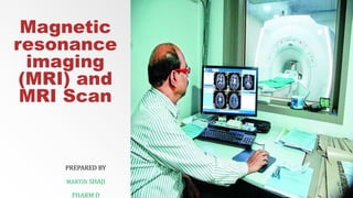
- 1. Magnetic resonance imaging (MRI) and MRI Scan PREPARED BY MARTIN SHAJI PHARM D
- 2. Diagnostics and therapy MRI (magnetic resonance imaging) scans – INTRODUCTION ▪ An MRI (magnetic resonance imaging) makes use of a big magnet, radio waves, and computer systems to provide high-quality detailed pictures of inside body parts. The MRI scan doesn't cause any pain. Patients don't really feel the magnetic field or radio waves. ▪ Doctors use an MRI to look at the brain, backbone, joints (knee, shoulder, hip, wrist, and ankle), abdomen, pelvis, breasts, blood vessels, heart, and different elements of the body. MRI scans can be used to help diagnose disease, including cancer, to monitor treatment progress and monitor remission in sufferers who've accomplished their treatment.
- 3. ▪ The MRI doesn't include ionizing radiation. Highlighter can be utilized to enhance the quality of the images. ▪ MRI machines use strong magnetic fields and radio waves (radiofrequency energy) to create pictures. During an MRI scan, the affected person is placed inside a strong magnetic field. Then radio waves are sent from a transmitter/receiver contained in the machine and received from it to examine the part of the body being studied, and these signals are used to create digital pictures of the area that have been examined.
- 4. What happens during an MRI scan? - PROCEDURE ▪ An affected person in an MRI position for her leg with a pediatric life specialist and two MRI technologists. ▪ A typical MRI takes 20 to 90 minutes, depending on which a part of the body is being imaged. ▪ The MRI appointment begins by registering the arrival of the affected person and his father at the registration office and waiting to be referred to as for an examination. It is essential that you just arrive minutes before your appointment to be able to check-in.
- 5. PROCEDURE ▪ When it's time for an examination, an MRI technologist or nurse will take the affected person and guardian into the room that contains the MRI machine or "radiography machine." ▪ The MRI machine seems to be like a big ring that has a socket in the middle. The machine has a padded table (sometimes called a bed) that slides out and in of the tube. The affected person lies on the table for the scan and should stay still during the MRI scan.
- 6. PROCEDURE ▪ Movement during the test will distort the resulting picture, and the examination will need to be repeated. A pediatric life specialist can train sufferers in relaxation techniques. Patients may also listen to music or watch a film with particular glasses. Patients who've difficulty maintaining stability could also be sedated so that they'll calm down or fall asleep during the procedure. ▪ The MRI scan doesn't cause any pain, and the affected person doesn't really feel the magnetic field or radio waves. But the scan makes lots of noise. Patients shall be given earplugs or headphones to block out noise and protect their listening to.
- 7. PROCEDURE ▪ During the examination, the technologist will move to an adjacent room and have the ability to see, hear and discuss to the affected person. The affected person can talk with the technologist through a two-way intercom system. Each children's centre has completely different policies, but normally one parent is allowed to stay in the next room with the technologist. ▪ A typical MRI takes 20 to 90 minutes, depending on which a part of the body is being imaged. ▪ After radiography, sufferers can resume their normal activities if analgesia has not yet been introduced. Patients who've received a sedative will need to regain consciousness first.
- 9. Indicator material ▪ In some instances, people will receive a brightener to help the MRI pictures appear clearer and brighter. The marker is injected through an IV. If the affected person doesn't have an intravenous line or an outlet, the nurse will install it. ▪ Usually, a brightener called gadolinium is used during an MRI. Gadolinium shouldn't be given to sufferers who're pregnant, have previously experienced an allergic reaction to this substance, or who's extreme kidney disease.
- 10. Indicator material ▪ Parents also need to inform the medical staff if the affected person has a history of heart illness, bronchial asthma, diabetes or thyroid issues. Some people could feel a temporary metallic taste in their mouth after the indicating substance is injected. ▪ A small number of people develop side effects from the indicating substance, including nausea and pain. In very rare instances, sufferers become allergic to the indicating substance and have reactions of hives, itchy eyes, or different reactions. In this case, a radiologist or other physician will be available to provide immediate assistance.
- 11. Magnetic resonance imaging of the abdomen and pelvis together ▪ If the physician schedules an MRI of the abdomen and pelvis, the affected person could receive a drug called hyoscyamine (Levsin) intravenously (intravenously). This medicine will reduce bowel movement during an MRI and help make the MRI picture clearer. ▪ Possible side effects of hyoscyamine (Levsin) include red skin, increased heart rate, delicate stomach pain and constipation. These side effects are normally minor. Patients won't receive hyoscyamine (Levsin) if they've certain medical conditions or if they are taking a drug that increases the chance of a bad reaction.
- 12. How to prepare for an MRI scan ▪ If sufferers are to be sedated, they shouldn't have any meals or drink for several hours earlier than the examination. Patients and families will receive particular instructions at their pediatric centre. ▪ Because of the strong magnet used for MRI, it needs to be patient and parents (when present) should remove any metallic objects before entering the MRI area. ▪ Before entering the affected person or parent into the MRI area, staff will ask the parent to fill out an examination form that includes questions on any metallic materials in the affected person’s body or clothes.
- 13. How to prepare for an MRI scan ▪ It is preferable to wear clothes that don't contain metal fasteners, zippers, or rivets. Parents should inform the medical staff of any medical devices implanted in the affected person. Patients may receive a hospital gown to wear. ▪ The father of pediatric cancer affected person gives his cell phone to the MRI technologist after a metal detector scan. ▪ Anyone entering the MRI area must go through a metal detector that's more sensitive than those found at most airports.
- 14. Items to remove include: Handbag, pockets, cash clip, bank card, magnetic stripe playing cards Electronic devices such as cell phones Hearing enhancement devices Metal jewellery, watches Pens, paper clips, keys, coins Hair clips, hairpins Any item of clothes with zippers, buttons, clips, metal fasteners, underwires, or metallic strings. Shoes, belt buckles, fixing pins Treatment patches
- 15. In most pediatric centres, anyone entering the MRI area should go through a metal detector that's more sensitive than those at most airports. If the detection device shows the presence of a metal object on or inside the individual's body, that individual should remove the metal object and undergo the examination again. If the metal object cannot be removed, the individual shouldn't enter the area until a qualified magnetic resonance safety professional determines that the metal is "safe for use in the MRI environment."
- 16. Here are some subjects that may affect picture quality if they are near the area being photographed: The metal bar for the spine Dishes, pins, nails, or metal mesh that may be used to fix a bone or joint Joint replacement or artificial limbs Metal ornaments, including those used for body piercing. Some kinds of tattoos or tattooed eyeliners (there is also a possibility of skin irritation or swelling) Cosmetics, nail polish, or different beauty objects that contain a mineral Dental fillings, braces and braces Magnetic resonance imaging showing a metallic distortion caused by a stent Magnetic resonance imaging of the thigh bone showing a metallic distortion
- 17. How do sufferers get the test results? ▪ A radiologist, a physician who has obtained particular training in reading an MRI, will analyze the photographs and prepare a report for presentation to the referring doctor. The referring doctor will share the results of the examination during the affected person's next visit. ▪ Childhood cancer affected person in a full-body MRI setting with her father and two MRI technicians. ▪ The MRI scan does not cause any pain, and the affected person does not feel the magnetic field or radio waves.
- 18. MRI front view