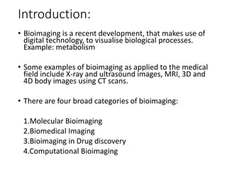
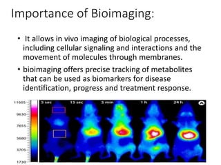
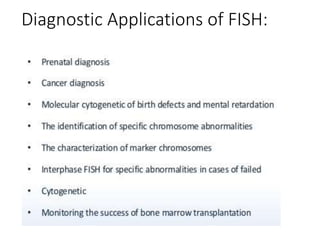
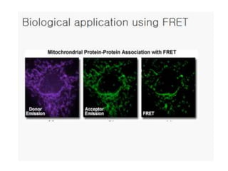
MOLECULAR BIOIMAGING
Presented by:
RUPAL AGRAWAL
M.Sc. Microbiology
16SOSMB21001
School of Science(SOS)
R.K. UNIVERSITY
Introduction:
• Bioimaging is a recent development, that makes use of
digital technology, to visualise biological processes.
Example: metabolism
• Some examples of bioimaging as applied to the medical
field include X-ray and ultrasound images, MRI, 3D and
4D body images using CT scans.
• There are four broad categories of bioimaging:
1.Molecular Bioimaging
2.Biomedical Imaging
3.Bioimaging in Drug discovery
4.Computational Bioimaging
Importance of Bioimaging:
• It allows in vivo imaging of biological processes,
including cellular signaling and interactions and the
movement of molecules through membranes.
• bioimaging offers precise tracking of metabolites
that can be used as biomarkers for disease
identification, progress and treatment response.
Shortfalls:
• There in no method in bioimaging today that
quantitatively tells a clinician by how much a tumor
grew or shrank in response to a drug.
• bioimaging cannot distinguish between benign and
malignant tumors.
Bioimage of tumor cells
taken from (CLSM) Confocal
lazer scanning microscope.
Some methods used in bioimaging:
1. Green Fluorescence protein ( GFP):
• Derived from jelly fish Aequorea Victoria.
• GFP is a protein composed of 238 amino acid
residues (26.9 kDa) that exhibits bright green
fluorescence when exposed to light in the blue to
ultraviolet range.
• gfp gene has been cloned from jelly fish and has
been successfully expressed in various plants.
2. Fluorescence in situ
hybridization ( FISH):
• It is a kind of cytogenetic technique which uses
fluorescent probes to detect and localize the
presence or absence of specific DNA sequences on
chromosomes.
• fluorescent probes will bind to only those parts of
the chromosome with a high degree of sequence
complementarity.
Fig: protocol of FISH technique outline
Diagnostic Applications of FISH:
3. GUS Reporter system:
• Derived from E.coli.
• Uid A gene code for 12-β- glucuronidase-enzyme
• enzymatic cleavage of X- Gluc ( 5-bromo-chloro-3-
indolylβ-D-glucuronide) undergoes an oxidative
dimerization to yield an indigo blue precipitate.
• An organism is suitable for a GUS assay if it has no
β-glucuronidase or if the activity is very low.
• Since there is no detectable GUS activity in higher
plants, mosses, algae, ferns, fungi and most
bacteria it perfectly suites for these organisms.
Rice embryo showing GUS
expression.
GUS gene expression on the trichomes of
Arabidopsis plant.
4. Fluorescence resonance energy
transfer (FRET):
• The mechanism of FRET involves a donor fluorophore
in an excited electronic state, which may transfer its
excitation energy to a nearby acceptor chromophore.
• The absorption spectrum of
the acceptor must overlap
fluorescence emission spectrum
of the donor.
Molecular bioimaging

Molecular bioimaging

  • 1.
    MOLECULAR BIOIMAGING Presented by: RUPALAGRAWAL M.Sc. Microbiology 16SOSMB21001 School of Science(SOS) R.K. UNIVERSITY
  • 2.
    Introduction: • Bioimaging isa recent development, that makes use of digital technology, to visualise biological processes. Example: metabolism • Some examples of bioimaging as applied to the medical field include X-ray and ultrasound images, MRI, 3D and 4D body images using CT scans. • There are four broad categories of bioimaging: 1.Molecular Bioimaging 2.Biomedical Imaging 3.Bioimaging in Drug discovery 4.Computational Bioimaging
  • 3.
    Importance of Bioimaging: •It allows in vivo imaging of biological processes, including cellular signaling and interactions and the movement of molecules through membranes. • bioimaging offers precise tracking of metabolites that can be used as biomarkers for disease identification, progress and treatment response.
  • 4.
    Shortfalls: • There inno method in bioimaging today that quantitatively tells a clinician by how much a tumor grew or shrank in response to a drug. • bioimaging cannot distinguish between benign and malignant tumors. Bioimage of tumor cells taken from (CLSM) Confocal lazer scanning microscope.
  • 5.
    Some methods usedin bioimaging: 1. Green Fluorescence protein ( GFP): • Derived from jelly fish Aequorea Victoria. • GFP is a protein composed of 238 amino acid residues (26.9 kDa) that exhibits bright green fluorescence when exposed to light in the blue to ultraviolet range. • gfp gene has been cloned from jelly fish and has been successfully expressed in various plants.
  • 7.
    2. Fluorescence insitu hybridization ( FISH): • It is a kind of cytogenetic technique which uses fluorescent probes to detect and localize the presence or absence of specific DNA sequences on chromosomes. • fluorescent probes will bind to only those parts of the chromosome with a high degree of sequence complementarity.
  • 8.
    Fig: protocol ofFISH technique outline
  • 10.
  • 13.
    3. GUS Reportersystem: • Derived from E.coli. • Uid A gene code for 12-β- glucuronidase-enzyme • enzymatic cleavage of X- Gluc ( 5-bromo-chloro-3- indolylβ-D-glucuronide) undergoes an oxidative dimerization to yield an indigo blue precipitate. • An organism is suitable for a GUS assay if it has no β-glucuronidase or if the activity is very low. • Since there is no detectable GUS activity in higher plants, mosses, algae, ferns, fungi and most bacteria it perfectly suites for these organisms.
  • 14.
    Rice embryo showingGUS expression. GUS gene expression on the trichomes of Arabidopsis plant.
  • 15.
    4. Fluorescence resonanceenergy transfer (FRET):
  • 16.
    • The mechanismof FRET involves a donor fluorophore in an excited electronic state, which may transfer its excitation energy to a nearby acceptor chromophore. • The absorption spectrum of the acceptor must overlap fluorescence emission spectrum of the donor.