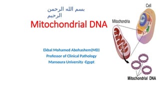
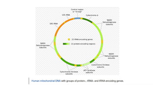
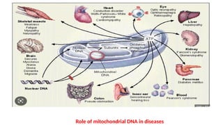
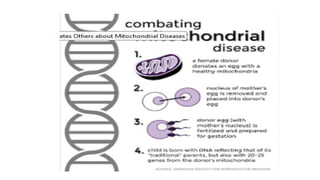
Mitochondrial DNA
Ekbal Mohamed Abohashem(MD)
Professor of Clinical Pathology
Mansoura University -Egypt
‫الرحمن‬ ‫الله‬ ‫بسم‬
‫الرحيم‬
Mitochondria are small, mobile, and plastic organelles located in
the cytoplasm of most eukaryotic cells. These organelles are
responsible for important cellular processes, such as regulation
of apoptosis, calcium homeostasis, and reactive oxygen species
(ROS) production. However, the main function of mitochondria
is energy production through oxidative phosphorylation
(OXPHOS), which takes place in the mitochondrial respiratory
chain (MRC).
Introduction: Mitochondria, the Powerhouses of the Cell
Mitochondria contain their own DNA, the mitochondrial DNA
(mtDNA), which is circular and double stranded. The
mitochondrial genome consists of 16,569 nucleotide pairs
that encode 13 proteins, two ribosomal RNA components,
and 22 transfer RNAs (tRNAs) .
Regarding mitochondrial structure, these organelles are composed by two
membranes, the inner and the outer mitochondrial membranes (IMMs and
OMMs, respectively) that delimit two main compartments: the internal
matrix and the intermembrane space. The IMM contains many folds named
cristae that protrude into the matrix and enlarge the IMM surface. This
membrane can be subdivided into two compartments, the inner boundary
membrane (IBM) and the cristae membrane (CM), that are connected via
cristae junctions.
Mitochondrial structure
 Although IMM is considered a continuous membrane, lateral diffusion of
membrane proteins is restricted and IBM and CM exhibit an asymmetric
protein distribution. This heterogeneity is important for efficient OXPHOS,
mitochondrial biogenesis, and remodeling.
 Defects in mitochondrial function have been linked not only to genetic
mitochondrial diseases but also to cardiovascular diseases ,and
neurodegenerative disorders such as Huntington’s and Parkinson’s diseases .
History of mitochondrial DNA :
Mitochondrial DNA was discovered in the 1960s by Margit M. K. Nass and
Sylvan Nass by electron microscopy as DNase-sensitive threads inside
mitochondria, and by Ellen Haslbrunner, Hans Tuppy and Gottfried Schatz
by biochemical assays on highly purified mitochondrial fractions
 Mitochondrial DNA (mtDNA or mDNA) is the DNA located in mitochondria, It
represents only a small portion of the DNA in an eukaryotic cell; most of the
DNA is found in the cell nucleus .
 Human mitochondrial DNA was the first significant part of the human genome
to be sequenced. This sequencing revealed that the human mtDNA includes
16,569 base pairs and encodes 13 proteins.
 Since animal mtDNA evolves faster than nuclear genetic markers, it represents
a mainstay of phylogenetics and evolutionary biology. It also permits an
examination of the relatedness of populations, and so has become important
in anthropology and biogeography.
Mitochondrial DNA:
 The two strands of the human mitochondrial DNA are distinguished as the heavy
strand and the light strand. The heavy strand is rich in guanine and encodes 12
subunits of the oxidative phosphorylation system, two ribosomal RNAs (12S and
16S), and 14 transfer RNAs (tRNAs). The light strand encodes one subunit, and 8
tRNAs. So, altogether mtDNA encodes for two rRNAs, 22 tRNAs, and 13
protein subunits, all of which are involved in the oxidative phosphorylation
process .
Genes on the human mtDNA and their transcription
 The promoters for the initiation of the transcription of the heavy and light
strands are located in the main non-coding region of the mtDNA called the
displacement loop, the D-loop. There is evidence that the transcription of the
mitochondrial rRNAs is regulated by the heavy-strand promoter 1 (HSP1), and
the transcription of the polycistronic transcripts coding for the protein subunits
are regulated by HSP2 .
Regulation of transcription
Map of human mtDNA
Map of human mtDNAThe genome encodes for 13 mRNA (green), 22 tRNA (violet), and 2 rRNA (pale blue)
molecules. There is also a major noncoding region (NCR), which is shown enlarged at the top. The major
NCR contains the heavy strand promoter (HSP), the light strand promoter (LSP), three conserved sequence
boxes (CSB1-3, orange), the H-strand origin of replication (OH), and the termination-associated sequence
(TAS, yellow). The triple-stranded displacement-loop (D-loop) structure is formed by premature
termination of nascent H-strand DNA synthesis at TAS. The short H-strand replication product formed in
this manner is termed 7S DNA. A minor NCR, located approximately 11,000 bp downstream of OH, contains
the L-strand origin of re
Mitochondrial inheritance
 In most multicellular organisms, mtDNA is inherited from the mother
(maternally inherited). Mechanisms for this include
* simple dilution (an egg contains on average 200,000 mtDNA molecules,
whereas a healthy human sperm has been reported to contain on average 5
molecules),
*
degradation of sperm mtDNA in the male genital tract and in the fertilized egg;
and,
* at least in a few organisms, failure of sperm mtDNA to enter the egg.
Whatever the mechanism, this single parent (uniparental inheritance) pattern of
mtDNA inheritance is found in most animals, most plants and also in fungi.
Heteroplasmy
 Heteroplasmy is the presence of more than one type of organellar
genome (mitochondrial DNA ) within a cell or individual. It is an
important factor in considering the severity of mitochondrial diseases.
Because most eukaryotic cells contain many hundreds of
mitochondria with hundreds of copies of mitochondrial DNA, it is
common for mutations to affect only some mitochondria, leaving
most unaffected.
Female inheritance
 In sexual reproduction, mitochondria are normally inherited exclusively from
the mother; the mitochondria in mammalian sperm are usually destroyed by
the egg cell after fertilization. Also, mitochondria are only in the sperm tail,
which is used for propelling the sperm cells and sometimes the tail is lost during
fertilization. In 1999 it was reported that paternal sperm mitochondria
(containing mtDNA) are marked with ubiquitin to select them for later
destruction inside the embryo. Some in vitro fertilization techniques,
particularly injecting a sperm into an oocyte, may interfere with this
Similar to the nuclear genome, the mitochondrial genome is built of
double-stranded DNA, and it encodes genes . However, the mitochondrial
genome differs from the nuclear genome in several ways :
•The mitochondrial genome is circular, whereas the nuclear genome is
linear .
•The mitochondrial genome is built of 16,569 DNA base pairs, whereas the
nuclear genome is made of 3.3 billion DNA base pairs.
Mitochondrial vs. Nuclear DNA
•The mitochondrial genome contains 37 genes that encode 13
proteins, 22 tRNAs, and 2 rRNAs.
•The 13 mitochondrial gene-encoded proteins all instruct cells to
produce protein subunits of the enzyme complexes of the oxidative
phosphorylation system, which enables mitochondria to act as the
powerhouses of our cells.
•The small mitochondrial genome is not able to independently produce
all of the proteins needed for functionality; thus, mitochondria rely
heavily on imported nuclear gene products.
•One mitochondrion contains dozens of copies of its mitochondrial
genome. In addition, each cell contains numerous mitochondria.
Therefore, a given cell can contain several thousand copies of its
mitochondrial genome, but only one copy of its nuclear genome.
•The mitochondrial genome is not enveloped, and is it not packaged
into chromatin.
•The mitochondrial genome contains few, if any, noncoding DNA
sequences. (Three percent of the mitochondrial genome is noncoding
DNA, whereas 93% of the nuclear genome is noncoding DNA).
•Some mitochondrial coding sequences (triplet codons) do not follow
the universal codon usage rules when they are translated into proteins.
•Some mitochondrial nucleotide bases exhibit functional overlap between
two genes; in other words, the same nucleotide can sometimes function as
both the last base of one gene and the first base of the next gene.
•The mitochondrial mode of inheritance is strictly maternal, whereas
nuclear genomes are inherited equally from both parents. Therefore,
mitochondria-associated disease mutations are also always inherited
maternally.
•Mitochondrial genes on both DNA strands are transcribed in a
polycistronic manner: Large mitochondrial mRNAs contain the
instructions to build many different proteins, which are encoded
one after the next along the mRNA. In contrast, nuclear genes are
usually transcribed one at a time from their own mRNA
 Each cell contains numerous mitochondria, and each mitochondrion
contains dozens of copies of the mitochondrial genome. Moreover, the
mitochondrial genome has a higher mutation rate (about 100-fold higher)
than the nuclear genome. This leads to a heterogeneous population of
mitochondrial DNA within the same cell, and even within the same
mitochondrion; as a result, mitochondria are considered heteroplasmic.
Mitochondrial DNA Mutations
 When a cell divides, its mitochondria are partitioned between
the two daughter cells. However, the process of mitochondrial
segregation occurs in a random manner and is much less
organized than the highly accurate process of
nuclear chromosome segregation during mitosis. As a result,
daughter cells receive similar, but not identical, copies of their
mitochondrial DNA
Due to the multicopy nature of mtDNA, these mutations can be
homoplasmic or heteroplasmic. Thus, MELAS and MERRF syndromes
are heteroplasmic, which means that mutant and wild-type mtDNA
copies coexist within the same cell . Mitochondrial disease may
become clinically apparent once the number of affected
mitochondria reaches a certain level; this phenomenon is
called "threshold expression"
 Mitochondrial diseases are a heterogeneous group of maternally inherited
rare genetic disorders caused by a partial or total dysfunction of mitochondria.
These illnesses can be caused by mutations in nDNA or mtDNA. These
mutations affect not only genes encoding for mitochondrial respiratory
chain( MRC) components but also those that are involved in protein translation
and assembly, mtDNA stability, as well as mutations in those nDNA-encoded
proteins involved in the maintenance of mitochondrial nucleotide pools,
nucleotide transport, mtDNA replication, RNA transcription, and mitochondrial
dynamics .
Mitochondrial Diseases :
 More than 400 pathogenic mutations in mitochondrial transfer RNA have been
characterized . Some frequent mitochondrial disorders caused by a point mutation
in mtDNA are MELAS and MERRF syndromes.
 In most of the cases, MELAS syndrome is caused by a transition from adenine to
guanine in the position 3243 in the mt-tRNALeu(UUR)
(MT-TL1) gene .This affects mt-
tRNA structure stabilization, methylation, aminoacylation, and triplet recognition .
 In the case of MERRF syndrome, the m.8344A > G mutation in the mt-tRNALys
(MT-
TK) gene is the most frequently associated with the disease .It affects both AAA
and AAG codon translation, causing a defect of whole mitochondrial protein
synthesis.
How common is mitochondrial disease?
An estimated 1 in 5,000 people has a genetic mitochondrial disease. It’s
common for mitochondrial diseases to receive a misdiagnosis due to the
number and type of symptoms and organ systems involved, so this number may
be underestimated.
What are the types of mitochondrial disease?
There are many types of mitochondrial diseases. Some of the most common include:
•Mitochondrial encephalopathy, lactic acidosis and stroke-like episodes (MELAS) synd
rome
.
•Leber hereditary optic neuropathy (LHON).
•Leigh syndrome.
•Kearns-Sayre syndrome (KSS).
•Myoclonic epilepsy and ragged-red fiber disease (MERRF).
 Mitochondrial diseases are clinically heterogeneous; they may occur at any
age, and patients manifest a wide variety of symptoms . However, all of them
share morphological and biochemical features. As a consequence of the MRC
deficiency, cells manifest a reduced enzymatic function of MRC components,
a reduction in oxygen consumption and ATP synthesis, and ROS
overproduction.
 Patients suffer from lactic acidosis and elevated serum pyruvate levels at
rest and, specially, after moderate exercise. Additionally, patients’ muscle
biopsies usually show ragged red fibers that reflect the proliferation of
oxidative phosphorylation ( OXPHOS-defective mitochondria .)
 Both MELAS and MERRF syndromes are associated with
neurological symptoms. MELAS syndrome affects several organs,
and some of its manifestations include stroke-like episodes,
dementia, epilepsy, lactic acidemia, myopathy, recurrent
headaches, hearing impairment, diabetes, and short stature .
 In the case of MERRF syndrome, the first symptom is usually
myoclonus that is followed by generalized epilepsy, ataxia,
weakness, and dementia. Other findings are hearing loss, short
stature, optic atrophy, and cardiomyopathy .
Role of mitochondrial DNA in diseases
 ). Mitochondrial dysfunctions contribute to diabetes, obesity, and
metabolic syndromes
 ). Mitochondria play diverse roles in cancer, such as providing energy and
biosynthetic products for rapid proliferation, supporting metabolic
adaptation to the tumor microenvironment, and regulating oncogenic
signaling and apoptosis
 ). Mitochondria support cellular immune functions. However,
dysfunctions in mitochondria or trauma could release immunogenic
mtDNA and mtRNA in the cytosol or circulation to cause severe or
chronic inflammation
 ). Mitochondrial genetics, metabolism, and inflammation
significantly impact age-associated pathologies
 ) and neurodegenerative diseases such as Alzheimer's and
Parkinson's diseases
The development of useful therapies for mitochondrial diseases is challenging
due to :
*the difficulty of correcting the lack or dysfunction of essential mitochondrial
proteins,
* the phenotypical heterogeneity of the diseases, and
*multisystem alteration.
Furthermore, the brain, one of the most affected organs, is difficult to reach by
potential therapies because it is protected by the blood–brain barrier. For
those reasons, there are no effective treatments available for mitochondrial
diseases and management of these diseases is mainly symptomatic.
Therapeutic Management of Mitochondrial Diseases:
 Pharmacological treatment options are generally focused on targeting cellular
pathways, such as mitochondrial biogenesis or autophagy, or preventing
oxidative damage. For these reasons, AMP-activated protein kinase (AMPK) and
mammalian target of rapamycin complex 1 (mTORC1) signaling have been the
main targets of these strategies.
 Particularly in mtDNA mutations, several supplements as antioxidants and
cofactors are being used.
 Given the diversity of mutations and the different therapeutic options,
a personalized therapeutic approach is required in mitochondrial
diseases. For this reason, the development of cellular models derived
from patients can be useful for both the evaluation of new drugs and
the repositioning of existing ones.
 Gene therapy is a promising alternative for treating mitochondrial
diseases. Since pathogenic mtDNA mutations are usually
heteroplasmic, reducing mutational load can be used as a therapeutic
approach. There are several tools that could target mtDNA, but only
two of them have been demonstrated to be successful: zinc-finger
nucleases (ZFNs) and transcription activator-like effector nucleases
(TALENs). Both tools are delivered into mitochondria using a
mitochondria localization signal, and they selectively target mtDNA
sequences to create double-strand breaks.
 TALENs are artificial restriction enzymes and can cut DNA strands at
any desired sequence,which makes them ideal for genetic
engineering and targeted genome editing . They have been widely
used for genetic manipulation in different organisms. This tool has
been demonstrated to reduce mutant mtDNA load and improve the
pathophysiology in a cellular model of MERRF syndrome as well as to
eliminate the m.3243A > G mutation in MELAS inexperimental
models and porcine oocytes.
 Zinc-finger nucleases have been demonstrated to reduce mutant mtDNA
and consequently restore mitochondrial respiratory function in cytoplasmic
hybrid (cybrid) cell models .
In addition, TALENs have been able to reduce mutant mtDNA load in a mouse
model harboring a mutation in a mt-tRNA, reverting disease-related phenotypes .
 The only option available is transferring embryos below the
threshold of clinical expression in order to avoid or at least reduce
the risk of transmission of mtDNA mutations. The selection of these
embryos is based on preimplantation genetic diagnosis (PGD) .
 In addition, there is a new strategy, the mitochondrial donation, that
consists of the substitution of mutant maternal mitochondria using
enucleated donor oocytes .However, this technique has raised ethical
issues and remains controversial).
Prevention of mitochondrial diseases,
THANK YOU

Mitochondrial DNA ,structure,function,and related diseases.

  • 1.
    Mitochondrial DNA Ekbal MohamedAbohashem(MD) Professor of Clinical Pathology Mansoura University -Egypt ‫الرحمن‬ ‫الله‬ ‫بسم‬ ‫الرحيم‬
  • 2.
    Mitochondria are small,mobile, and plastic organelles located in the cytoplasm of most eukaryotic cells. These organelles are responsible for important cellular processes, such as regulation of apoptosis, calcium homeostasis, and reactive oxygen species (ROS) production. However, the main function of mitochondria is energy production through oxidative phosphorylation (OXPHOS), which takes place in the mitochondrial respiratory chain (MRC). Introduction: Mitochondria, the Powerhouses of the Cell
  • 3.
    Mitochondria contain theirown DNA, the mitochondrial DNA (mtDNA), which is circular and double stranded. The mitochondrial genome consists of 16,569 nucleotide pairs that encode 13 proteins, two ribosomal RNA components, and 22 transfer RNAs (tRNAs) .
  • 4.
    Regarding mitochondrial structure,these organelles are composed by two membranes, the inner and the outer mitochondrial membranes (IMMs and OMMs, respectively) that delimit two main compartments: the internal matrix and the intermembrane space. The IMM contains many folds named cristae that protrude into the matrix and enlarge the IMM surface. This membrane can be subdivided into two compartments, the inner boundary membrane (IBM) and the cristae membrane (CM), that are connected via cristae junctions.
  • 5.
  • 6.
     Although IMMis considered a continuous membrane, lateral diffusion of membrane proteins is restricted and IBM and CM exhibit an asymmetric protein distribution. This heterogeneity is important for efficient OXPHOS, mitochondrial biogenesis, and remodeling.  Defects in mitochondrial function have been linked not only to genetic mitochondrial diseases but also to cardiovascular diseases ,and neurodegenerative disorders such as Huntington’s and Parkinson’s diseases .
  • 7.
    History of mitochondrialDNA : Mitochondrial DNA was discovered in the 1960s by Margit M. K. Nass and Sylvan Nass by electron microscopy as DNase-sensitive threads inside mitochondria, and by Ellen Haslbrunner, Hans Tuppy and Gottfried Schatz by biochemical assays on highly purified mitochondrial fractions
  • 8.
     Mitochondrial DNA(mtDNA or mDNA) is the DNA located in mitochondria, It represents only a small portion of the DNA in an eukaryotic cell; most of the DNA is found in the cell nucleus .  Human mitochondrial DNA was the first significant part of the human genome to be sequenced. This sequencing revealed that the human mtDNA includes 16,569 base pairs and encodes 13 proteins.  Since animal mtDNA evolves faster than nuclear genetic markers, it represents a mainstay of phylogenetics and evolutionary biology. It also permits an examination of the relatedness of populations, and so has become important in anthropology and biogeography. Mitochondrial DNA:
  • 9.
     The twostrands of the human mitochondrial DNA are distinguished as the heavy strand and the light strand. The heavy strand is rich in guanine and encodes 12 subunits of the oxidative phosphorylation system, two ribosomal RNAs (12S and 16S), and 14 transfer RNAs (tRNAs). The light strand encodes one subunit, and 8 tRNAs. So, altogether mtDNA encodes for two rRNAs, 22 tRNAs, and 13 protein subunits, all of which are involved in the oxidative phosphorylation process . Genes on the human mtDNA and their transcription
  • 10.
     The promotersfor the initiation of the transcription of the heavy and light strands are located in the main non-coding region of the mtDNA called the displacement loop, the D-loop. There is evidence that the transcription of the mitochondrial rRNAs is regulated by the heavy-strand promoter 1 (HSP1), and the transcription of the polycistronic transcripts coding for the protein subunits are regulated by HSP2 . Regulation of transcription
  • 11.
  • 12.
    Map of humanmtDNAThe genome encodes for 13 mRNA (green), 22 tRNA (violet), and 2 rRNA (pale blue) molecules. There is also a major noncoding region (NCR), which is shown enlarged at the top. The major NCR contains the heavy strand promoter (HSP), the light strand promoter (LSP), three conserved sequence boxes (CSB1-3, orange), the H-strand origin of replication (OH), and the termination-associated sequence (TAS, yellow). The triple-stranded displacement-loop (D-loop) structure is formed by premature termination of nascent H-strand DNA synthesis at TAS. The short H-strand replication product formed in this manner is termed 7S DNA. A minor NCR, located approximately 11,000 bp downstream of OH, contains the L-strand origin of re
  • 15.
    Mitochondrial inheritance  Inmost multicellular organisms, mtDNA is inherited from the mother (maternally inherited). Mechanisms for this include * simple dilution (an egg contains on average 200,000 mtDNA molecules, whereas a healthy human sperm has been reported to contain on average 5 molecules), * degradation of sperm mtDNA in the male genital tract and in the fertilized egg; and, * at least in a few organisms, failure of sperm mtDNA to enter the egg. Whatever the mechanism, this single parent (uniparental inheritance) pattern of mtDNA inheritance is found in most animals, most plants and also in fungi.
  • 16.
    Heteroplasmy  Heteroplasmy isthe presence of more than one type of organellar genome (mitochondrial DNA ) within a cell or individual. It is an important factor in considering the severity of mitochondrial diseases. Because most eukaryotic cells contain many hundreds of mitochondria with hundreds of copies of mitochondrial DNA, it is common for mutations to affect only some mitochondria, leaving most unaffected.
  • 17.
    Female inheritance  Insexual reproduction, mitochondria are normally inherited exclusively from the mother; the mitochondria in mammalian sperm are usually destroyed by the egg cell after fertilization. Also, mitochondria are only in the sperm tail, which is used for propelling the sperm cells and sometimes the tail is lost during fertilization. In 1999 it was reported that paternal sperm mitochondria (containing mtDNA) are marked with ubiquitin to select them for later destruction inside the embryo. Some in vitro fertilization techniques, particularly injecting a sperm into an oocyte, may interfere with this
  • 18.
    Similar to thenuclear genome, the mitochondrial genome is built of double-stranded DNA, and it encodes genes . However, the mitochondrial genome differs from the nuclear genome in several ways : •The mitochondrial genome is circular, whereas the nuclear genome is linear . •The mitochondrial genome is built of 16,569 DNA base pairs, whereas the nuclear genome is made of 3.3 billion DNA base pairs. Mitochondrial vs. Nuclear DNA
  • 19.
    •The mitochondrial genomecontains 37 genes that encode 13 proteins, 22 tRNAs, and 2 rRNAs. •The 13 mitochondrial gene-encoded proteins all instruct cells to produce protein subunits of the enzyme complexes of the oxidative phosphorylation system, which enables mitochondria to act as the powerhouses of our cells.
  • 20.
    •The small mitochondrialgenome is not able to independently produce all of the proteins needed for functionality; thus, mitochondria rely heavily on imported nuclear gene products. •One mitochondrion contains dozens of copies of its mitochondrial genome. In addition, each cell contains numerous mitochondria. Therefore, a given cell can contain several thousand copies of its mitochondrial genome, but only one copy of its nuclear genome.
  • 21.
    •The mitochondrial genomeis not enveloped, and is it not packaged into chromatin. •The mitochondrial genome contains few, if any, noncoding DNA sequences. (Three percent of the mitochondrial genome is noncoding DNA, whereas 93% of the nuclear genome is noncoding DNA). •Some mitochondrial coding sequences (triplet codons) do not follow the universal codon usage rules when they are translated into proteins.
  • 22.
    •Some mitochondrial nucleotidebases exhibit functional overlap between two genes; in other words, the same nucleotide can sometimes function as both the last base of one gene and the first base of the next gene. •The mitochondrial mode of inheritance is strictly maternal, whereas nuclear genomes are inherited equally from both parents. Therefore, mitochondria-associated disease mutations are also always inherited maternally.
  • 23.
    •Mitochondrial genes onboth DNA strands are transcribed in a polycistronic manner: Large mitochondrial mRNAs contain the instructions to build many different proteins, which are encoded one after the next along the mRNA. In contrast, nuclear genes are usually transcribed one at a time from their own mRNA
  • 24.
     Each cellcontains numerous mitochondria, and each mitochondrion contains dozens of copies of the mitochondrial genome. Moreover, the mitochondrial genome has a higher mutation rate (about 100-fold higher) than the nuclear genome. This leads to a heterogeneous population of mitochondrial DNA within the same cell, and even within the same mitochondrion; as a result, mitochondria are considered heteroplasmic. Mitochondrial DNA Mutations
  • 25.
     When acell divides, its mitochondria are partitioned between the two daughter cells. However, the process of mitochondrial segregation occurs in a random manner and is much less organized than the highly accurate process of nuclear chromosome segregation during mitosis. As a result, daughter cells receive similar, but not identical, copies of their mitochondrial DNA
  • 26.
    Due to themulticopy nature of mtDNA, these mutations can be homoplasmic or heteroplasmic. Thus, MELAS and MERRF syndromes are heteroplasmic, which means that mutant and wild-type mtDNA copies coexist within the same cell . Mitochondrial disease may become clinically apparent once the number of affected mitochondria reaches a certain level; this phenomenon is called "threshold expression"
  • 27.
     Mitochondrial diseasesare a heterogeneous group of maternally inherited rare genetic disorders caused by a partial or total dysfunction of mitochondria. These illnesses can be caused by mutations in nDNA or mtDNA. These mutations affect not only genes encoding for mitochondrial respiratory chain( MRC) components but also those that are involved in protein translation and assembly, mtDNA stability, as well as mutations in those nDNA-encoded proteins involved in the maintenance of mitochondrial nucleotide pools, nucleotide transport, mtDNA replication, RNA transcription, and mitochondrial dynamics . Mitochondrial Diseases :
  • 28.
     More than400 pathogenic mutations in mitochondrial transfer RNA have been characterized . Some frequent mitochondrial disorders caused by a point mutation in mtDNA are MELAS and MERRF syndromes.  In most of the cases, MELAS syndrome is caused by a transition from adenine to guanine in the position 3243 in the mt-tRNALeu(UUR) (MT-TL1) gene .This affects mt- tRNA structure stabilization, methylation, aminoacylation, and triplet recognition .  In the case of MERRF syndrome, the m.8344A > G mutation in the mt-tRNALys (MT- TK) gene is the most frequently associated with the disease .It affects both AAA and AAG codon translation, causing a defect of whole mitochondrial protein synthesis.
  • 29.
    How common ismitochondrial disease? An estimated 1 in 5,000 people has a genetic mitochondrial disease. It’s common for mitochondrial diseases to receive a misdiagnosis due to the number and type of symptoms and organ systems involved, so this number may be underestimated. What are the types of mitochondrial disease? There are many types of mitochondrial diseases. Some of the most common include: •Mitochondrial encephalopathy, lactic acidosis and stroke-like episodes (MELAS) synd rome . •Leber hereditary optic neuropathy (LHON). •Leigh syndrome. •Kearns-Sayre syndrome (KSS). •Myoclonic epilepsy and ragged-red fiber disease (MERRF).
  • 30.
     Mitochondrial diseasesare clinically heterogeneous; they may occur at any age, and patients manifest a wide variety of symptoms . However, all of them share morphological and biochemical features. As a consequence of the MRC deficiency, cells manifest a reduced enzymatic function of MRC components, a reduction in oxygen consumption and ATP synthesis, and ROS overproduction.
  • 31.
     Patients sufferfrom lactic acidosis and elevated serum pyruvate levels at rest and, specially, after moderate exercise. Additionally, patients’ muscle biopsies usually show ragged red fibers that reflect the proliferation of oxidative phosphorylation ( OXPHOS-defective mitochondria .)
  • 33.
     Both MELASand MERRF syndromes are associated with neurological symptoms. MELAS syndrome affects several organs, and some of its manifestations include stroke-like episodes, dementia, epilepsy, lactic acidemia, myopathy, recurrent headaches, hearing impairment, diabetes, and short stature .  In the case of MERRF syndrome, the first symptom is usually myoclonus that is followed by generalized epilepsy, ataxia, weakness, and dementia. Other findings are hearing loss, short stature, optic atrophy, and cardiomyopathy .
  • 34.
    Role of mitochondrialDNA in diseases
  • 35.
     ). Mitochondrialdysfunctions contribute to diabetes, obesity, and metabolic syndromes  ). Mitochondria play diverse roles in cancer, such as providing energy and biosynthetic products for rapid proliferation, supporting metabolic adaptation to the tumor microenvironment, and regulating oncogenic signaling and apoptosis  ). Mitochondria support cellular immune functions. However, dysfunctions in mitochondria or trauma could release immunogenic mtDNA and mtRNA in the cytosol or circulation to cause severe or chronic inflammation
  • 36.
     ). Mitochondrialgenetics, metabolism, and inflammation significantly impact age-associated pathologies  ) and neurodegenerative diseases such as Alzheimer's and Parkinson's diseases
  • 38.
    The development ofuseful therapies for mitochondrial diseases is challenging due to : *the difficulty of correcting the lack or dysfunction of essential mitochondrial proteins, * the phenotypical heterogeneity of the diseases, and *multisystem alteration. Furthermore, the brain, one of the most affected organs, is difficult to reach by potential therapies because it is protected by the blood–brain barrier. For those reasons, there are no effective treatments available for mitochondrial diseases and management of these diseases is mainly symptomatic. Therapeutic Management of Mitochondrial Diseases:
  • 39.
     Pharmacological treatmentoptions are generally focused on targeting cellular pathways, such as mitochondrial biogenesis or autophagy, or preventing oxidative damage. For these reasons, AMP-activated protein kinase (AMPK) and mammalian target of rapamycin complex 1 (mTORC1) signaling have been the main targets of these strategies.  Particularly in mtDNA mutations, several supplements as antioxidants and cofactors are being used.
  • 40.
     Given thediversity of mutations and the different therapeutic options, a personalized therapeutic approach is required in mitochondrial diseases. For this reason, the development of cellular models derived from patients can be useful for both the evaluation of new drugs and the repositioning of existing ones.  Gene therapy is a promising alternative for treating mitochondrial diseases. Since pathogenic mtDNA mutations are usually heteroplasmic, reducing mutational load can be used as a therapeutic approach. There are several tools that could target mtDNA, but only two of them have been demonstrated to be successful: zinc-finger nucleases (ZFNs) and transcription activator-like effector nucleases (TALENs). Both tools are delivered into mitochondria using a mitochondria localization signal, and they selectively target mtDNA sequences to create double-strand breaks.
  • 41.
     TALENs areartificial restriction enzymes and can cut DNA strands at any desired sequence,which makes them ideal for genetic engineering and targeted genome editing . They have been widely used for genetic manipulation in different organisms. This tool has been demonstrated to reduce mutant mtDNA load and improve the pathophysiology in a cellular model of MERRF syndrome as well as to eliminate the m.3243A > G mutation in MELAS inexperimental models and porcine oocytes.
  • 42.
     Zinc-finger nucleaseshave been demonstrated to reduce mutant mtDNA and consequently restore mitochondrial respiratory function in cytoplasmic hybrid (cybrid) cell models . In addition, TALENs have been able to reduce mutant mtDNA load in a mouse model harboring a mutation in a mt-tRNA, reverting disease-related phenotypes .
  • 43.
     The onlyoption available is transferring embryos below the threshold of clinical expression in order to avoid or at least reduce the risk of transmission of mtDNA mutations. The selection of these embryos is based on preimplantation genetic diagnosis (PGD) .  In addition, there is a new strategy, the mitochondrial donation, that consists of the substitution of mutant maternal mitochondria using enucleated donor oocytes .However, this technique has raised ethical issues and remains controversial). Prevention of mitochondrial diseases,
  • 45.