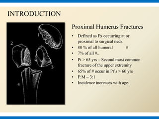
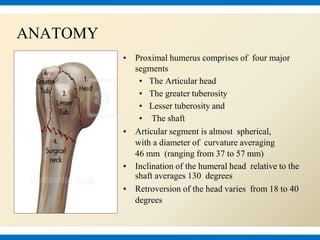
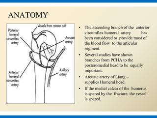
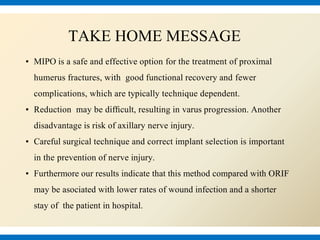
Bangalore Call Girl Whatsapp Number 100% Complete Your Sexual Needs
MIPO TECHNIQUE FOR PROXIMAL HUMERUS FRACTURE
1. Result of MIPO Technique in
Proximal Humerus Fracture
Fixation
By: Dr. Arvind Kumar
PGT, Peerless Hospital, Kolkata
2. Proximal Humerus Fractures
▪ Defined as Fx occurring at or
proximal to surgical neck
▪ 80 % of all humeral #
▪ 7% of all #..
▪ Pt > 65 yrs – Second most common
fracture of the upper extremity
▪ 65% of # occur in Pt’s > 60 yrs
▪ F:M – 3:1
▪ Incidence increases with age.
INTRODUCTION
3. ANATOMY
▪ Proximal humerus comprises of four major
segments
• The Articular head
• The greater tuberosity
• Lesser tuberosity and
• The shaft
▪ Articular segment is almost spherical,
with a diameter of curvature averaging
46 mm (ranging from 37 to 57 mm)
▪ Inclination of the humeral head relative to the
shaft averages 130 degrees
▪ Retroversion of the head varies from 18 to 40
degrees
4. ANATOMY
▪ The ascending branch of the anterior
circumflex humeral artery has
been considered to provide most of
the blood flow to the articular
segment.
▪ Several studies have shown
branches from PCHA to the
posteromedial head to be equally
important.
▪ Arcuate artery of Liang –
supplies Humeral head.
▪ If the medial calcar of the humerus
is spared by the fracture, the vessel
is spared.
5. AXILLARY NERVE
COURSE:
▪ It passes inferiorly and
laterally along the posterior
wall of the axilla to exit the
axilla.
▪ Then, it passes posteriorly
around the surgical neck of the
humerus , (clinical importance).
▪ It is accompanied by the
posterior circumflex humeral
vessels.
6. DEFORMING FORCES OF PHF
▪ The greater tuberosity is pulled
posteromedially by the effect of
the supra- and infraspinatus
tendons.
▪ The lesser tuberosity is pulled
anteriorly by the
subscapularis tendon.
▪ The shaft segment is pulled
anteromedially by the pectoralis
major tendon.
8. What is optimal management for displaced
Proximal Humerus Fractures?
CONTROVERSIAL
9. ▪ Non Operative Treatment
▪ Operative Treatment
➢ CRIF With Percutaneous K Wire Fixation
➢ Intramedullary Nailing
➢ ORIF with LCP
➢ Shoulder Arthroplasty
MANAGEMENT
10. ▪ The treatment of displaced proximal humeral
▪ # is complex & requires careful assessment
▪ Pt factors (age & activity level)
▪ Fracture-related factors (bone quality, fracture pattern, degree of
comminution, & vascular status).
▪ The goal of treatment is a pain-free shoulder with restoration of
pre-injury function.
OPERATIVE TREATMENT
11. Minimally Invasive
▪ Access to the bone through soft-tissue windows
▪ Minimal trauma to the soft tissue and the bone by indirect reduction
▪ Minimal additional trauma at the fracture site when direct reduction
is necessary
▪ Tools which cause “small footprints”
DEFINITION
12. PATIENT POSITIONING & SURGICAL
APPROACH
Proximally a 5-6cm longitudnal incision from the tip of acromion down the lateral aspect
of upper arm. The length and position of distal incision depends on the site of fracture and
length of implant used.
16. TAKE HOME MESSAGE
▪ MIPO is a safe and effective option for the treatment of proximal
humerus fractures, with good functional recovery and fewer
complications, which are typically technique dependent.
▪ Reduction may be difficult, resulting in varus progression. Another
disadvantage is risk of axillary nerve injury.
▪ Careful surgical technique and correct implant selection is important
in the prevention of nerve injury.
▪ Furthermore our results indicate that this method compared with ORIF
may be asociated with lower rates of wound infection and a shorter
stay of the patient in hospital.
Ellderly inflict freacture due to direct impact in low enegy trauma
Branches: 1) Motor to the deltoid and teres minor muscles. (2) Sensory- superior lateral cutaneous nerve of arm that loops around the posterior margin of the deltoid muscle to innervate the skin over that region.
Indications- 1. stable nondispaced or minimally displaced fractures. 2. Patients not fit for surgery. 3. Elderly patients with low functional demand.
Relative Contraindications- displaced fractures with loss of bony contact
There is no inter-nervous plane. Deep surgical dissection is made through the proximal window in an epi-periosteal plane. Care must be taken not to retract the deltoid muscle fibres proximally or distally in an aggressive manner.