Downloaded 156 times

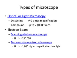
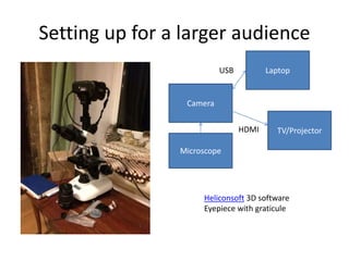

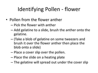
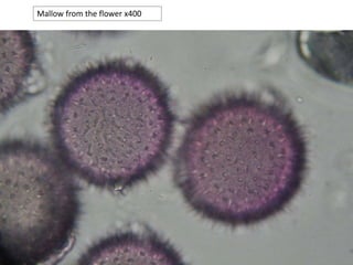
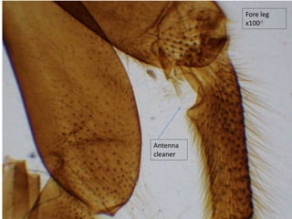
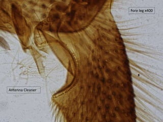
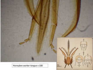
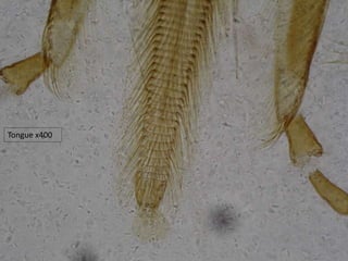
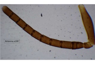
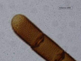
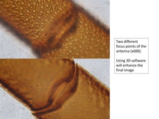
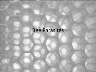

This document provides an overview of microscopy for beekeepers. It discusses what microscopy is and the main types of microscopes, including light microscopes, electron microscopes, and scanning probe microscopes. It focuses on setting up and using light microscopes to examine pollen, bee biology, and bee parasites. Specific topics covered include how to prepare slides of pollen from flowers and honey, examining bee anatomy like legs and wings, and looking at bee parasites like tracheal mites. The document recommends areas to examine under the microscope and references for further information.


![Polymer [ बहुलक ] Chemistry Notes PDF - Irfanullah Mehar - JJ Sir Chemistry.pdf](https://cdn.slidesharecdn.com/ss_thumbnails/polymerchemistrynotespdf-irfanullahmehar-jjsirchemistry-260210172118-3f9b37f7-thumbnail.jpg?width=640&height=640&fit=bounds)

