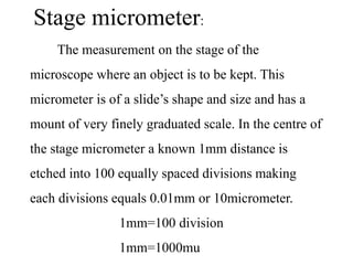
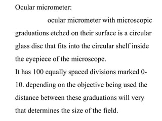
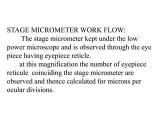
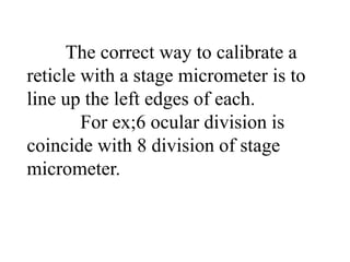
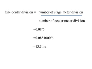
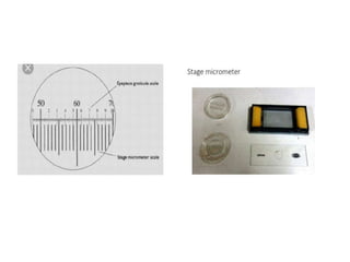
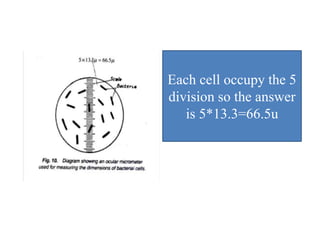
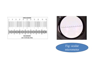
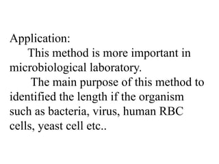
MICROMETRY is a technique used to measure the size of microscopic objects.It is based on principle of Calibration of the ocular micrometer using the stage micrometer. If you change microscope, the calibration process must be done again for each of the objective lenses that you are using because the magnification is different on different microscopes.
Microscopy - Magnification, Resolving power, Principles, Types and ApplicationsNethravathi Siri
Magnification, Resolving power, Principles and Applications of Simple, Compound, Stereozoom, Phase contrast, Fluorescent and Electron microscopes (TEM & SEM).
Microscopy is the technical field that uses microscopes to observe samples which are not in the resolution range of the normal-unaided eye.
microorganisms are microscopic objects that are visible only with the help of a microscope. micrometry refers to the measurement of dimensions of the desired microorganisms. ocular micrometer and stage micrometer.
This presentation include information about electron microscope & types of electron microscope i.e. SEM (Scanning electron microscope) & TEM (Transmission electron microscope).
An electron microscope is a microscope that uses a beam of scattered electrons as a source of illumination. It is used to get information about structure, topology, morphology & composition of materials. It has many advantages. Basically there are 4 types of electron microscope but here we will discuss only 2 types.
Transmission electron microscopy is a microscopy technique in which a beam of electrons is transmitted through an ultra-thin specimen, interacting with the specimen as it passes through it. Its resolution & magnification is about 10,000,000x. There are 5 types of transmission electron microscope i.e. BFTEM (Bright field transmision electron microscope), DFTEM (Dark field transmission electron microscope), HRTEM (High resolution transmission electron microscope), EFTEM (Energy filtered transmission electron microscope), ED (Electron diffraction). there are 4 techniques of TEM i.e. negative staining, shadow casting, Freeze fracture replication, freeze etching. It has many applications e.g, for the study of Cancer research, virology, chemical industry, electronic structure etc.
A scanning electron microscope is a type of electron microscope that produces images of a sample by scanning it with a focused beam of electrons. Types of signals produce by SEM include secondary electrons, back scattered electrons, X-rays, light rays. There are many advantages of SEM e.g, Btter resolution, fast imaging easy to operate, work with low voltage etc.
1. MICROSCOPY - introduction + principle (Basics)Nethravathi Siri
Basics only
Microscopy is the technical field that uses microscopes to observe samples which are
not in the resolution range of the normal-unaided eye.
Microscope is a scientific-instrument consisting of magnifying lens that enables an
observer to view the minute features distinctly.
In greek, micro = small
skopein = to view.
LIGHT MICROSCOPY by SIVASANGARI SHANMUGAM
The optical microscope, The functions of a light microscope is based on its ability to focus a beam of light through, which is very small and transparent, to produce an image.
BRIGHT FIELD MICROSCOPY by SIVASANGARI SHANMUGAM
bRIGHT FIELD MICROSCOPY is also called a compound microscope. The name bright - field is derived from the fact that the specimen is dark and contrasted by the surrounding bright viewing field.
An isotope is one of two or more atoms having the same atomic number but different mass numbers.
Unstable isotopes are called Radioisotopes.
uses of radioisotopes are many which are discussed in this slide.
Cyanobacteria are important in the nitrogen cycle.
Cyanobacteria are very important organisms for the health and growth of many plants. They are one of very few groups of organisms that can convert inert atmospheric nitrogen into an organic form, such as nitrate or ammonia.
Centrifugation principle and types by Dr. Anurag YadavDr Anurag Yadav
concept of cnetrifugation,
basic Principle
centrifugal force
types of centrifugation based on use and rotor type
application of the each type of centrifuge
Ultracentrifuge in detail
application in general
i am HAFIZ M WASEEM from mailsi vehari
BSc in science college Multan Pakistan
MSC university of education Lahore Pakistan
I love Pakistan and my teachers
Travis Hills' Endeavors in Minnesota: Fostering Environmental and Economic Pr...Travis Hills MN
Travis Hills of Minnesota developed a method to convert waste into high-value dry fertilizer, significantly enriching soil quality. By providing farmers with a valuable resource derived from waste, Travis Hills helps enhance farm profitability while promoting environmental stewardship. Travis Hills' sustainable practices lead to cost savings and increased revenue for farmers by improving resource efficiency and reducing waste.
Observation of Io’s Resurfacing via Plume Deposition Using Ground-based Adapt...Sérgio Sacani
Since volcanic activity was first discovered on Io from Voyager images in 1979, changes
on Io’s surface have been monitored from both spacecraft and ground-based telescopes.
Here, we present the highest spatial resolution images of Io ever obtained from a groundbased telescope. These images, acquired by the SHARK-VIS instrument on the Large
Binocular Telescope, show evidence of a major resurfacing event on Io’s trailing hemisphere. When compared to the most recent spacecraft images, the SHARK-VIS images
show that a plume deposit from a powerful eruption at Pillan Patera has covered part
of the long-lived Pele plume deposit. Although this type of resurfacing event may be common on Io, few have been detected due to the rarity of spacecraft visits and the previously low spatial resolution available from Earth-based telescopes. The SHARK-VIS instrument ushers in a new era of high resolution imaging of Io’s surface using adaptive
optics at visible wavelengths.
Nucleophilic Addition of carbonyl compounds.pptxSSR02
Nucleophilic addition is the most important reaction of carbonyls. Not just aldehydes and ketones, but also carboxylic acid derivatives in general.
Carbonyls undergo addition reactions with a large range of nucleophiles.
Comparing the relative basicity of the nucleophile and the product is extremely helpful in determining how reversible the addition reaction is. Reactions with Grignards and hydrides are irreversible. Reactions with weak bases like halides and carboxylates generally don’t happen.
Electronic effects (inductive effects, electron donation) have a large impact on reactivity.
Large groups adjacent to the carbonyl will slow the rate of reaction.
Neutral nucleophiles can also add to carbonyls, although their additions are generally slower and more reversible. Acid catalysis is sometimes employed to increase the rate of addition.
Nutraceutical market, scope and growth: Herbal drug technologyLokesh Patil
As consumer awareness of health and wellness rises, the nutraceutical market—which includes goods like functional meals, drinks, and dietary supplements that provide health advantages beyond basic nutrition—is growing significantly. As healthcare expenses rise, the population ages, and people want natural and preventative health solutions more and more, this industry is increasing quickly. Further driving market expansion are product formulation innovations and the use of cutting-edge technology for customized nutrition. With its worldwide reach, the nutraceutical industry is expected to keep growing and provide significant chances for research and investment in a number of categories, including vitamins, minerals, probiotics, and herbal supplements.
Toxic effects of heavy metals : Lead and Arsenicsanjana502982
Heavy metals are naturally occuring metallic chemical elements that have relatively high density, and are toxic at even low concentrations. All toxic metals are termed as heavy metals irrespective of their atomic mass and density, eg. arsenic, lead, mercury, cadmium, thallium, chromium, etc.
Remote Sensing and Computational, Evolutionary, Supercomputing, and Intellige...University of Maribor
Slides from talk:
Aleš Zamuda: Remote Sensing and Computational, Evolutionary, Supercomputing, and Intelligent Systems.
11th International Conference on Electrical, Electronics and Computer Engineering (IcETRAN), Niš, 3-6 June 2024
Inter-Society Networking Panel GRSS/MTT-S/CIS Panel Session: Promoting Connection and Cooperation
https://www.etran.rs/2024/en/home-english/
hematic appreciation test is a psychological assessment tool used to measure an individual's appreciation and understanding of specific themes or topics. This test helps to evaluate an individual's ability to connect different ideas and concepts within a given theme, as well as their overall comprehension and interpretation skills. The results of the test can provide valuable insights into an individual's cognitive abilities, creativity, and critical thinking skills
Comparing Evolved Extractive Text Summary Scores of Bidirectional Encoder Rep...University of Maribor
Slides from:
11th International Conference on Electrical, Electronics and Computer Engineering (IcETRAN), Niš, 3-6 June 2024
Track: Artificial Intelligence
https://www.etran.rs/2024/en/home-english/
ANAMOLOUS SECONDARY GROWTH IN DICOT ROOTS.pptxRASHMI M G
Abnormal or anomalous secondary growth in plants. It defines secondary growth as an increase in plant girth due to vascular cambium or cork cambium. Anomalous secondary growth does not follow the normal pattern of a single vascular cambium producing xylem internally and phloem externally.
The use of Nauplii and metanauplii artemia in aquaculture (brine shrimp).pptxMAGOTI ERNEST
Although Artemia has been known to man for centuries, its use as a food for the culture of larval organisms apparently began only in the 1930s, when several investigators found that it made an excellent food for newly hatched fish larvae (Litvinenko et al., 2023). As aquaculture developed in the 1960s and ‘70s, the use of Artemia also became more widespread, due both to its convenience and to its nutritional value for larval organisms (Arenas-Pardo et al., 2024). The fact that Artemia dormant cysts can be stored for long periods in cans, and then used as an off-the-shelf food requiring only 24 h of incubation makes them the most convenient, least labor-intensive, live food available for aquaculture (Sorgeloos & Roubach, 2021). The nutritional value of Artemia, especially for marine organisms, is not constant, but varies both geographically and temporally. During the last decade, however, both the causes of Artemia nutritional variability and methods to improve poorquality Artemia have been identified (Loufi et al., 2024).
Brine shrimp (Artemia spp.) are used in marine aquaculture worldwide. Annually, more than 2,000 metric tons of dry cysts are used for cultivation of fish, crustacean, and shellfish larva. Brine shrimp are important to aquaculture because newly hatched brine shrimp nauplii (larvae) provide a food source for many fish fry (Mozanzadeh et al., 2021). Culture and harvesting of brine shrimp eggs represents another aspect of the aquaculture industry. Nauplii and metanauplii of Artemia, commonly known as brine shrimp, play a crucial role in aquaculture due to their nutritional value and suitability as live feed for many aquatic species, particularly in larval stages (Sorgeloos & Roubach, 2021).
2. Introduction:
Microorganisms are microscopic
objects that are visible only with the help of
a microscope. Sometimes it is necessary to
measure its dimensions for its identification
process. But determination of the size of a
microorganism is not an easy process.
3. Micrometry:
Micrometry refers to the measurement of
dimensions of the desired microorganisms
under a microscope which uses two micro-
scale known as micrometers. These are two
types
4. Types of Micrometry:
There are usually two types of
micrometers
Stage micrometer.
Ocular meter or ocular micrometer.
5. Stage micrometer:
The measurement on the stage of the
microscope where an object is to be kept. This
micrometer is of a slide’s shape and size and has a
mount of very finely graduated scale. In the centre of
the stage micrometer a known 1mm distance is
etched into 100 equally spaced divisions making
each divisions equals 0.01mm or 10micrometer.
1mm=100 division
1mm=1000mu
6. Ocular micrometer:
ocular micrometer with microscopic
graduations etched on their surface is a circular
glass disc that fits into the circular shelf inside
the eyepiece of the microscope.
It has 100 equally spaced divisions marked 0-
10. depending on the objective being used the
distance between these graduations will very
that determines the size of the field.
7. STAGE MICROMETER WORK FLOW:
The stage micrometer kept under the low
power microscope and is observed through the eye
piece having eyepiece reticle.
at this magnification the number of eyepiece
reticule coinciding the stage micrometer are
observed and thence calculated for microns per
ocular divisions.
8. The correct way to calibrate a
reticle with a stage micrometer is to
line up the left edges of each.
For ex;6 ocular division is
coincide with 8 division of stage
micrometer.
9. One ocular division = number of stage meter division
number of ocular meter division
=0.08/6
=0.08*1000/6
=13.3mu
10.
11. Ocular micrometer works flow:
Insert the ocular meter in the eye piece of
your calibrated microscope by unscrewing its
upper eye lens.
Keep the spore or bacterial slide to be
measured on the stage of the microscope and
observe through the eyepiece containing ocular
meter.
The number of ocular divisions covered by
the microbe is counted by viewing through he
eye piece.
12. The size of the microorganism is
determined by multiplying the
number of ocular divisions covered
by the microbe with the calibration
factor.
15. Application:
This method is more important in
microbiological laboratory.
The main purpose of this method to
identified the length if the organism
such as bacteria, virus, human RBC
cells, yeast cell etc..