Downloaded 226 times

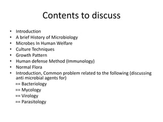
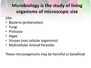
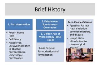
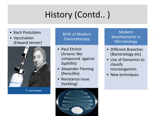
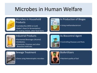
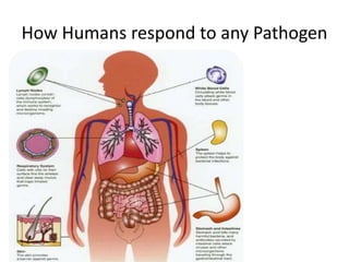
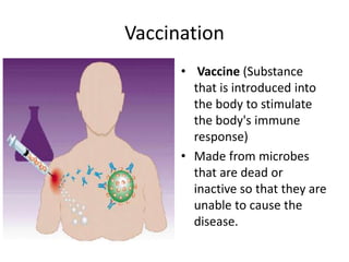
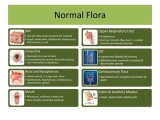
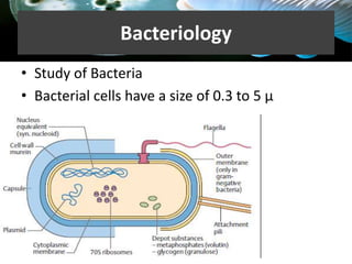
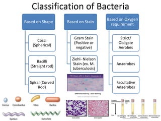
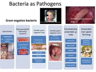
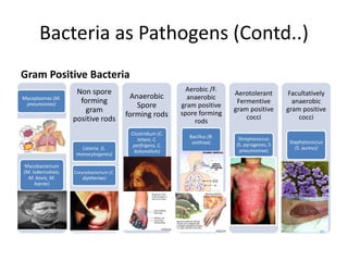
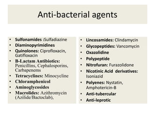
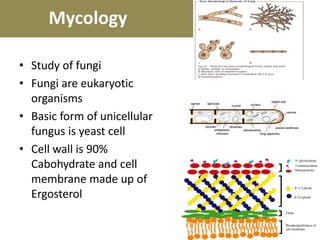
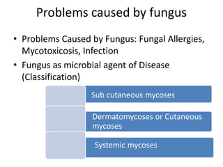

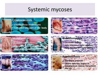
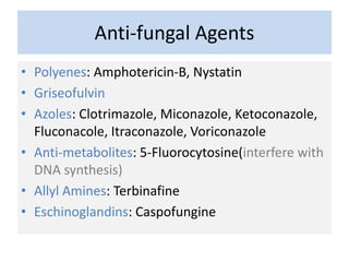

Microbiology is the study of microscopic organisms. This document provides an overview of microbiology, including a brief history, classification of microbes, their role in human welfare and disease. It discusses techniques for studying bacteria, fungi, viruses and parasites. It also outlines several common pathogenic microbes and the antimicrobial treatments used to combat infections. In summary, the document introduces the key topics and organisms within microbiology, from early discoveries to current classification and treatment of infectious diseases.


