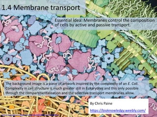
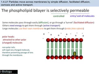
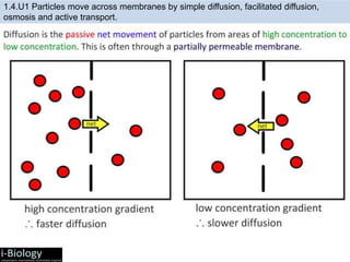
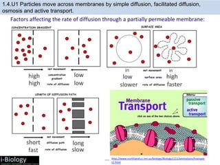
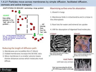
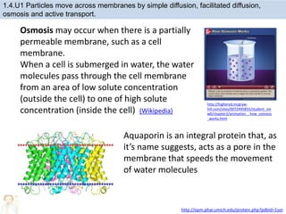
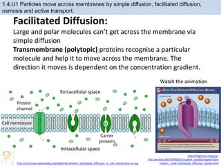
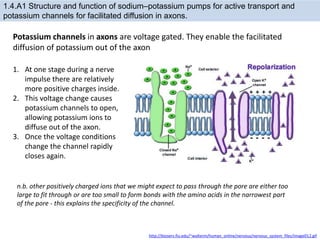
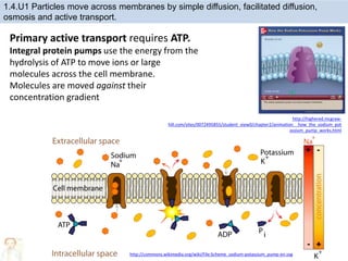
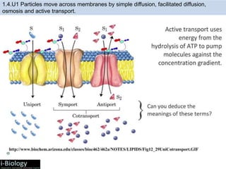
1. Membranes control the composition of cells through active and passive transport. Particles can move across membranes via simple diffusion, facilitated diffusion, osmosis, and active transport.
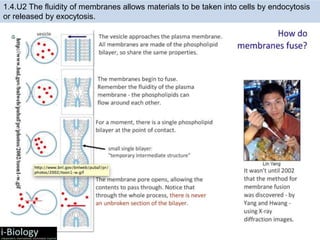
2. Vesicles transport materials within cells and the fluidity of membranes allows materials to enter and exit cells through endocytosis and exocytosis.
3. Tissues and organs used in medical procedures must be bathed in a solution of the same osmolarity as cytoplasm to prevent damage from osmosis.