4. Nerve net
Hydra (cnidarian)
Radial
nerve
Nerve
ring
Sea star (echinoderm)
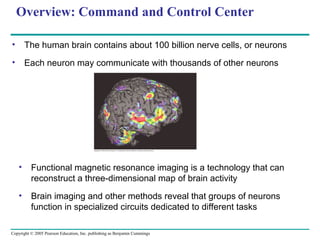
The simplest animals with
nervous systems, the cnidarians,
have neurons arranged in nerve
nets
Sea stars have a nerve net in
each arm connected by radial
nerves to a central nerve ring
6. LE 48-3
Sensor
Sensory input
Motor output
Integration
Effector
Peripheral nervous
system (PNS)
Central nervous
system (CNS)
In vertebrates, the central nervous system consists of a brain and
dorsal spinal cord
The PNS connects to the CNS
Nervous systems process information in three stages: sensory input,
integration, and motor output
8. LE 48-4
Quadriceps
muscle
Cell body of
sensory neuron in
dorsal root
ganglion
Sensory neuron
Spinal cord
(cross section)
White
matter
Hamstring
muscle
Gray
matter
Motor neuron
Interneuron
13. In the CNS,
astrocytes provide
structural support
for neurons and
regulate
extracellular
concentrations of
ions and
neurotransmitters
14. LE 48-8
Axon Nodes of
Ranvier
Schwann
cell
Myelin sheath
Nucleus of
Schwann cell
Schwann
cell
Nodes of Ranvier
Layers of myelin
Axon
0.1 µm
Oligodendrocytes (in the CNS) and Schwann cells (in the
PNS) form the myelin sheaths around axons of many
vertebrate neurons
25. Hyperpolarizations
Graded potential hyperpolarizations Graded potential depolarizations
5
Time (msec)
Resting
potential
43210
Threshold
–100
–50
0
Membranepotential(mV)
Stimuli
+50
Depolarizations
5
Time (msec)
Resting
potential
43210
Threshold
–100
–50
0
Membranepotential(mV)
Stimuli
+50
Action potential
5
Time (msec)
Resting
potential
43210
Threshold
–100
–50
0
Membranepotential(mV)
Stronger depolarizing stimulus
+50
Action
potential
6
If a cell has gated ion channels, its membrane potential may change
in response to stimuli that open or close those channels
Some stimuli trigger a hyperpolarization, an increase in magnitude
of the membrane potential
Other stimuli trigger a depolarization, a reduction in the magnitude of
the membrane potential
30. An action potential is generated as Na+
flows inward
across the membrane at one location.
Na+
Action
potential
Axon
Na+
Action
potentialK+
The depolarization of the action potential spreads to the
neighboring region of the membrane, re-initiating the
action potential there. To the left of this region, the
membrane is repolarizing as K+
flows outward.
K+
Na+
Action
potentialK+
The depolarization-repolarization process is repeated in the
next region of the membrane. In this way, local currents of
ions across the plasma membrane cause the action
potential to be propagated along the length of the axon.
K+
Conduction of Action Potentials
•An action potential can travel
long distances by regenerating
itself along the axon
•At the site where the action
potential is generated, usually the
axon hillock, an electrical current
depolarizes the neighboring
region of the axon membrane
32. LE 48-15
Cell body
Schwann cell
Depolarized region
(node of Ranvier)
Myelin
sheath
Axon
Na+ and K+ channels are concentrated at the
nodes to initiate a series of action potentials
35. LE 48-17
Postsynaptic cellPresynaptic
cell
Synaptic vesicles
containing
neurotransmitter
Presynaptic
membrane
Voltage-gated
Ca2+
channel
Ca2+
Postsynaptic
membrane
Postsynaptic
membrane
Neuro-
transmitter
Ligand-
gated
ion channel
Na+
K+
Ligand-gated
ion channels
Synaptic cleft
When an action potential reaches a terminal, the final result
is release of neurotransmitters into the synaptic cleft
•Direct synaptic transmission involves binding of neurotransmitters to
ligand-gated ion channels
38. Postsynaptic
neuron
Terminal branch
of presynaptic
neuron
E1
E1
Axon
hillock
E1
E2
E1
I
Action
potential
E1E1 + E2
Spatial summation
of EPSP and IPSP
Spatial summation
I E1 + I
Action
potential
E1
Temporal summation
E1
Threshold of axon of
postsynaptic neuron
E1
Subthreshold, no
summation
E1
Resting
potential
Membranepotential(mV)
–70
0
•If two EPSPs are produced in rapid succession, an effect called
temporal summation occurs
•In spatial summation, EPSPs produced nearly simultaneously by
different synapses on the same postsynaptic neuron add together
•Through summation, an IPSP can counter the effect of an EPSP
The team constructed Brainbow using a two-step process: first, a specific genetic construct was generated that could be recombined in multiple arrangements to produce one of either three or four colors based on the particular fluorescent proteins (XFPs) being implemented.[2] Next, multiple copies of the same transgenic construct were inserted into the genome of the target species, resulting in the random expression of different XFP ratios and subsequently causing different cells to exhibit a variety of colorful hues.[2]
Now we can label individual neurons
Basics
Just know there are different organizations of nervous systems. No need to memorize
Clusters of nervous systems are referred to as ganglia
Information Processed
Sensory neurons receive sensory stimuli, touch, or taste
Integrated in the central nervous system
Comes out as an output (motor neuron)
Sensory Neurons
Interneurons
Motor Neurons
Region between two neurons is called the synapse
Receptors are at the end of dendrites
Presynaptic sends the signal
Postsynaptic receives the signal
Neurons have different shapes and sizes. Structure correlates to function
Astrocytes and radial glia provide structure.
Astrocytes brings neurons back to resting states
Oligodendrocytes and Schwann cells are myelin sheets producing cells
The extracellular matrix is regulated by supporting cells.
DevBio9e-Fig-09-28-0.jpg
Pretend there is a stimulus and write what happens at every step in the nerve cell.
From signal to response in a muscle
When there is a charge difference there is potential.
Resting potential When it is not transmitting signal
Sodium is higher outside than inside the cell
Potassium is higher inside the cell
A neuron that’s not signaling has open potassium channels but very few open potassium channels
The ion channels in the membrane of a cell can be opened in response to being stretched, ligand, or voltage (gated).
Stimuli can open and close those channels.
Hyperpolarization increases the difference
Graded potentials: they are not going to hit the action potential because they don’t pass the action potential
Need frequent or intense signals to activate action potential
Voltage-Gated: a certain change in membrane potential causes the sodium or potassium channels to close
1. The sodium and potassium channels are closed
2. Voltage gated channels open
3. If action potential is meet the neuron fires
4. Causes potassium channels to open releasing the positive charges
5. Potassium overshoots