Meiosis is the process of cell division that produces gametes (sex cells) like sperm and eggs, which have half the number of chromosomes as regular body cells. During meiosis, homologous chromosomes pair up and may exchange sections through crossing over, introducing genetic variation. This results in four daughter cells from two divisions for males and one viable egg cell for females. Meiosis reduces the chromosome number so fertilization restores the diploid number, maintaining genetic diversity between generations.
About how cellular respiration occurs in Mitochondria, it discusses first the parts and functions of mitochondrion then the types of respiration and the 3 processes occurs in aerobic respiration.
Codominance is the situation when the effect of both genes are observed. This presentation describes how codominant traits work to determine human blood types and flower coloring .
Presentation on Sex influenced traits. Very informative for Biology students. This presentation include the basic terminologies and have the information that how sex influenced traits are different from sex linked traits. This presentation contains information that how these traits are transferred to next generations.
Observation of Io’s Resurfacing via Plume Deposition Using Ground-based Adapt...Sérgio Sacani
Since volcanic activity was first discovered on Io from Voyager images in 1979, changes
on Io’s surface have been monitored from both spacecraft and ground-based telescopes.
Here, we present the highest spatial resolution images of Io ever obtained from a groundbased telescope. These images, acquired by the SHARK-VIS instrument on the Large
Binocular Telescope, show evidence of a major resurfacing event on Io’s trailing hemisphere. When compared to the most recent spacecraft images, the SHARK-VIS images
show that a plume deposit from a powerful eruption at Pillan Patera has covered part
of the long-lived Pele plume deposit. Although this type of resurfacing event may be common on Io, few have been detected due to the rarity of spacecraft visits and the previously low spatial resolution available from Earth-based telescopes. The SHARK-VIS instrument ushers in a new era of high resolution imaging of Io’s surface using adaptive
optics at visible wavelengths.
Seminar of U.V. Spectroscopy by SAMIR PANDASAMIR PANDA
Spectroscopy is a branch of science dealing the study of interaction of electromagnetic radiation with matter.
Ultraviolet-visible spectroscopy refers to absorption spectroscopy or reflect spectroscopy in the UV-VIS spectral region.
Ultraviolet-visible spectroscopy is an analytical method that can measure the amount of light received by the analyte.
THE IMPORTANCE OF MARTIAN ATMOSPHERE SAMPLE RETURN.Sérgio Sacani
The return of a sample of near-surface atmosphere from Mars would facilitate answers to several first-order science questions surrounding the formation and evolution of the planet. One of the important aspects of terrestrial planet formation in general is the role that primary atmospheres played in influencing the chemistry and structure of the planets and their antecedents. Studies of the martian atmosphere can be used to investigate the role of a primary atmosphere in its history. Atmosphere samples would also inform our understanding of the near-surface chemistry of the planet, and ultimately the prospects for life. High-precision isotopic analyses of constituent gases are needed to address these questions, requiring that the analyses are made on returned samples rather than in situ.
(May 29th, 2024) Advancements in Intravital Microscopy- Insights for Preclini...Scintica Instrumentation
Intravital microscopy (IVM) is a powerful tool utilized to study cellular behavior over time and space in vivo. Much of our understanding of cell biology has been accomplished using various in vitro and ex vivo methods; however, these studies do not necessarily reflect the natural dynamics of biological processes. Unlike traditional cell culture or fixed tissue imaging, IVM allows for the ultra-fast high-resolution imaging of cellular processes over time and space and were studied in its natural environment. Real-time visualization of biological processes in the context of an intact organism helps maintain physiological relevance and provide insights into the progression of disease, response to treatments or developmental processes.
In this webinar we give an overview of advanced applications of the IVM system in preclinical research. IVIM technology is a provider of all-in-one intravital microscopy systems and solutions optimized for in vivo imaging of live animal models at sub-micron resolution. The system’s unique features and user-friendly software enables researchers to probe fast dynamic biological processes such as immune cell tracking, cell-cell interaction as well as vascularization and tumor metastasis with exceptional detail. This webinar will also give an overview of IVM being utilized in drug development, offering a view into the intricate interaction between drugs/nanoparticles and tissues in vivo and allows for the evaluation of therapeutic intervention in a variety of tissues and organs. This interdisciplinary collaboration continues to drive the advancements of novel therapeutic strategies.
Professional air quality monitoring systems provide immediate, on-site data for analysis, compliance, and decision-making.
Monitor common gases, weather parameters, particulates.
The increased availability of biomedical data, particularly in the public domain, offers the opportunity to better understand human health and to develop effective therapeutics for a wide range of unmet medical needs. However, data scientists remain stymied by the fact that data remain hard to find and to productively reuse because data and their metadata i) are wholly inaccessible, ii) are in non-standard or incompatible representations, iii) do not conform to community standards, and iv) have unclear or highly restricted terms and conditions that preclude legitimate reuse. These limitations require a rethink on data can be made machine and AI-ready - the key motivation behind the FAIR Guiding Principles. Concurrently, while recent efforts have explored the use of deep learning to fuse disparate data into predictive models for a wide range of biomedical applications, these models often fail even when the correct answer is already known, and fail to explain individual predictions in terms that data scientists can appreciate. These limitations suggest that new methods to produce practical artificial intelligence are still needed.
In this talk, I will discuss our work in (1) building an integrative knowledge infrastructure to prepare FAIR and "AI-ready" data and services along with (2) neurosymbolic AI methods to improve the quality of predictions and to generate plausible explanations. Attention is given to standards, platforms, and methods to wrangle knowledge into simple, but effective semantic and latent representations, and to make these available into standards-compliant and discoverable interfaces that can be used in model building, validation, and explanation. Our work, and those of others in the field, creates a baseline for building trustworthy and easy to deploy AI models in biomedicine.
Bio
Dr. Michel Dumontier is the Distinguished Professor of Data Science at Maastricht University, founder and executive director of the Institute of Data Science, and co-founder of the FAIR (Findable, Accessible, Interoperable and Reusable) data principles. His research explores socio-technological approaches for responsible discovery science, which includes collaborative multi-modal knowledge graphs, privacy-preserving distributed data mining, and AI methods for drug discovery and personalized medicine. His work is supported through the Dutch National Research Agenda, the Netherlands Organisation for Scientific Research, Horizon Europe, the European Open Science Cloud, the US National Institutes of Health, and a Marie-Curie Innovative Training Network. He is the editor-in-chief for the journal Data Science and is internationally recognized for his contributions in bioinformatics, biomedical informatics, and semantic technologies including ontologies and linked data.
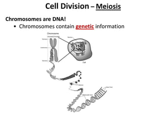
1. Cell Division – Meiosis
Chromosomes are DNA!
• Chromosomes contain genetic information
2. Cell Division – Mitosis (Review)
– Division of a somatic cell that results in 2 genetically
identical daughter cells
• Cells must divide for growth, repair of tissues, and
asexual reproduction
• Cell division begins in Interphase when the
chromosomes duplicate
3. Parent cell
Chromosomes
duplicate 2 new daughter
cells identical to
parent cell
• Daughter cells are genetically identical to parent cell –
same kind and number of chromosomes
• Mitosis occurs in somatic or body cells
Ex: liver, heart, skin, stomach
• Every organism has its own unique number of
chromosomes. Humans have 46.
This is called its diploid number or the total number of
chromosomes in a somatic cell.
Diploid means “2 sets” and is written as “2N”.
4. • Body cells of adult organisms have 2 sets of
homologous (matching) chromosomes – 1 set from
female parent and 1 set from male parent
5. Cell Division –Meiosis
– the process in which the number of chromosomes in the
original cell is reduced by HALF through the separation
of homologous chromosomes
• Meiosis occurs in sex organs only
• Males (XY) – sex organs are the testes in humans
• Females (XX) – sex organs are the ovaries in humans
• Meiosis also occurs in the sex organs of other
animals, plants, fungi, etc…
8. Meiosis produces sex cells –
cells with ½ the number of
chromosomes as the original
cell
• Males – meiosis produces
4 sperm
• Females – meiosis
produces 1 (viable) egg
The other 3 cells are
called polar bodies – they
give up their cytoplasm to
nourish the 1 good egg.
• Egg and sperm (sex cells)
are also called gametes
9. • Gametes have ½ the number of chromosomes as
somatic (body) cells. We call this the haploid number.
Haploid means “1 set” and is written as “N”.
If human diploid number is 46, what is its haploid
number? 23
Diploid # of a dog – 78 Haploid # of a dog – 39
Diploid # of a fly – 8 Haploid # of a fly – 4
10. • When does meiosis occur in humans?
1. Males beginning at puberty
2. Females before birth – all eggs are produced before
birth and at puberty eggs mature
11. Chromosome Number
• Remember, chromosome number is unique to each
kind of organism and all cells (except sex cells) in an
organism have the same kind and number of
chromosomes.
Ex: All humans have 46 chromosomes and all cells in
the human body (except sperm and egg) have 46
chromosomes.
• This is why the chromosome number in sex cells must
be reduced in half by meiosis
Ex: Humans have 46 chromosomes in their somatic
cells, but 23 chromosomes in their sex cells
(egg and sperm)
12.
13. 23
23
46
Zygote develops into
embryo and finally adult
organism by mitosis
Fertilization – process by which an egg and sperm unite
Zygote – fertilized egg
Embryo – organism in early stage of development
Fertilized egg –
zygote
15. Unique events in Meiosis
• Homologous (matching) chromosomes pair up before
1st cell division
Homologous chromosomes:
-look alike
-code for same traits
-receive one from each parent
16. • During 1st division, homologous chromosomes exchange
genes during process called “crossing over”
• These homologous chromosomes separate during 2nd
division of meiosis – so chromosomes in gametes are
different from each other due to crossing over
• Crossing over increases genetic variation and is the
reason why siblings look different
17. No crossing over
– daughter cells
are identical to
parent cells
Crossing over
occurs –causes
genetic variation
(Daughter cells
are NOT identical
to parent cell)
20. Mitosis Meiosis
What kind of cells? Somatic cells
Male (XY) = Sperm
Female (XX) = Egg
When does this occur? Any time
Male (XY) = puberty
Female (XX) = before birth
# of Divisions
(Draw picture)
1 2
# of Daughter cells 2
Male (XY) = 4 sperm
Female (XX) = 1 viable egg
# of Chromosomes
Same as parent cell
diploid or 2N
In humans 46
Half as many as parent cell
haploid or N
In humans 23
Type of Reproduction Asexual Sexual
Genetic Composition
Daughter cells
identical / not identical
to parent cell
Daughter cells
identical / not identical
to parent cell
Genetic variation
Pairing of Homologous
Chromosomes
YES / NO
YES / NO
Crossing over of genes
Function/Importance
Growth, repair; development of adult
from zygote
Production of gametes:
eggs and sperm
Sex Cells