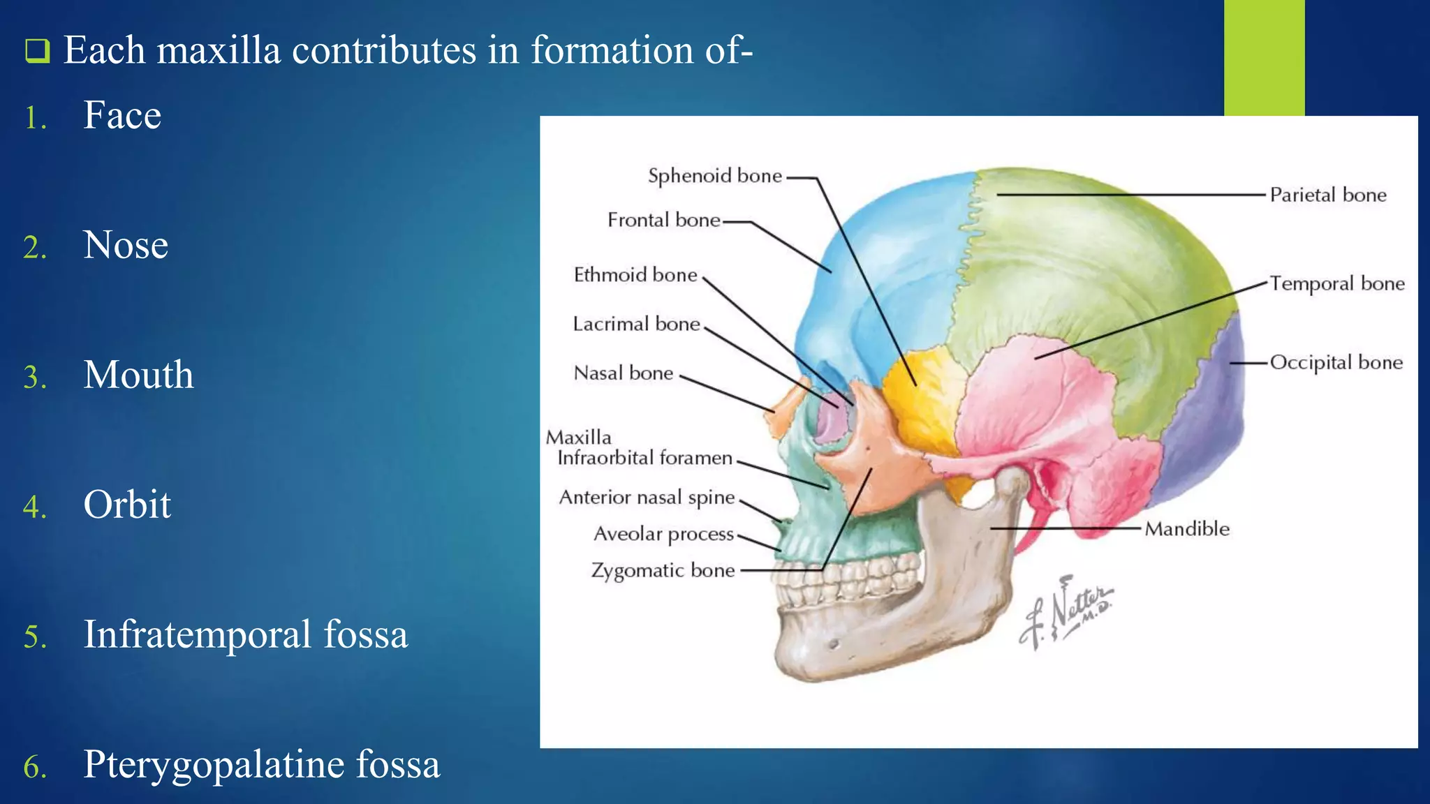
SlideShare a Scribd company logo
1 of 58
MAXILLA: BASIC
AND APPLIED
ANATOMY
MODERATED BY: DR MAYANK SINGHAL
PRESENTED BY: DR RAJIV KUMAR SINGH
(PG 1ST YR STUDENT)
DEPARTMENT OF ORALAND MAXILLOFACIAL SURGERY
SANTOSH DENTAL COLLEGE,GHAZIABAD
CONTENTS
 INTRODUCTION
 DEVELOPMENT
 FEATURES OF MAXILLA
 MAXILLARY SINUS
 AGE CHANGES IN MAXILLA
 VASCULATURE
 RESOURCES
INTRODUCTION
• 2nd largest bone of the face.
• Paired bone
• 2 maxilla forms whole of upper jaw
• Hollowed out by the formation of maxillary sinus and nasal cavity
 Each maxilla contributes in formation of-
1. Face
2. Nose
3. Mouth
4. Orbit
5. Infratemporal fossa
6. Pterygopalatine fossa
NORMA FRONTALIS
BONE FRONTAL
NASAL
MAXILLA
ZYGOMATIC
MANDIBLE
SUTURES FRONTONASAL
FRONTOZYGOMATIC
ZYGOMATICOMAXILLARY
METOPIC
DEVELOPMENT OF MAXILLA
 MAXILLA ossifies from 3 centers in the membrane –
1) 1 center for maxilla proper – 6th week of IUL, above the canine fossa
2) 2 centers for premaxilla
 Of 2 premaxillary centers –
• Main center above the incisive fossa- 7th week of IUL
• Second center – ventral margin of nasal septum – 10th week of IUL and soon fuses
with palatal process of maxilla.
 Premaxilla begin to fuse with alveolar process almost immediately after the
ossification begins.
Embryology
 Maxillofacial development starts at the fourth week of gestation with the
formation of the five facial prominences around the stomodeum, the primordial
mouth and the topographical center of the face during embryonic development.
 First pharyngeal arch and neural crest cells contribute to form the five facial
prominences: paired maxillary, paired mandibular and frontonasal prominence.
 The stomodeum is demarcated cranially by the frontonasal prominence,
laterally by the maxillary prominence and inferolaterally by the mandibular
prominence.
 Maxillary prominences give rise to the secondary palate, the majority of the
maxilla and the lateral upper lip.
 By the end of the fourth week, the lower half of the frontonasal prominence
gives rise to the nasal placodes, which divide into paired lateral and medial
nasal processes, with the nasal groove dividing them.
 At the end of week six the medial nasal processes fuse to form the philtrum,
and at the end of the eighth week, they fuse with both maxillary processes to
form the intermaxillary segment to form the upper lip and the primary palate.
 Secondary palate begins to develop during the 6th week of development
 During the seventh week, as the jaw elongates and the tongue descends, the
palatal shelves acquire a horizontal position.
 Fusion of the palate is completed by the tenth week and fully forms by the
twelfth week of embryonic development
SIDE DETERMINATION
 Anterior surface ends medially into a deeply concave border nasal notch.
 Posterior surface is convex.
 Alveolar border with sockets faces downwards with its convexity directed
outwards.
 Frontal process is the longest process which is directed upwards.
 Medial surface is large irregular opening, the maxillary hiatus/antrum of
Highmore for maxillary air sinus.
FEATURES
 Each maxilla has –
1. A body
2. 4 processes - frontal
zygomatic
alveolar
palatine
.
Body of maxilla
 Shape – Pyramidal
 It has 4 surfaces-
1. Anterior or facial
2. Posterior or infratemporal
3. Superior or orbital
4. Medial or nasal
 Encloses a large cavity - Maxillary sinus
Anterior or Facial Surface
 Directed forwards and laterally.
 Incisive fossa gives origin to depressor septi and orbicularis oris.
 Canine fossa gives origin to levator anguli oris.
 Above the canine fossa, there is infraorbital foramen, which transmits
infraorbital nerve and vessels.
 Levator labii superioris arises between the infraorbital margin and infraorbital
foramen.
 Medially - nasal notch
anterior nasal spine
Posterior or Infratemporal Surface
 Posterior surface is convex
 Directed backwards and laterally.
 Forms the anterior wall of infratemporal fossa
 Separated from anterior surface by the zygomatic process and a rounded ridge
which descends from the process to the first molar tooth.
 Near the center of the surface open 2-3 alveolar canals for posterior superior
alveolar nerve and vessels.
 Posteroinferiorly, there is a rounded eminence, the maxillary tuberosity, which
articulates superomedially with pyramidal process of palatine bone, and gives
origin laterally to the superficial head of medial pterygoid muscle.
 Above the maxillary tuberosity, the smooth surface forms anterior wall of
pterygopalatine fossa, and is grooved by maxillary nerve
Superior or Orbital Surface
 Superior surface is smooth, triangular and slightly concave
 Forms the greater part of the floor of orbit.
Anterior border
Forms a part of infraorbital margin. Medially, it is continuous with the lacrimal
crest of the frontal process.
Posterior border
 Smooth and rounded
 Forms most of the anterior margin of inferior orbital fissure.
 In middle, it is notched by the infraorbital groove.
Medial border
 presents anteriorly lacrimal notch converted into nasolacrimal canal.
 Behind the notch, the border articulates with
lacrimal, labyrinth of ethmoid, and the orbital process of palatine bone.
 The surface presents infraorbital groove leading forwards to infraorbital canal
which opens on the anterior surface as infraorbital foramen. The groove, canal
and foramen transmit the infraorbital nerve and vessels. Near the midpoint, the
canal gives off laterally a branch, the canalis sinuous, for the passage of anterior
superior alveolar nerve and vessels.
 Inferior oblique muscle of eyeball arises from a depression just lateral to
lacrimal notch at the anteromedial angle of the surface.
Medial or Nasal Surface
 Forms a part of the lateral wall of nose.
 Posterosuperiorly, it displays a large irregular opening of the maxillary sinus,
the maxillary hiatus.
 Above the hiatus, there are parts of air sinuses which are completed by the
ethmoid and lacrimal bones.
 Below the hiatus, the smooth concave surface forms a part of inferior meatus
of nose.
 Behind the hiatus
Surface articulates with perpendicular plate of palatine bone, enclosing the greater
palatine canal which runs downwards and forwards, and transmits greater
palatine vessels and the anterior, middle and posterior palatine nerves.
 In front of the hiatus
There is nasolacrimal groove, which is converted into the nasolacrimal canal by
articulation with the descending process of lacrimal bone and the lacrimal process
of inferior nasal concha. The canal transmits nasolacrimal duct to the inferior
meatus of nose.
 More anteriorly, an oblique ridge forms the conchal crest for articulation with the
inferior nasal concha.
 Above the conchal crest, the shallow depression forms a part of the atrium of
middle meatus of nose.
Processes of Maxilla
 Zygomatic Process
 Frontal Process
 Alveolar Process
 Palatine Process
FRONTAL VIEW OF SKULL
Zygomatic Process
 Pyramidal lateral projection
 Anterior, posterior, and superior surfaces of
maxilla converge.
 In front and behind, it is continuous with the
corresponding surfaces of the body, but
superiorly it is rough for articulation with the
zygomatic bone.
Frontal Process
 Projects upwards and backwards to articulate above with the nasal margin of
frontal bone, in front with nasal bone, and behind with lacrimal bone.
 Lateral surface is divided by a vertical ridge, the anterior lacrimal crest, into a
smooth anterior part and a grooved posterior part.
 The lacrimal crest gives attachment to lacrimal fascia and the medial palpebral
ligament, and is continuous below with the infraorbital margin.
 The anterior smooth area gives origin to orbicularis oculi and levator labii
superioris alaeque nasi. The posterior grooved area forms the anterior half of
the floor of lacrimal groove.
 Medial surface forms a part of the lateral wall of nose. The surface presents
following features:
1. Uppermost area is rough for articulation with ethmoid to close the anterior
ethmoidal sinuses.
 Ethmoidal crest is a horizontal ridge about the middle of the process. Posterior
part of the crest articulates with middle nasal concha, and the anterior part lies
beneath the agger nasi.
 The area below the ethmoidal crest is hollowed out to form the atrium of the
middle meatus.
 Below the atrium is the conchal crest which articulates with inferior nasal
concha.
 Below the conchal crest, there lies the inferior meatus of the nose with
nasolacrimal groove ending just behind the crest.
Alveolar Process
 Forms half of the alveolar arch
 Bears sockets for maxillary teeth.
 In adults, there are eight sockets
 It has a horseshoe configuration, with the curved portion facing anteriorly.
 Canine socket is deepest; molar sockets are widest and divided into three
minor sockets by septa; the incisor and second premolar sockets are single;
and the first premolar socket is sometimes divided into two.
 Buccinator arises from the posterior part of its outer surface up to the first
molar tooth.
 A rough ridge, the maxillary torus, is sometimes present on the inner surface
opposite the molar sockets.
Palatine Process
 Palatine process is a thick horizontal plate projecting medially from the lowest
part of the nasal surface. It forms a large part of the roof of mouth and the floor
of nasal cavity.
 Inferior surface is concave, and the two palatine processes form anterior three-
fourths of the bony palate. It presents numerous vascular foramina and pits for
palatine glands.
The Maxilla, The bony palate and alveolar
arch.
 Posterolaterally, it is marked by two anteroposterior grooves for the greater
palatine vessels and anterior palatine nerves.
 Superior surface is concave from side-to-side, and forms greater part of the
floor of nasal cavity.
The Maxilla, The bony palate and alveolar
arch.
 Medial border is thicker in front than behind. It is raised superiorly into the
nasal crest. Groove between the nasal crests of two maxillae receives lower
border of vomer; anterior part of the ridge is high and is known as incisor crest
which terminates anteriorly into the anterior nasal spine.
 Incisive canal traverses near the anterior part of the medial border.
 Posterior border articulates with horizontal plate of palatine bone.
 Lateral border is continuous with the alveolar process
ARTICULATIONS OF MAXILLA
 Superiorly, it articulates with three bones—the nasal, frontal and lacrimal.
 Medially, it articulates with five bones—the ethmoid, inferior nasal concha,
vomer, palatine and opposite maxilla.
 Laterally, it articulates with one bone—the zygomatic
AGE CHANGES IN MAXILLA
AT BIRTH –
1. Transverse and anteroposterior diameter > vertical diameter
2. Well marked frontal process
3. Body consists of little more than alveolar process
4. Tooth socket – close to orbit
5. Maxillary sinus is a mere furrow on the lateral wall of nose
Anterior surface of Maxilla at Birth
IN ADULTS-
 Vertical diameter is more due to –
1. Developed alveolar process
2. Increased size of maxillary sinus
IN OLD –
1. Infantile condition
2. Resorption of alveolar bone
Adults
Clinical correlation
Fractures of maxilla
A. Unilateral fracture involves the alveolar process of the maxilla.
B. Bilateral fractures are classified into following three types–
1. Le Fort I: It extends posteriorly, on a horizontal plane from the pyriform
aperture through the zygomaticoalveolar crest to maxillary tuberosities into the
pterygopalatine fossa.
This type of fracture separates the hard palate and alveolar process from the facial
skull.
Le Fort II: Fractures are pyramidal in shape. It extends from the nasofrontal
suture to the fronto maxillary suture, through the orbital floor and maxillary sinus,
extending inferiorly to the zygomaticoalveolar crest bilaterally. Inferiorly, proceeds
caudally to the maxillary tuberosities into the pterygoid process. Superiorly, the
fractures extend caudally sectioning through the nasal septum.
This fracture dissociates the nasal bones, the nasal septum, and maxilla from the
cranial skull and the lateral midface.
Le Fort III (craniofacial dysfunction): fractures extend from the
nasofrontal suture down through the medial wall and orbital floor to the inferior
orbital fissure. It proceeds laterally, interrupts the zygomaticofrontal suture and
continues inferiorly through the zygomatic arches bilaterally. From the
nasofrontal suture, it extends caudally through the ethmoid and perpendicular
plate of the palatine bone, fracturing the pterygoid process and vomer,
terminating in the palatine fossa.
This fracture completely separates the facial skeleton from the cranial skul
MAXILLARY SINUS
 Largest of the paranasal sinuses.
 Also called as ANTRUM OF
HIGHMORE
 Completely fill the bodies of the
maxillae
 Thin walls
 Primary lymphatic drainage is to the
submandibular lymph nodes
 Pyramidal in shape with the apex directed laterally and the base deep to the
lateral wall of the adjacent nasal cavity
 The medial wall or base of the maxillary sinus is formed by the maxilla, and by
parts of the inferior concha and palatine bone that overlie the maxillary hiatus
 Vertical: 3.5 cm.
 Transverse: 2.5 cm.
 Anteroposterior: 3.25 cm
FUNCTIONS OF MAXILLARY SINUS
 Speech and voice resonance
 Reduce the weight of the skull
 Warmth inhaled oxygen
 Filtration of the inspired air
 Immunological barrier
 Regulation of intra nasal pressure
Arterial Supply
 It is by the anterior, middle, and posterior superior alveolar arteries from
maxillary and infraorbital arteries.
Lymphatic Drainage
 The sinus drains into submandibular lymph nodes.
Nerve Supply
 Maxillary sinuses are supplied by the anterior, middle, and posterior superior
alveolar nerves from the maxillary and infraorbital nerves.
Relationships of the maxillary sinus
Superior: Orbit, Infraorbital nerve and vessels
Inferior: Roots of molars and premolars
Medial: Nasal cavity
Lateral and anterior: Cheek
Posterior: Infratemporal fossa, Pterygopalatine fossa and contents
Relations of Maxillary sinus
CORONAL SECTION OF FACE
Clinical correlation
Maxillary sinusitis –
 Infection can reach into sinus from infected nose (viral rhinitis), carious upper
premolar and molar teeth, especially molars, and infected frontal and anterior
ethmoidal sinuses.
 Being most dependent part, it acts as a secondary reservoir for pus from frontal
air sinus through frontonasal duct and hiatus semilunaris.
 Pain of maxillary sinusitis is referred to the upper teeth and infraorbital skin due
to common innervation by the maxillary nerve.
Axial view of CT of Face
Drainage of maxillary sinus -
 Antral puncture (antrostomy) by using trocar and cannula, which are passed
below the inferior nasal concha in an outward and backward direction.
 Fenestrating the antrum through canine fossa in the gingivolabial sulcus
(Caldwell–Luc operation).
Caldwell –Luc operation
Carcinoma of maxillary sinus-
 The medial invasion encroaches the nasal cavity causing obstruction and
epistaxis. The obstruction of nasolacrimal duct in this wall produces epiphora
(overflow of tears).
 The upward invasion into the orbit displaces the eyeball causing proptosis
(protrusion of eyeball) and diplopia (double vision).
 Involvement of infraorbital nerve produces pain and anesthesia in the skin over
the face below the orbit.
 The downward invasion into the floor produces visible bulge or even ulceration
of palatal roof of the oral cavity.
 The lateral invasion produces swelling on the face and palpable mass in the
gingivolabial fold (groove).
 Backward (posterior) invasion may involve the palatine nerves leading to severe
referred pain to the upper teeth.
References
 Gray’s Anatomy 41st edition
 BD chaurasia’s Human Anatomy 8th edition
 Netter’s Head and neck anatomy for dentistry 2nd edition
 Vishram singh Text book of anatomy 2nd edition
 https://www.ncbi.nlm.nih.gov/books/NBK538527/ ( Osteology of maxilla)
 https://www.ncbi.nlm.nih.gov/pmc/articles/PMC4809813/pdf/12070_2015_
Article_883.pdf ( Cald-well luc operation)
THANK YOU

More Related Content

Similar to Maxilla basics and applied anatomy DR RAJIV.pptx

Nose and paranasal sinuses
Nose and paranasal sinusesNose and paranasal sinuses
Nose and paranasal sinusesAnil Narayanam
 
nose and paranasal sinuses EXCELLENT SLIDES TO CULL FROM NOSE SINUSES [Autosa...
nose and paranasal sinuses EXCELLENT SLIDES TO CULL FROM NOSE SINUSES [Autosa...nose and paranasal sinuses EXCELLENT SLIDES TO CULL FROM NOSE SINUSES [Autosa...
nose and paranasal sinuses EXCELLENT SLIDES TO CULL FROM NOSE SINUSES [Autosa...TariqJamilFaridi
 
Anatomy of oropharynx maxilla mandible neck nodes
Anatomy of oropharynx maxilla mandible neck nodesAnatomy of oropharynx maxilla mandible neck nodes
Anatomy of oropharynx maxilla mandible neck nodesSamik Sharma
 
Anatomy of Nose and PNS
Anatomy of Nose and PNSAnatomy of Nose and PNS
Anatomy of Nose and PNSVictoria2021
 
Surgical anatomy of nose
Surgical anatomy of noseSurgical anatomy of nose
Surgical anatomy of noseAugustine raj
 
Anatomy of nose and para nasal sinuses . by DR. MD. KHURSHID PERVEJ. GMC PATIALA
Anatomy of nose and para nasal sinuses . by DR. MD. KHURSHID PERVEJ. GMC PATIALAAnatomy of nose and para nasal sinuses . by DR. MD. KHURSHID PERVEJ. GMC PATIALA
Anatomy of nose and para nasal sinuses . by DR. MD. KHURSHID PERVEJ. GMC PATIALAkbristi12
 
EMBERYOLOGY AND ANATOMY OF EXTERNAL NOSE.pptx
EMBERYOLOGY AND ANATOMY OF EXTERNAL NOSE.pptxEMBERYOLOGY AND ANATOMY OF EXTERNAL NOSE.pptx
EMBERYOLOGY AND ANATOMY OF EXTERNAL NOSE.pptxAhlam Alzuway
 
ANATOMY OF NOSE AND PNS.pptx
ANATOMY OF NOSE AND PNS.pptxANATOMY OF NOSE AND PNS.pptx
ANATOMY OF NOSE AND PNS.pptxSatishray9
 
Growth and Development of Nasomaxillary complex PPT.ppt
Growth and Development of Nasomaxillary complex PPT.pptGrowth and Development of Nasomaxillary complex PPT.ppt
Growth and Development of Nasomaxillary complex PPT.pptchandrashekarpatil15
 
Dentist in pune.(BDS. MDS) - Dr. Amit T. Suryawanshi. Nose & Paranasal sinuses.
Dentist in pune.(BDS. MDS) - Dr. Amit T. Suryawanshi. Nose & Paranasal sinuses.Dentist in pune.(BDS. MDS) - Dr. Amit T. Suryawanshi. Nose & Paranasal sinuses.
Dentist in pune.(BDS. MDS) - Dr. Amit T. Suryawanshi. Nose & Paranasal sinuses.All Good Things
 
Development of the Face, Tongue, Palate, Thyroid gland
Development of the Face, Tongue, Palate, Thyroid gland Development of the Face, Tongue, Palate, Thyroid gland
Development of the Face, Tongue, Palate, Thyroid gland profgoodnewszion
 
Nasal septum and its diseases[1]
Nasal septum and its diseases[1]Nasal septum and its diseases[1]
Nasal septum and its diseases[1]Priyanka Shastri
 
Facial region of Head by Adiev and Shaikh Masood.pptx
Facial region of Head by Adiev and Shaikh Masood.pptxFacial region of Head by Adiev and Shaikh Masood.pptx
Facial region of Head by Adiev and Shaikh Masood.pptxshahajipawale0
 
nasal polyp ,ayurvedic view
nasal polyp ,ayurvedic viewnasal polyp ,ayurvedic view
nasal polyp ,ayurvedic viewSwathi Anand
 
anatomyofnoseandparanasalsinuses-copy-230629065517-07a23e0b.pdf
anatomyofnoseandparanasalsinuses-copy-230629065517-07a23e0b.pdfanatomyofnoseandparanasalsinuses-copy-230629065517-07a23e0b.pdf
anatomyofnoseandparanasalsinuses-copy-230629065517-07a23e0b.pdfAliyahJohanis1
 
Anatomy of Nose And Paranasal Sinuses - Copy.pptx
Anatomy of Nose And Paranasal Sinuses - Copy.pptxAnatomy of Nose And Paranasal Sinuses - Copy.pptx
Anatomy of Nose And Paranasal Sinuses - Copy.pptxHtet Ko
 

Similar to Maxilla basics and applied anatomy DR RAJIV.pptx (20)

Nose and paranasal sinuses
Nose and paranasal sinusesNose and paranasal sinuses
Nose and paranasal sinuses
 
nose and paranasal sinuses EXCELLENT SLIDES TO CULL FROM NOSE SINUSES [Autosa...
nose and paranasal sinuses EXCELLENT SLIDES TO CULL FROM NOSE SINUSES [Autosa...nose and paranasal sinuses EXCELLENT SLIDES TO CULL FROM NOSE SINUSES [Autosa...
nose and paranasal sinuses EXCELLENT SLIDES TO CULL FROM NOSE SINUSES [Autosa...
 
Anatomy of oropharynx maxilla mandible neck nodes
Anatomy of oropharynx maxilla mandible neck nodesAnatomy of oropharynx maxilla mandible neck nodes
Anatomy of oropharynx maxilla mandible neck nodes
 
seminar on maxilla.pptx
seminar on maxilla.pptxseminar on maxilla.pptx
seminar on maxilla.pptx
 
seminar on maxilla.pptx
seminar on maxilla.pptxseminar on maxilla.pptx
seminar on maxilla.pptx
 
1.osteology of zmc and nose
1.osteology of zmc and nose1.osteology of zmc and nose
1.osteology of zmc and nose
 
Anatomy of Nose and PNS
Anatomy of Nose and PNSAnatomy of Nose and PNS
Anatomy of Nose and PNS
 
Surgical anatomy of nose
Surgical anatomy of noseSurgical anatomy of nose
Surgical anatomy of nose
 
Anatomy of nose and para nasal sinuses . by DR. MD. KHURSHID PERVEJ. GMC PATIALA
Anatomy of nose and para nasal sinuses . by DR. MD. KHURSHID PERVEJ. GMC PATIALAAnatomy of nose and para nasal sinuses . by DR. MD. KHURSHID PERVEJ. GMC PATIALA
Anatomy of nose and para nasal sinuses . by DR. MD. KHURSHID PERVEJ. GMC PATIALA
 
EMBERYOLOGY AND ANATOMY OF EXTERNAL NOSE.pptx
EMBERYOLOGY AND ANATOMY OF EXTERNAL NOSE.pptxEMBERYOLOGY AND ANATOMY OF EXTERNAL NOSE.pptx
EMBERYOLOGY AND ANATOMY OF EXTERNAL NOSE.pptx
 
ANATOMY OF NOSE AND PNS.pptx
ANATOMY OF NOSE AND PNS.pptxANATOMY OF NOSE AND PNS.pptx
ANATOMY OF NOSE AND PNS.pptx
 
Growth and Development of Nasomaxillary complex PPT.ppt
Growth and Development of Nasomaxillary complex PPT.pptGrowth and Development of Nasomaxillary complex PPT.ppt
Growth and Development of Nasomaxillary complex PPT.ppt
 
Anatomy of nose
Anatomy of noseAnatomy of nose
Anatomy of nose
 
Dentist in pune.(BDS. MDS) - Dr. Amit T. Suryawanshi. Nose & Paranasal sinuses.
Dentist in pune.(BDS. MDS) - Dr. Amit T. Suryawanshi. Nose & Paranasal sinuses.Dentist in pune.(BDS. MDS) - Dr. Amit T. Suryawanshi. Nose & Paranasal sinuses.
Dentist in pune.(BDS. MDS) - Dr. Amit T. Suryawanshi. Nose & Paranasal sinuses.
 
Development of the Face, Tongue, Palate, Thyroid gland
Development of the Face, Tongue, Palate, Thyroid gland Development of the Face, Tongue, Palate, Thyroid gland
Development of the Face, Tongue, Palate, Thyroid gland
 
Nasal septum and its diseases[1]
Nasal septum and its diseases[1]Nasal septum and its diseases[1]
Nasal septum and its diseases[1]
 
Facial region of Head by Adiev and Shaikh Masood.pptx
Facial region of Head by Adiev and Shaikh Masood.pptxFacial region of Head by Adiev and Shaikh Masood.pptx
Facial region of Head by Adiev and Shaikh Masood.pptx
 
nasal polyp ,ayurvedic view
nasal polyp ,ayurvedic viewnasal polyp ,ayurvedic view
nasal polyp ,ayurvedic view
 
anatomyofnoseandparanasalsinuses-copy-230629065517-07a23e0b.pdf
anatomyofnoseandparanasalsinuses-copy-230629065517-07a23e0b.pdfanatomyofnoseandparanasalsinuses-copy-230629065517-07a23e0b.pdf
anatomyofnoseandparanasalsinuses-copy-230629065517-07a23e0b.pdf
 
Anatomy of Nose And Paranasal Sinuses - Copy.pptx
Anatomy of Nose And Paranasal Sinuses - Copy.pptxAnatomy of Nose And Paranasal Sinuses - Copy.pptx
Anatomy of Nose And Paranasal Sinuses - Copy.pptx
 

Recently uploaded

CELL CYCLE Division Science 8 quarter IV.pptx
CELL CYCLE Division Science 8 quarter IV.pptxCELL CYCLE Division Science 8 quarter IV.pptx
CELL CYCLE Division Science 8 quarter IV.pptxJiesonDelaCerna
 
Proudly South Africa powerpoint Thorisha.pptx
Proudly South Africa powerpoint Thorisha.pptxProudly South Africa powerpoint Thorisha.pptx
Proudly South Africa powerpoint Thorisha.pptxthorishapillay1
 
DATA STRUCTURE AND ALGORITHM for beginners
DATA STRUCTURE AND ALGORITHM for beginnersDATA STRUCTURE AND ALGORITHM for beginners
DATA STRUCTURE AND ALGORITHM for beginnersSabitha Banu
 
Organic Name Reactions for the students and aspirants of Chemistry12th.pptx
Organic Name Reactions  for the students and aspirants of Chemistry12th.pptxOrganic Name Reactions  for the students and aspirants of Chemistry12th.pptx
Organic Name Reactions for the students and aspirants of Chemistry12th.pptxVS Mahajan Coaching Centre
 
Alper Gobel In Media Res Media Component
Alper Gobel In Media Res Media ComponentAlper Gobel In Media Res Media Component
Alper Gobel In Media Res Media ComponentInMediaRes1
 
Hierarchy of management that covers different levels of management
Hierarchy of management that covers different levels of managementHierarchy of management that covers different levels of management
Hierarchy of management that covers different levels of managementmkooblal
 
भारत-रोम व्यापार.pptx, Indo-Roman Trade,
भारत-रोम व्यापार.pptx, Indo-Roman Trade,भारत-रोम व्यापार.pptx, Indo-Roman Trade,
भारत-रोम व्यापार.pptx, Indo-Roman Trade,Virag Sontakke
 
Introduction to AI in Higher Education_draft.pptx
Introduction to AI in Higher Education_draft.pptxIntroduction to AI in Higher Education_draft.pptx
Introduction to AI in Higher Education_draft.pptxpboyjonauth
 
Roles & Responsibilities in Pharmacovigilance
Roles & Responsibilities in PharmacovigilanceRoles & Responsibilities in Pharmacovigilance
Roles & Responsibilities in PharmacovigilanceSamikshaHamane
 
Incoming and Outgoing Shipments in 1 STEP Using Odoo 17
Incoming and Outgoing Shipments in 1 STEP Using Odoo 17Incoming and Outgoing Shipments in 1 STEP Using Odoo 17
Incoming and Outgoing Shipments in 1 STEP Using Odoo 17Celine George
 
Types of Journalistic Writing Grade 8.pptx
Types of Journalistic Writing Grade 8.pptxTypes of Journalistic Writing Grade 8.pptx
Types of Journalistic Writing Grade 8.pptxEyham Joco
 
Capitol Tech U Doctoral Presentation - April 2024.pptx
Capitol Tech U Doctoral Presentation - April 2024.pptxCapitol Tech U Doctoral Presentation - April 2024.pptx
Capitol Tech U Doctoral Presentation - April 2024.pptxCapitolTechU
 
Earth Day Presentation wow hello nice great
Earth Day Presentation wow hello nice greatEarth Day Presentation wow hello nice great
Earth Day Presentation wow hello nice greatYousafMalik24
 
Pharmacognosy Flower 3. Compositae 2023.pdf
Pharmacognosy Flower 3. Compositae 2023.pdfPharmacognosy Flower 3. Compositae 2023.pdf
Pharmacognosy Flower 3. Compositae 2023.pdfMahmoud M. Sallam
 
Biting mechanism of poisonous snakes.pdf
Biting mechanism of poisonous snakes.pdfBiting mechanism of poisonous snakes.pdf
Biting mechanism of poisonous snakes.pdfadityarao40181
 
CARE OF CHILD IN INCUBATOR..........pptx
CARE OF CHILD IN INCUBATOR..........pptxCARE OF CHILD IN INCUBATOR..........pptx
CARE OF CHILD IN INCUBATOR..........pptxGaneshChakor2
 
call girls in Kamla Market (DELHI) 🔝 >༒9953330565🔝 genuine Escort Service 🔝✔️✔️
call girls in Kamla Market (DELHI) 🔝 >༒9953330565🔝 genuine Escort Service 🔝✔️✔️call girls in Kamla Market (DELHI) 🔝 >༒9953330565🔝 genuine Escort Service 🔝✔️✔️
call girls in Kamla Market (DELHI) 🔝 >༒9953330565🔝 genuine Escort Service 🔝✔️✔️9953056974 Low Rate Call Girls In Saket, Delhi NCR
 

Recently uploaded (20)

CELL CYCLE Division Science 8 quarter IV.pptx
CELL CYCLE Division Science 8 quarter IV.pptxCELL CYCLE Division Science 8 quarter IV.pptx
CELL CYCLE Division Science 8 quarter IV.pptx
 
Proudly South Africa powerpoint Thorisha.pptx
Proudly South Africa powerpoint Thorisha.pptxProudly South Africa powerpoint Thorisha.pptx
Proudly South Africa powerpoint Thorisha.pptx
 
DATA STRUCTURE AND ALGORITHM for beginners
DATA STRUCTURE AND ALGORITHM for beginnersDATA STRUCTURE AND ALGORITHM for beginners
DATA STRUCTURE AND ALGORITHM for beginners
 
Organic Name Reactions for the students and aspirants of Chemistry12th.pptx
Organic Name Reactions  for the students and aspirants of Chemistry12th.pptxOrganic Name Reactions  for the students and aspirants of Chemistry12th.pptx
Organic Name Reactions for the students and aspirants of Chemistry12th.pptx
 
Alper Gobel In Media Res Media Component
Alper Gobel In Media Res Media ComponentAlper Gobel In Media Res Media Component
Alper Gobel In Media Res Media Component
 
OS-operating systems- ch04 (Threads) ...
OS-operating systems- ch04 (Threads) ...OS-operating systems- ch04 (Threads) ...
OS-operating systems- ch04 (Threads) ...
 
Hierarchy of management that covers different levels of management
Hierarchy of management that covers different levels of managementHierarchy of management that covers different levels of management
Hierarchy of management that covers different levels of management
 
भारत-रोम व्यापार.pptx, Indo-Roman Trade,
भारत-रोम व्यापार.pptx, Indo-Roman Trade,भारत-रोम व्यापार.pptx, Indo-Roman Trade,
भारत-रोम व्यापार.pptx, Indo-Roman Trade,
 
Introduction to AI in Higher Education_draft.pptx
Introduction to AI in Higher Education_draft.pptxIntroduction to AI in Higher Education_draft.pptx
Introduction to AI in Higher Education_draft.pptx
 
TataKelola dan KamSiber Kecerdasan Buatan v022.pdf
TataKelola dan KamSiber Kecerdasan Buatan v022.pdfTataKelola dan KamSiber Kecerdasan Buatan v022.pdf
TataKelola dan KamSiber Kecerdasan Buatan v022.pdf
 
Model Call Girl in Bikash Puri Delhi reach out to us at 🔝9953056974🔝
Model Call Girl in Bikash Puri  Delhi reach out to us at 🔝9953056974🔝Model Call Girl in Bikash Puri  Delhi reach out to us at 🔝9953056974🔝
Model Call Girl in Bikash Puri Delhi reach out to us at 🔝9953056974🔝
 
Roles & Responsibilities in Pharmacovigilance
Roles & Responsibilities in PharmacovigilanceRoles & Responsibilities in Pharmacovigilance
Roles & Responsibilities in Pharmacovigilance
 
Incoming and Outgoing Shipments in 1 STEP Using Odoo 17
Incoming and Outgoing Shipments in 1 STEP Using Odoo 17Incoming and Outgoing Shipments in 1 STEP Using Odoo 17
Incoming and Outgoing Shipments in 1 STEP Using Odoo 17
 
Types of Journalistic Writing Grade 8.pptx
Types of Journalistic Writing Grade 8.pptxTypes of Journalistic Writing Grade 8.pptx
Types of Journalistic Writing Grade 8.pptx
 
Capitol Tech U Doctoral Presentation - April 2024.pptx
Capitol Tech U Doctoral Presentation - April 2024.pptxCapitol Tech U Doctoral Presentation - April 2024.pptx
Capitol Tech U Doctoral Presentation - April 2024.pptx
 
Earth Day Presentation wow hello nice great
Earth Day Presentation wow hello nice greatEarth Day Presentation wow hello nice great
Earth Day Presentation wow hello nice great
 
Pharmacognosy Flower 3. Compositae 2023.pdf
Pharmacognosy Flower 3. Compositae 2023.pdfPharmacognosy Flower 3. Compositae 2023.pdf
Pharmacognosy Flower 3. Compositae 2023.pdf
 
Biting mechanism of poisonous snakes.pdf
Biting mechanism of poisonous snakes.pdfBiting mechanism of poisonous snakes.pdf
Biting mechanism of poisonous snakes.pdf
 
CARE OF CHILD IN INCUBATOR..........pptx
CARE OF CHILD IN INCUBATOR..........pptxCARE OF CHILD IN INCUBATOR..........pptx
CARE OF CHILD IN INCUBATOR..........pptx
 
call girls in Kamla Market (DELHI) 🔝 >༒9953330565🔝 genuine Escort Service 🔝✔️✔️
call girls in Kamla Market (DELHI) 🔝 >༒9953330565🔝 genuine Escort Service 🔝✔️✔️call girls in Kamla Market (DELHI) 🔝 >༒9953330565🔝 genuine Escort Service 🔝✔️✔️
call girls in Kamla Market (DELHI) 🔝 >༒9953330565🔝 genuine Escort Service 🔝✔️✔️
 

Maxilla basics and applied anatomy DR RAJIV.pptx

  • 1. MAXILLA: BASIC AND APPLIED ANATOMY MODERATED BY: DR MAYANK SINGHAL PRESENTED BY: DR RAJIV KUMAR SINGH (PG 1ST YR STUDENT) DEPARTMENT OF ORALAND MAXILLOFACIAL SURGERY SANTOSH DENTAL COLLEGE,GHAZIABAD
  • 2. CONTENTS  INTRODUCTION  DEVELOPMENT  FEATURES OF MAXILLA  MAXILLARY SINUS  AGE CHANGES IN MAXILLA  VASCULATURE  RESOURCES
  • 3. INTRODUCTION • 2nd largest bone of the face. • Paired bone • 2 maxilla forms whole of upper jaw • Hollowed out by the formation of maxillary sinus and nasal cavity
  • 4.  Each maxilla contributes in formation of- 1. Face 2. Nose 3. Mouth 4. Orbit 5. Infratemporal fossa 6. Pterygopalatine fossa
  • 5.
  • 6. NORMA FRONTALIS BONE FRONTAL NASAL MAXILLA ZYGOMATIC MANDIBLE SUTURES FRONTONASAL FRONTOZYGOMATIC ZYGOMATICOMAXILLARY METOPIC
  • 7. DEVELOPMENT OF MAXILLA  MAXILLA ossifies from 3 centers in the membrane – 1) 1 center for maxilla proper – 6th week of IUL, above the canine fossa 2) 2 centers for premaxilla  Of 2 premaxillary centers – • Main center above the incisive fossa- 7th week of IUL • Second center – ventral margin of nasal septum – 10th week of IUL and soon fuses with palatal process of maxilla.
  • 8.  Premaxilla begin to fuse with alveolar process almost immediately after the ossification begins.
  • 9. Embryology  Maxillofacial development starts at the fourth week of gestation with the formation of the five facial prominences around the stomodeum, the primordial mouth and the topographical center of the face during embryonic development.  First pharyngeal arch and neural crest cells contribute to form the five facial prominences: paired maxillary, paired mandibular and frontonasal prominence.  The stomodeum is demarcated cranially by the frontonasal prominence, laterally by the maxillary prominence and inferolaterally by the mandibular prominence.
  • 10.  Maxillary prominences give rise to the secondary palate, the majority of the maxilla and the lateral upper lip.  By the end of the fourth week, the lower half of the frontonasal prominence gives rise to the nasal placodes, which divide into paired lateral and medial nasal processes, with the nasal groove dividing them.  At the end of week six the medial nasal processes fuse to form the philtrum, and at the end of the eighth week, they fuse with both maxillary processes to form the intermaxillary segment to form the upper lip and the primary palate.  Secondary palate begins to develop during the 6th week of development
  • 11.  During the seventh week, as the jaw elongates and the tongue descends, the palatal shelves acquire a horizontal position.  Fusion of the palate is completed by the tenth week and fully forms by the twelfth week of embryonic development
  • 12. SIDE DETERMINATION  Anterior surface ends medially into a deeply concave border nasal notch.  Posterior surface is convex.  Alveolar border with sockets faces downwards with its convexity directed outwards.
  • 13.  Frontal process is the longest process which is directed upwards.  Medial surface is large irregular opening, the maxillary hiatus/antrum of Highmore for maxillary air sinus.
  • 14. FEATURES  Each maxilla has – 1. A body 2. 4 processes - frontal zygomatic alveolar palatine .
  • 15. Body of maxilla  Shape – Pyramidal  It has 4 surfaces- 1. Anterior or facial 2. Posterior or infratemporal 3. Superior or orbital 4. Medial or nasal  Encloses a large cavity - Maxillary sinus
  • 16. Anterior or Facial Surface  Directed forwards and laterally.  Incisive fossa gives origin to depressor septi and orbicularis oris.  Canine fossa gives origin to levator anguli oris.  Above the canine fossa, there is infraorbital foramen, which transmits infraorbital nerve and vessels.
  • 17.  Levator labii superioris arises between the infraorbital margin and infraorbital foramen.  Medially - nasal notch anterior nasal spine
  • 18. Posterior or Infratemporal Surface  Posterior surface is convex  Directed backwards and laterally.  Forms the anterior wall of infratemporal fossa  Separated from anterior surface by the zygomatic process and a rounded ridge which descends from the process to the first molar tooth.
  • 19.  Near the center of the surface open 2-3 alveolar canals for posterior superior alveolar nerve and vessels.  Posteroinferiorly, there is a rounded eminence, the maxillary tuberosity, which articulates superomedially with pyramidal process of palatine bone, and gives origin laterally to the superficial head of medial pterygoid muscle.  Above the maxillary tuberosity, the smooth surface forms anterior wall of pterygopalatine fossa, and is grooved by maxillary nerve
  • 20. Superior or Orbital Surface  Superior surface is smooth, triangular and slightly concave  Forms the greater part of the floor of orbit. Anterior border Forms a part of infraorbital margin. Medially, it is continuous with the lacrimal crest of the frontal process.
  • 21. Posterior border  Smooth and rounded  Forms most of the anterior margin of inferior orbital fissure.  In middle, it is notched by the infraorbital groove. Medial border  presents anteriorly lacrimal notch converted into nasolacrimal canal.  Behind the notch, the border articulates with lacrimal, labyrinth of ethmoid, and the orbital process of palatine bone.
  • 22.  The surface presents infraorbital groove leading forwards to infraorbital canal which opens on the anterior surface as infraorbital foramen. The groove, canal and foramen transmit the infraorbital nerve and vessels. Near the midpoint, the canal gives off laterally a branch, the canalis sinuous, for the passage of anterior superior alveolar nerve and vessels.  Inferior oblique muscle of eyeball arises from a depression just lateral to lacrimal notch at the anteromedial angle of the surface.
  • 23. Medial or Nasal Surface  Forms a part of the lateral wall of nose.  Posterosuperiorly, it displays a large irregular opening of the maxillary sinus, the maxillary hiatus.  Above the hiatus, there are parts of air sinuses which are completed by the ethmoid and lacrimal bones.  Below the hiatus, the smooth concave surface forms a part of inferior meatus of nose.
  • 24.  Behind the hiatus Surface articulates with perpendicular plate of palatine bone, enclosing the greater palatine canal which runs downwards and forwards, and transmits greater palatine vessels and the anterior, middle and posterior palatine nerves.  In front of the hiatus There is nasolacrimal groove, which is converted into the nasolacrimal canal by articulation with the descending process of lacrimal bone and the lacrimal process of inferior nasal concha. The canal transmits nasolacrimal duct to the inferior meatus of nose.
  • 25.  More anteriorly, an oblique ridge forms the conchal crest for articulation with the inferior nasal concha.  Above the conchal crest, the shallow depression forms a part of the atrium of middle meatus of nose.
  • 26. Processes of Maxilla  Zygomatic Process  Frontal Process  Alveolar Process  Palatine Process FRONTAL VIEW OF SKULL
  • 27. Zygomatic Process  Pyramidal lateral projection  Anterior, posterior, and superior surfaces of maxilla converge.  In front and behind, it is continuous with the corresponding surfaces of the body, but superiorly it is rough for articulation with the zygomatic bone.
  • 28. Frontal Process  Projects upwards and backwards to articulate above with the nasal margin of frontal bone, in front with nasal bone, and behind with lacrimal bone.  Lateral surface is divided by a vertical ridge, the anterior lacrimal crest, into a smooth anterior part and a grooved posterior part.
  • 29.  The lacrimal crest gives attachment to lacrimal fascia and the medial palpebral ligament, and is continuous below with the infraorbital margin.  The anterior smooth area gives origin to orbicularis oculi and levator labii superioris alaeque nasi. The posterior grooved area forms the anterior half of the floor of lacrimal groove.  Medial surface forms a part of the lateral wall of nose. The surface presents following features: 1. Uppermost area is rough for articulation with ethmoid to close the anterior ethmoidal sinuses.
  • 30.  Ethmoidal crest is a horizontal ridge about the middle of the process. Posterior part of the crest articulates with middle nasal concha, and the anterior part lies beneath the agger nasi.  The area below the ethmoidal crest is hollowed out to form the atrium of the middle meatus.  Below the atrium is the conchal crest which articulates with inferior nasal concha.  Below the conchal crest, there lies the inferior meatus of the nose with nasolacrimal groove ending just behind the crest.
  • 31. Alveolar Process  Forms half of the alveolar arch  Bears sockets for maxillary teeth.  In adults, there are eight sockets  It has a horseshoe configuration, with the curved portion facing anteriorly.
  • 32.  Canine socket is deepest; molar sockets are widest and divided into three minor sockets by septa; the incisor and second premolar sockets are single; and the first premolar socket is sometimes divided into two.  Buccinator arises from the posterior part of its outer surface up to the first molar tooth.  A rough ridge, the maxillary torus, is sometimes present on the inner surface opposite the molar sockets.
  • 33. Palatine Process  Palatine process is a thick horizontal plate projecting medially from the lowest part of the nasal surface. It forms a large part of the roof of mouth and the floor of nasal cavity.  Inferior surface is concave, and the two palatine processes form anterior three- fourths of the bony palate. It presents numerous vascular foramina and pits for palatine glands. The Maxilla, The bony palate and alveolar arch.
  • 34.  Posterolaterally, it is marked by two anteroposterior grooves for the greater palatine vessels and anterior palatine nerves.  Superior surface is concave from side-to-side, and forms greater part of the floor of nasal cavity. The Maxilla, The bony palate and alveolar arch.
  • 35.  Medial border is thicker in front than behind. It is raised superiorly into the nasal crest. Groove between the nasal crests of two maxillae receives lower border of vomer; anterior part of the ridge is high and is known as incisor crest which terminates anteriorly into the anterior nasal spine.  Incisive canal traverses near the anterior part of the medial border.  Posterior border articulates with horizontal plate of palatine bone.  Lateral border is continuous with the alveolar process
  • 36. ARTICULATIONS OF MAXILLA  Superiorly, it articulates with three bones—the nasal, frontal and lacrimal.  Medially, it articulates with five bones—the ethmoid, inferior nasal concha, vomer, palatine and opposite maxilla.  Laterally, it articulates with one bone—the zygomatic
  • 37. AGE CHANGES IN MAXILLA AT BIRTH – 1. Transverse and anteroposterior diameter > vertical diameter 2. Well marked frontal process 3. Body consists of little more than alveolar process 4. Tooth socket – close to orbit 5. Maxillary sinus is a mere furrow on the lateral wall of nose Anterior surface of Maxilla at Birth
  • 38. IN ADULTS-  Vertical diameter is more due to – 1. Developed alveolar process 2. Increased size of maxillary sinus IN OLD – 1. Infantile condition 2. Resorption of alveolar bone Adults
  • 39. Clinical correlation Fractures of maxilla A. Unilateral fracture involves the alveolar process of the maxilla. B. Bilateral fractures are classified into following three types– 1. Le Fort I: It extends posteriorly, on a horizontal plane from the pyriform aperture through the zygomaticoalveolar crest to maxillary tuberosities into the pterygopalatine fossa. This type of fracture separates the hard palate and alveolar process from the facial skull.
  • 40.
  • 41. Le Fort II: Fractures are pyramidal in shape. It extends from the nasofrontal suture to the fronto maxillary suture, through the orbital floor and maxillary sinus, extending inferiorly to the zygomaticoalveolar crest bilaterally. Inferiorly, proceeds caudally to the maxillary tuberosities into the pterygoid process. Superiorly, the fractures extend caudally sectioning through the nasal septum. This fracture dissociates the nasal bones, the nasal septum, and maxilla from the cranial skull and the lateral midface.
  • 42. Le Fort III (craniofacial dysfunction): fractures extend from the nasofrontal suture down through the medial wall and orbital floor to the inferior orbital fissure. It proceeds laterally, interrupts the zygomaticofrontal suture and continues inferiorly through the zygomatic arches bilaterally. From the nasofrontal suture, it extends caudally through the ethmoid and perpendicular plate of the palatine bone, fracturing the pterygoid process and vomer, terminating in the palatine fossa. This fracture completely separates the facial skeleton from the cranial skul
  • 43. MAXILLARY SINUS  Largest of the paranasal sinuses.  Also called as ANTRUM OF HIGHMORE  Completely fill the bodies of the maxillae  Thin walls  Primary lymphatic drainage is to the submandibular lymph nodes
  • 44.  Pyramidal in shape with the apex directed laterally and the base deep to the lateral wall of the adjacent nasal cavity  The medial wall or base of the maxillary sinus is formed by the maxilla, and by parts of the inferior concha and palatine bone that overlie the maxillary hiatus  Vertical: 3.5 cm.  Transverse: 2.5 cm.  Anteroposterior: 3.25 cm
  • 45. FUNCTIONS OF MAXILLARY SINUS  Speech and voice resonance  Reduce the weight of the skull  Warmth inhaled oxygen  Filtration of the inspired air  Immunological barrier  Regulation of intra nasal pressure
  • 46. Arterial Supply  It is by the anterior, middle, and posterior superior alveolar arteries from maxillary and infraorbital arteries. Lymphatic Drainage  The sinus drains into submandibular lymph nodes. Nerve Supply  Maxillary sinuses are supplied by the anterior, middle, and posterior superior alveolar nerves from the maxillary and infraorbital nerves.
  • 47. Relationships of the maxillary sinus Superior: Orbit, Infraorbital nerve and vessels Inferior: Roots of molars and premolars Medial: Nasal cavity Lateral and anterior: Cheek Posterior: Infratemporal fossa, Pterygopalatine fossa and contents Relations of Maxillary sinus
  • 49. Clinical correlation Maxillary sinusitis –  Infection can reach into sinus from infected nose (viral rhinitis), carious upper premolar and molar teeth, especially molars, and infected frontal and anterior ethmoidal sinuses.  Being most dependent part, it acts as a secondary reservoir for pus from frontal air sinus through frontonasal duct and hiatus semilunaris.  Pain of maxillary sinusitis is referred to the upper teeth and infraorbital skin due to common innervation by the maxillary nerve.
  • 50.
  • 51. Axial view of CT of Face
  • 52. Drainage of maxillary sinus -  Antral puncture (antrostomy) by using trocar and cannula, which are passed below the inferior nasal concha in an outward and backward direction.  Fenestrating the antrum through canine fossa in the gingivolabial sulcus (Caldwell–Luc operation). Caldwell –Luc operation
  • 53. Carcinoma of maxillary sinus-  The medial invasion encroaches the nasal cavity causing obstruction and epistaxis. The obstruction of nasolacrimal duct in this wall produces epiphora (overflow of tears).  The upward invasion into the orbit displaces the eyeball causing proptosis (protrusion of eyeball) and diplopia (double vision).  Involvement of infraorbital nerve produces pain and anesthesia in the skin over the face below the orbit.
  • 54.  The downward invasion into the floor produces visible bulge or even ulceration of palatal roof of the oral cavity.  The lateral invasion produces swelling on the face and palpable mass in the gingivolabial fold (groove).  Backward (posterior) invasion may involve the palatine nerves leading to severe referred pain to the upper teeth.
  • 55.
  • 56.
  • 57. References  Gray’s Anatomy 41st edition  BD chaurasia’s Human Anatomy 8th edition  Netter’s Head and neck anatomy for dentistry 2nd edition  Vishram singh Text book of anatomy 2nd edition  https://www.ncbi.nlm.nih.gov/books/NBK538527/ ( Osteology of maxilla)  https://www.ncbi.nlm.nih.gov/pmc/articles/PMC4809813/pdf/12070_2015_ Article_883.pdf ( Cald-well luc operation)

Editor's Notes

  1. incision is placed 3 mm above the line of reflection and starting at the canine ridge runs laterally for 3.5–4 cm parallel to the teeth. Elevation of periosteum over canine fossa till Infraorbital foramen, not to injure the nerve Antrum is opened at canine fossa either by gouge and hammer or cutting burr.
  2. The logic behind this surgery is to replace the diseased and scarred mucosa from maxillary sinus with new mucosa. In this study we have tried to analyze the indications and surgical procedure adapted and complications.