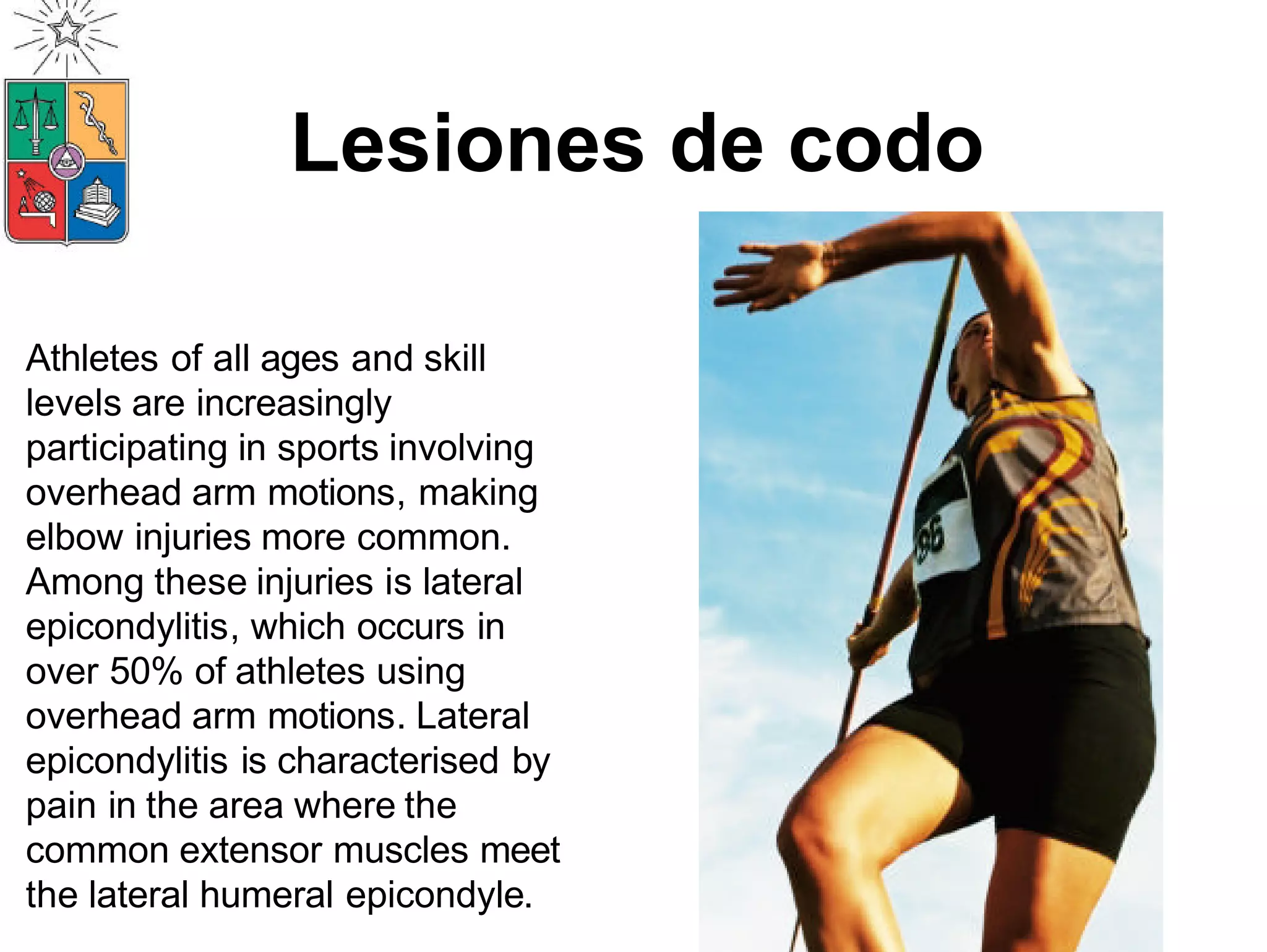
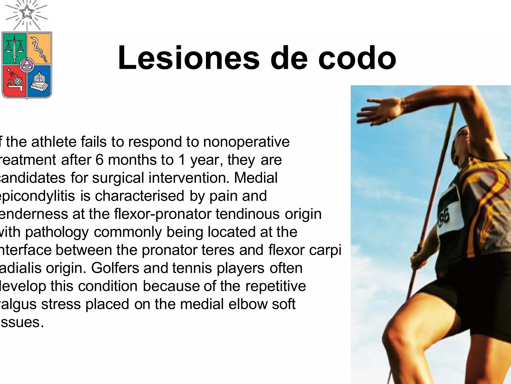
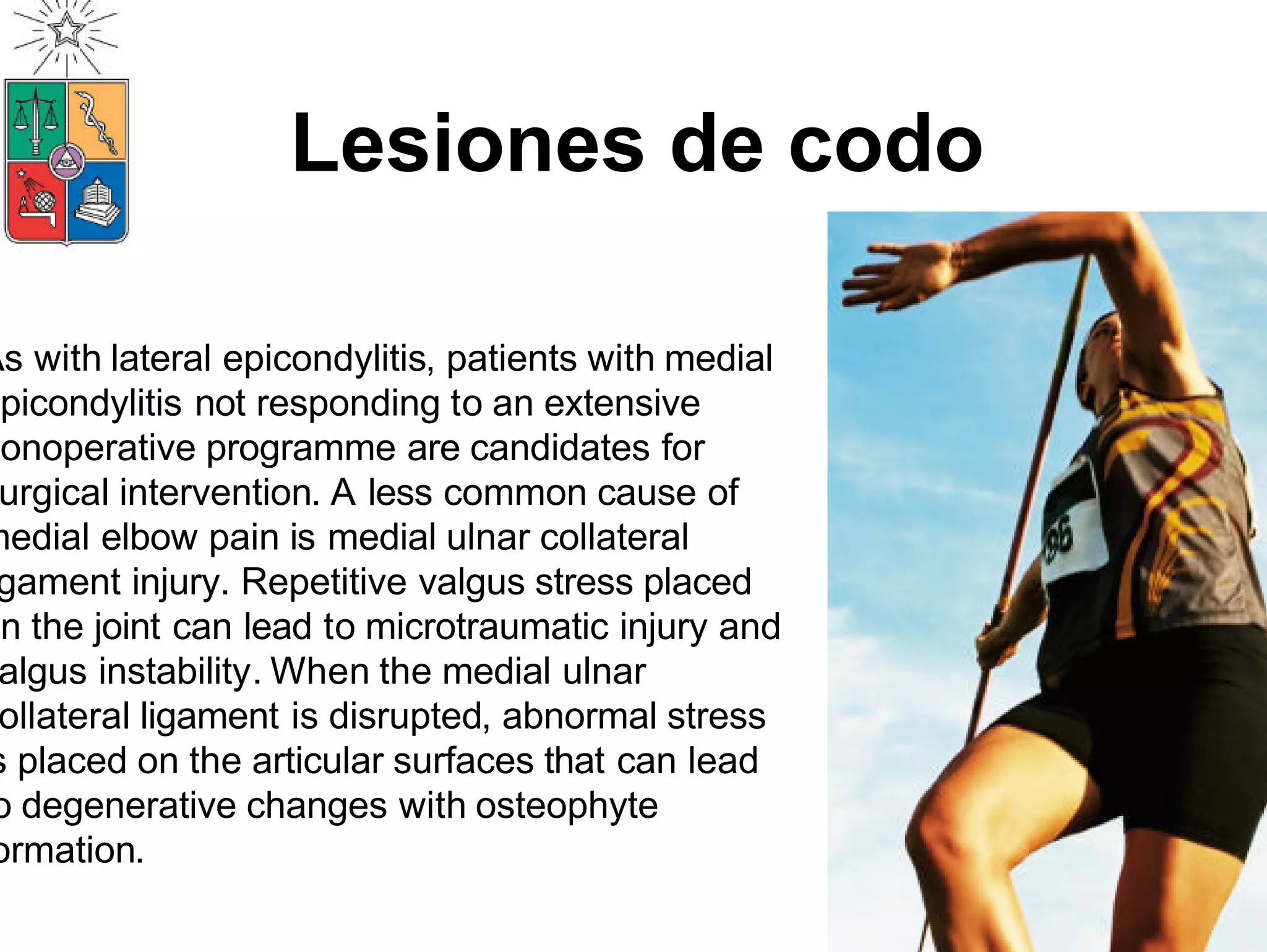
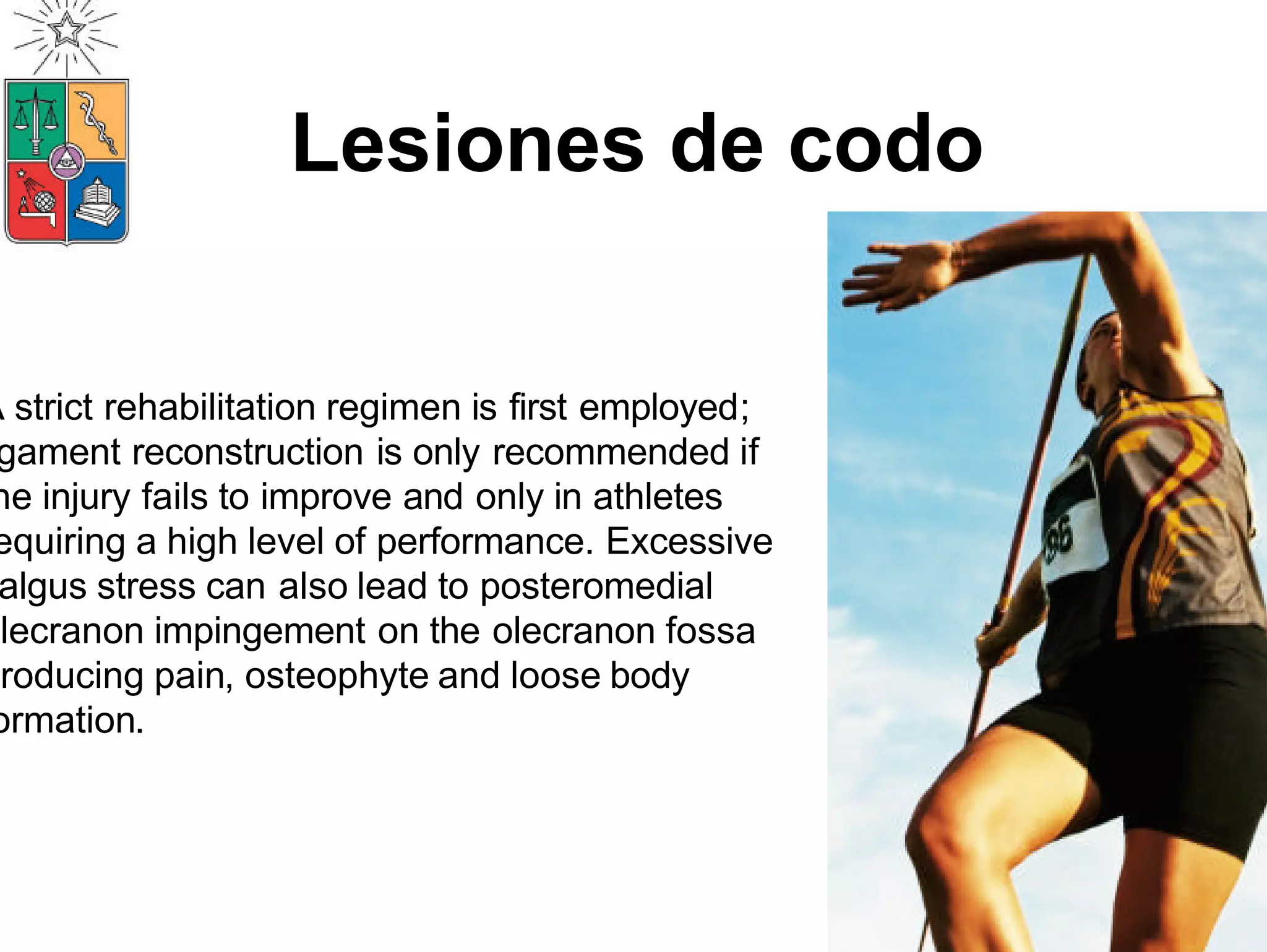
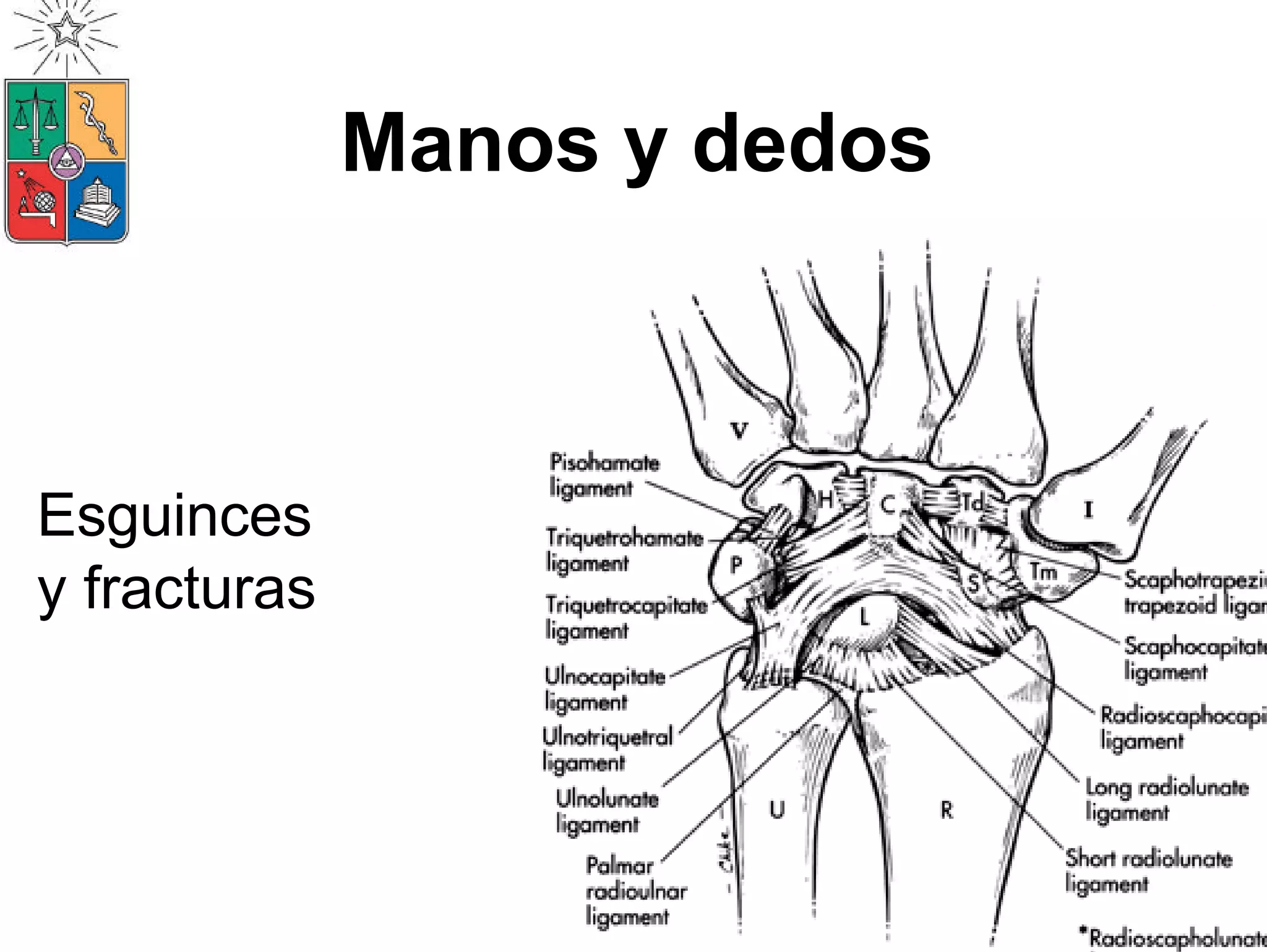
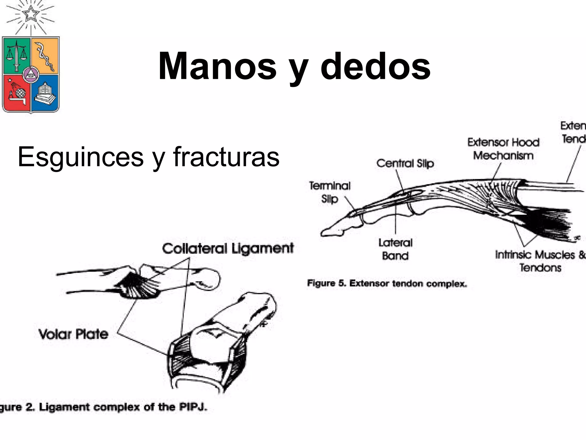
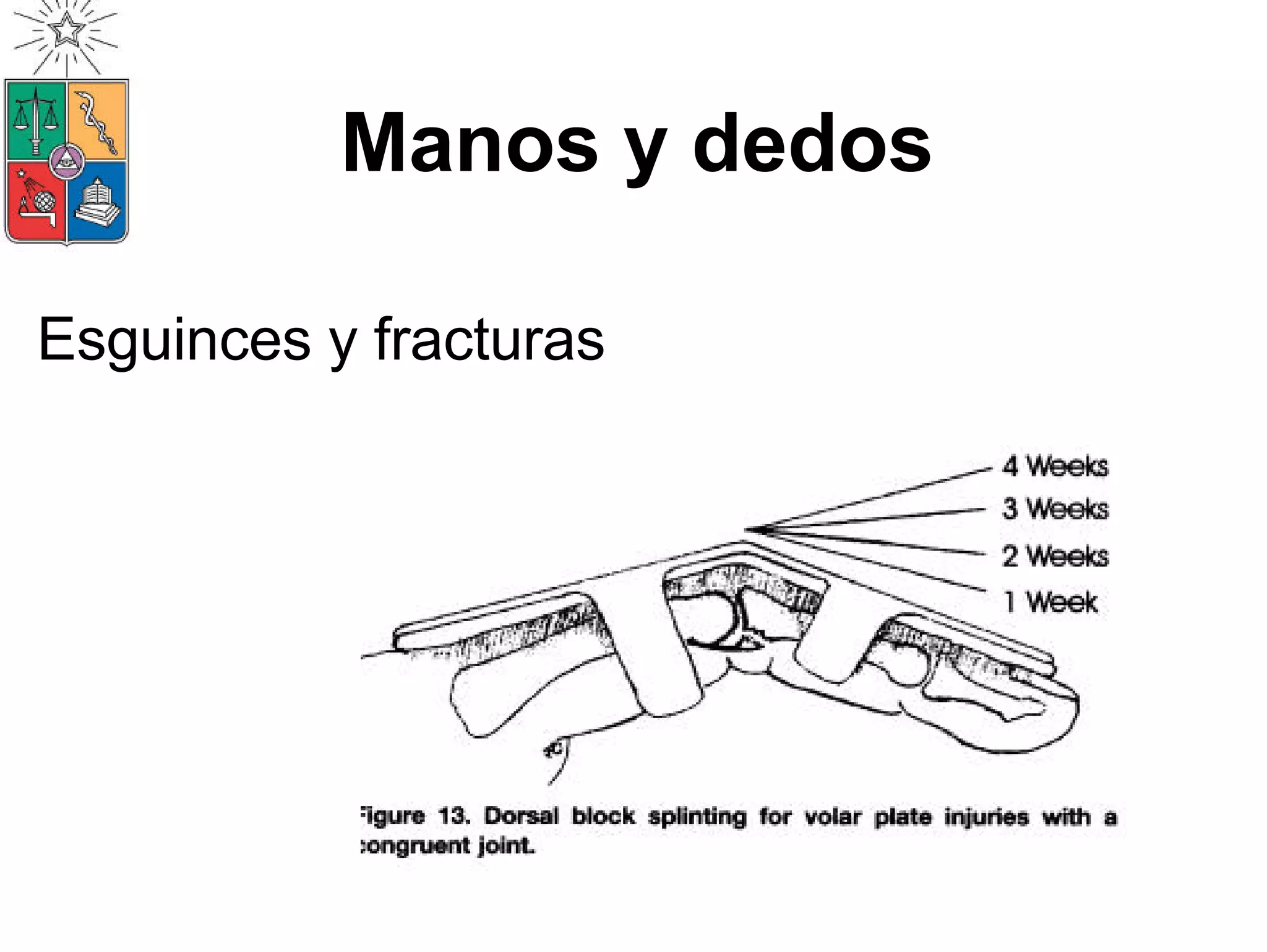
4. Lesiones de codo
Athletes of all ages and skill
levels are increasingly
participating in sports involving
overhead arm motions, making
elbow injuries more common.
Among these injuries is lateral
epicondylitis, which occurs in
over 50% of athletes using
overhead arm motions. Lateral
epicondylitis is characterised by
pain in the area where the
common extensor muscles meet
the lateral humeral epicondyle.
5. Lesiones de codo
The onset of this pathological condition
begins with the excessive use of the wrist
extensor musculature. Repetitive
microtraumatic injury can lead to mucinoid
degeneration of the extensor origin and
subsequent failure of the tendon. Lateral
epicondylitis can almost always be treated
nonoperatively with activity modification and
specific exercises.
6. Lesiones de codo
f the athlete fails to respond to nonoperative
reatment after 6 months to 1 year, they are
candidates for surgical intervention. Medial
epicondylitis is characterised by pain and
enderness at the flexor-pronator tendinous origin
with pathology commonly being located at the
nterface between the pronator teres and flexor carpi
adialis origin. Golfers and tennis players often
develop this condition because of the repetitive
valgus stress placed on the medial elbow soft
issues.
7. Lesiones de codo
As with lateral epicondylitis, patients with medial
epicondylitis not responding to an extensive
nonoperative programme are candidates for
urgical intervention. A less common cause of
medial elbow pain is medial ulnar collateral
gament injury. Repetitive valgus stress placed
on the joint can lead to microtraumatic injury and
algus instability. When the medial ulnar
ollateral ligament is disrupted, abnormal stress
s placed on the articular surfaces that can lead
o degenerative changes with osteophyte
ormation.
8. Lesiones de codo
A strict rehabilitation regimen is first employed;
gament reconstruction is only recommended if
he injury fails to improve and only in athletes
equiring a high level of performance. Excessive
algus stress can also lead to posteromedial
olecranon impingement on the olecranon fossa
producing pain, osteophyte and loose body
ormation.
12. Seminario de palpación
Con su compañer@ identifique las
siguientes
estructuras:
1.Todos los bordes de la escápula
2.Fosa supraespinosa
3.Articulación esternoclavicular (sienta en
los diferentes movimientos de clavícula)
4.Articulación acromioclavicular
5.Apófisis coracoides
13. Seminario de palpación
6. Tendón bicipital
7. Músculo infraespinoso
8. Líneas articulares de codo
9. Tendones extensores de muñeca y
dedos
10. Inserción proximal de tendones
flexores de muñeca y dedos.
14. Seminario Integración
Sepárese en 6 grupos y para el deporte y
lesión asignada identifique:
• Estructuras anatómicas comprometidas
• Elementos internos y externos que
pueden aumentar el riesgo que se
produzca la lesión
• Manejo inmediato y tardío de la lesión
(elementos más relevantes a tomar en
consideración)