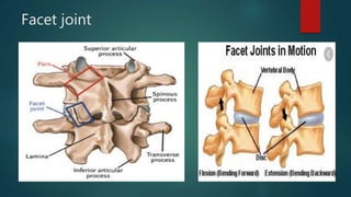
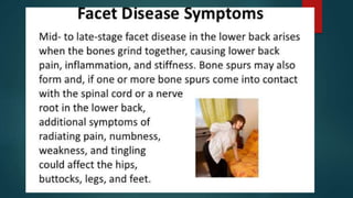
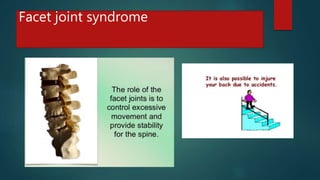
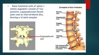
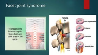
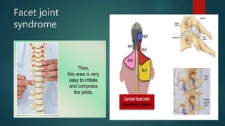
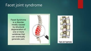
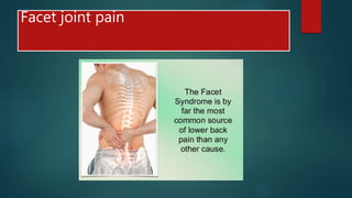
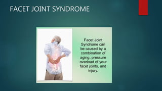
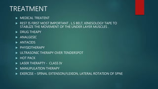
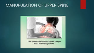
Facet joint syndrome
BY
DR SHAHIDUZ ZAFAR
DEPT OF PHYSIOTHERAPY
SMAS
GALGOTIAS UNIVERSITY
FACET JOINT SYNDROME
 Low back pain is one of the most common musculoskeletal complaints enc
ountered in clinical practice. It is the
leading cause of disability in the developed world and accounts for billions
of dollars in healthcare costs annually.
Although epidemiological studies vary, the incidence of low back pain is es
timated to be 5% to 10% with a lifetime
prevalence of 60% to 90%. Most occurrences of low back pain are self-
limited and will resolve without intervention
beyond brief periods of rest, activity modification, and physical therapy. A
pproximately 50% of cases will resolve
within 1 to 2 weeks. Ninety percent of cases will resolve in 6 to 12 weeks.
Etiology
 Lumbosacral facet syndrome can occur secondary to repetitive overuse and micro
trauma, spinal strains and torsional
forces, poor body mechanics, obesity, and intervertebral disk degeneration over th
e years. This notion is supported by
the strong association between the incidence of facet arthropathy and increasing
age.
In some instances, an inciting event such as trauma or whiplash injury can be iden
tified, although trauma patients tend
to develop cervical facet arthropathy more commonly than lumbosacral facet synd
rome. The role of trauma remains
controversial in the literature. The irritation of the degenerative zygapophyseal joi
nt over time leads to inflammation, which is perceived as low back pain.
Epidemiology
 Low back pain is the second greatest cause of absenteeism behind upper r
espiratory tract infections in the workplace.
Approximately 25 million people miss 1 or more days of work due to low b
ack pain, and more than five million are
disabled from it. Patients with chronic back pain account for 80% to 90% of
all healthcare expenditures.
Epidemiology
 The cited
prevalence of lumbosacral facet arthropathy is highly variable in the literature and has been listed as ranging
from 5%
to greater than 90%. Many of the studies investigating the prevalence of facetogenic pain used a combinatio
n of
history, physical exam, and radiologic imaging which has been shown to be relatively unreliable in establishin
g a
diagnosis. This likely accounts for the wide range seen in estimated prevalence. A trend that has been consist
ent
throughout studies, however, is that the prevalence of lumbosacral facet syndrome tends to increase with ag
e. A 2004
study by Manchikanti et al. found that of 397 patients screened, 198 (50%) obtained an initial positive respon
se to
medial branch block with lidocaine. Of those patients, 124 patients (31%) reported "definite pain relief" when
they received treatment with a repeat medial branch block using bupivacaine.
Pathophysiology
 The spine consists of seven cervical, 12 thoracic vertebrae, five lumbar, five sacral, and four
coccygeal bones. The
lumbar facets are composed of the inferior articular process of the vertebra above and the
superior articular process of
the vertebra below. The zygapophyseal joints serve to stabilize the spine and prevent injury
by limiting the spinal
range of motion. The facet joints are true synovial joints. The articular surface is contained i
n a cartilaginous sheath
over an encapsulated fibrous capsule. Like other cartilaginous joints, the lumbar facets are
prone to degradation over
time and can cause chronic low back pain when irritated. The sensory innervation of the fac
et is supplied by the
medial branch of the posterior rami of the spinal nerve at the level of, and the level above, t
he facet joint. For example, The L3-
L4 medial branch receives innervation from the L2 and L3 medial branch nerves. Noxious
stimulation of the medial branches is caused by degenerative changes in the facet joint, wh
ich results in facet pain and lumbosacral facet syndrome.
Facet joint
Facet joint
Facet joint syndrome
Facet joint
syndrome
Facet joint syndrome
Facet joint pain
Facet joint pain
FACET JOINT SYNDROME
FACET JOINT ARHRITIS
FACET JOINT DEFORMITY
Diagnosis
 X ray ap / lat view
 MRI
 CT SCAN
 ULTRASOUND SCAN
TREATMENT
 MEDICAL TREATENT
 REST IS FIRST MOST IMPORTANT , L.S BELT, KINESOLOGY TAPE TO
STABLIZE THE MOVEMENT OF THE UNDER LAYER MUSCLES .
 DRUG THEAPY
 ANALGESIC
 ANTACIDS
 PHYSIOTHERAPY
 ULTRASONIC THERAPY OVER TENDERSPOT
 HOT PACK
 LASER THERAPTY - CLASS IV
 MANUPULATION THERAPY
 EXERCISE – SPINAL EXTENSON,FLEXION, LATERAL ROTATION OF SPNE
FACET JOINT MANUPULATION
FACET JOINT SYNDOME
MANUPULATION OF UPPER SPINE
EXERCISE OF SPIN
EXERCISE
EXERCISE
EXERCISE
THANK YOU

Facet joint syndrome

  • 1.
    Facet joint syndrome BY DRSHAHIDUZ ZAFAR DEPT OF PHYSIOTHERAPY SMAS GALGOTIAS UNIVERSITY
  • 2.
    FACET JOINT SYNDROME Low back pain is one of the most common musculoskeletal complaints enc ountered in clinical practice. It is the leading cause of disability in the developed world and accounts for billions of dollars in healthcare costs annually. Although epidemiological studies vary, the incidence of low back pain is es timated to be 5% to 10% with a lifetime prevalence of 60% to 90%. Most occurrences of low back pain are self- limited and will resolve without intervention beyond brief periods of rest, activity modification, and physical therapy. A pproximately 50% of cases will resolve within 1 to 2 weeks. Ninety percent of cases will resolve in 6 to 12 weeks.
  • 3.
    Etiology  Lumbosacral facetsyndrome can occur secondary to repetitive overuse and micro trauma, spinal strains and torsional forces, poor body mechanics, obesity, and intervertebral disk degeneration over th e years. This notion is supported by the strong association between the incidence of facet arthropathy and increasing age. In some instances, an inciting event such as trauma or whiplash injury can be iden tified, although trauma patients tend to develop cervical facet arthropathy more commonly than lumbosacral facet synd rome. The role of trauma remains controversial in the literature. The irritation of the degenerative zygapophyseal joi nt over time leads to inflammation, which is perceived as low back pain.
  • 4.
    Epidemiology  Low backpain is the second greatest cause of absenteeism behind upper r espiratory tract infections in the workplace. Approximately 25 million people miss 1 or more days of work due to low b ack pain, and more than five million are disabled from it. Patients with chronic back pain account for 80% to 90% of all healthcare expenditures.
  • 5.
    Epidemiology  The cited prevalenceof lumbosacral facet arthropathy is highly variable in the literature and has been listed as ranging from 5% to greater than 90%. Many of the studies investigating the prevalence of facetogenic pain used a combinatio n of history, physical exam, and radiologic imaging which has been shown to be relatively unreliable in establishin g a diagnosis. This likely accounts for the wide range seen in estimated prevalence. A trend that has been consist ent throughout studies, however, is that the prevalence of lumbosacral facet syndrome tends to increase with ag e. A 2004 study by Manchikanti et al. found that of 397 patients screened, 198 (50%) obtained an initial positive respon se to medial branch block with lidocaine. Of those patients, 124 patients (31%) reported "definite pain relief" when they received treatment with a repeat medial branch block using bupivacaine.
  • 6.
    Pathophysiology  The spineconsists of seven cervical, 12 thoracic vertebrae, five lumbar, five sacral, and four coccygeal bones. The lumbar facets are composed of the inferior articular process of the vertebra above and the superior articular process of the vertebra below. The zygapophyseal joints serve to stabilize the spine and prevent injury by limiting the spinal range of motion. The facet joints are true synovial joints. The articular surface is contained i n a cartilaginous sheath over an encapsulated fibrous capsule. Like other cartilaginous joints, the lumbar facets are prone to degradation over time and can cause chronic low back pain when irritated. The sensory innervation of the fac et is supplied by the medial branch of the posterior rami of the spinal nerve at the level of, and the level above, t he facet joint. For example, The L3- L4 medial branch receives innervation from the L2 and L3 medial branch nerves. Noxious stimulation of the medial branches is caused by degenerative changes in the facet joint, wh ich results in facet pain and lumbosacral facet syndrome.
  • 7.
  • 8.
  • 13.
  • 15.
  • 16.
  • 17.
  • 18.
  • 19.
  • 20.
  • 21.
  • 22.
  • 24.
    Diagnosis  X rayap / lat view  MRI  CT SCAN  ULTRASOUND SCAN
  • 25.
    TREATMENT  MEDICAL TREATENT REST IS FIRST MOST IMPORTANT , L.S BELT, KINESOLOGY TAPE TO STABLIZE THE MOVEMENT OF THE UNDER LAYER MUSCLES .  DRUG THEAPY  ANALGESIC  ANTACIDS  PHYSIOTHERAPY  ULTRASONIC THERAPY OVER TENDERSPOT  HOT PACK  LASER THERAPTY - CLASS IV  MANUPULATION THERAPY  EXERCISE – SPINAL EXTENSON,FLEXION, LATERAL ROTATION OF SPNE
  • 28.
  • 29.
  • 30.
  • 31.
  • 33.
  • 34.
  • 35.
  • 36.
  • 38.