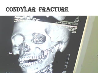
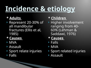
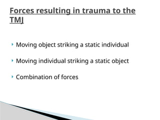
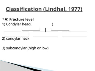
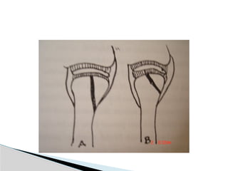
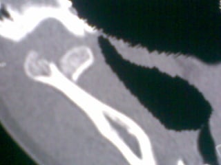
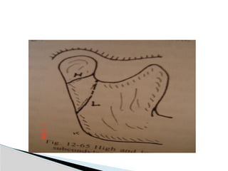
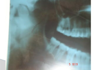
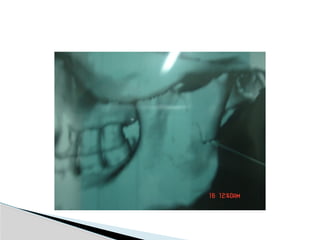
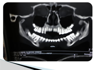
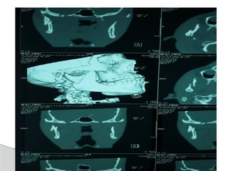
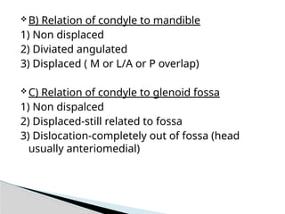
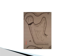
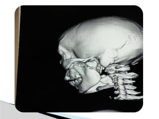
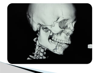
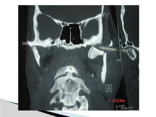
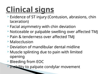
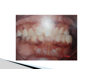
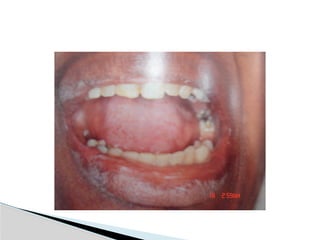
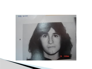
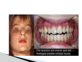
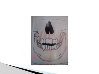
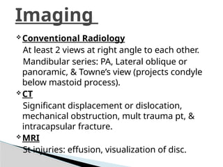
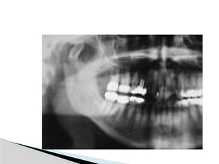
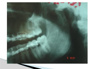
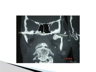
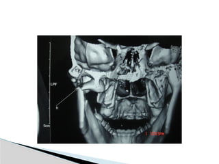
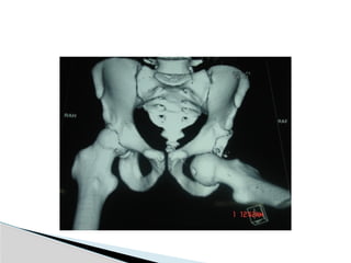
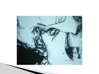
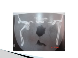
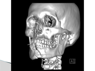
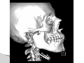
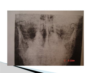
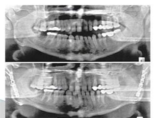
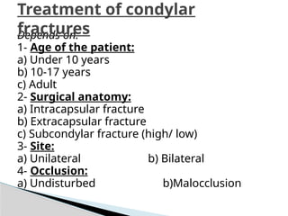
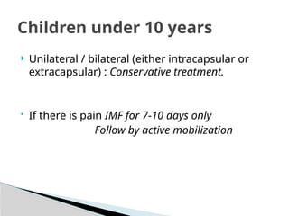
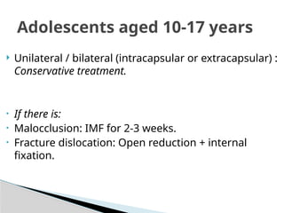
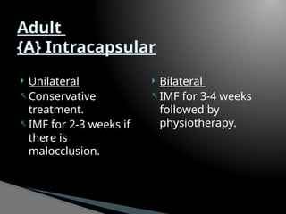
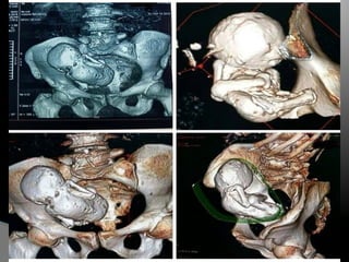
Condylar fracture
01/17/2026
Condylar fracture
Fractures involving the
mandibular condyle,
are the only facial
bone fractures which
involves a synovial
joint.
Mandible Anatomy
* Adults
 Represent 20-30% of
all mandibular
fractures (Ellis et al,
1985)
* Causes
 MVA
 Assault
 Sport relate injuries
 Falls
* Children
 Higher involvement
ranging from 40-
60% (Lehman &
Saddawi, 1976)
* Causes
 Falls
 MVA
 Sport related injuries
 Assault
Incidence & etiology
 Moving object striking a static individual
 Moving individual striking a static object
 Combination of forces
Forces resulting in trauma to the
TMJ
Classification (Lindhal, 1977)
 A) Fracture level
1) Condylar head( )
2) condylar neck
3) subcondylar (high or low)
 B) Relation of condyle to mandible
1) Non displaced
2) Diviated angulated
3) Displaced ( M or L/A or P overlap)
 C) Relation of condyle to glenoid fossa
1) Non dispalced
2) Displaced-still related to fossa
3) Dislocation-completely out of fossa (head
usually anteriomedial)
 Evidence of ST injury (Contusion, abrasions, chin
laceration)
 Facial asymmetry with chin deviation
 Noticeable or palpable swelling over affected TMJ
 Pain & tenderness over affected TMJ
 Malocclusion
 Deviation of mandibular dental midline
 Muscle splinting due to pain with limited
opening
 Bleeding from EOC
 Inability to palpate condylar movement
Clinical signs
Battle sign
Conventional Radiology
At least 2 views at right angle to each other.
Mandibular series: PA, Lateral oblique or
panoramic, & Towne’s view (projects condyle
below mastoid process).
CT
Significant displacement or dislocation,
mechanical obstruction, mult trauma pt, &
intracapsular fracture.
MRI
St injuries: effusion, visualization of disc.
Imaging
 1) Ankylosis:
Predisposing causes:
a) Age of the patient (<10 years).
c) Site & type of fracture:
 Intracapsular fracture.
 Close contact between the glenoid fossa &
condylar stamp.
c) Duration of immobilization.
(contributing factor in adults)
(causative factor in children)
d) Damage to meniscus.
Complications of condylar fracture
 2) Disturbance of mandibular growth
inchildren:
 Condylar growth center (Brosh 1930)
 Function matrix theory (Moss 1968)
 Damage to condylar cartilage in children lead
to hypoplasia
 Stable occlusion
 Restoration of interincisal opening
 Full range of mandibular excursive movements
 Minimize deviation
 Pain free
 Avoid growth disturbance
 Avoid ankylosis
Goals of treatment
TREATMENT
surgical conservative
 Unlike fractures of other bones, the exact
anatomic reapproximation of the fractured
segments may not be absolutely essential.
 There is no correlation between radiographic
findings & either preoperative symptoms or
postoperative function.
 Complications are uncommon with
conservative treatment.
Conservative treatment of condylar
fractuers
1) Normal occlusion with minimal discomfort soft
diet and maintain as near normal function as
possible.
2) Malocclusion, deviation with function, pain
period of immobilization (7-21 days) in the
form of arch bars or ivy loops, followed by
active mobilization and physical therapy.
Period of immobilization depends on age of
pt, level of fx, and degree of displacement.
 Perceived benefits
(Muller 1976)
 Early mobilization of
the mandible ensures
normal joint function
& action.
 Restoration of normal
mouth & jaw activity.
 Possible complications
(Eckelt 1984)
 Potential visible
scarring.
 Damage to facial
nerve.
 Intraoperative
bleeding from the
maxillary artery.
 Loss of blood supply
with avascular
necrosis of the
condyle.
Open reduction
 Absolute indications:
1- Inability to obtain
adequate occlusin with
closed reduction.
2- Displacement of the
condyle into the middle
cranial fossa.
3- Lateral extra capsular
displacement.
4- Foreign body in the joint
capsule.
 Relative indications:
1- condylar fx asso
comminuted mid facial
fx.
2- B condylar fxin an
edentulous pt splints are
unavailable or impossible
because of sever ridge
atrophy.
3- Displaced condyle fx in a
medically compromised
pt (sz d/o, psych pt, or
alcoholism) where IMF is
contraindicated.
Indications for open reduction of condylar
fractures in adults (Zide & Kent, 1983)
 Periauricular
 Endaural
 Submandibular
 Retromandibular
 Intra oral
 Combination
*Depending on the level of fracture, degree of
displacement or dislocation, & the planned
method of fixation.
Approaches for open reduction
RETROMANDIBULAR APPROACH
 Mini plates
 Lag screws
 Pin fixation
 Inter osseous wire
Methods of fixation
Depends on:
1- Age of the patient:
a) Under 10 years
b) 10-17 years
c) Adult
2- Surgical anatomy:
a) Intracapsular fracture
b) Extracapsular fracture
c) Subcondylar fracture (high/ low)
3- Site:
a) Unilateral b) Bilateral
4- Occlusion:
a) Undisturbed b)Malocclusion
Treatment of condylar
fractures
 Unilateral / bilateral (either intracapsular or
extracapsular) : Conservative treatment.
• If there is pain IMF for 7-10 days only
Follow by active mobilization
Children under 10 years
 Unilateral / bilateral (intracapsular or extracapsular) :
Conservative treatment.
• If there is:
• Malocclusion: IMF for 2-3 weeks.
• Fracture dislocation: Open reduction + internal
fixation.
Adolescents aged 10-17 years
 Unilateral
- Conservative
treatment.
- IMF for 2-3 weeks if
there is
malocclusion.
 Bilateral
- IMF for 3-4 weeks
followed by
physiotherapy.
Adult
{A} Intracapsular
 Unilateral
- Conservative treatment
if the occlusion is
undisturbed.
- Low condylar fracture
with malocclusion
(open reduction).
- High condylar fracture
with extensive
malocclusion:
- IMF for 3-4 weeks.
- Some surgeons
favors ORIF.
 Bilateral
- Malocclusion is
present.
- At least ORIF for one
side.
- IMF for 4-6 weeks
followed by elastic
bands for several
weeks.
{B} Extracapsular
Thank you

Condylar injuries presentation, oral surgery

  • 1.
  • 3.
  • 4.
  • 5.
    Fractures involving the mandibularcondyle, are the only facial bone fractures which involves a synovial joint.
  • 9.
  • 11.
    * Adults  Represent20-30% of all mandibular fractures (Ellis et al, 1985) * Causes  MVA  Assault  Sport relate injuries  Falls * Children  Higher involvement ranging from 40- 60% (Lehman & Saddawi, 1976) * Causes  Falls  MVA  Sport related injuries  Assault Incidence & etiology
  • 12.
     Moving objectstriking a static individual  Moving individual striking a static object  Combination of forces Forces resulting in trauma to the TMJ
  • 13.
    Classification (Lindhal, 1977) A) Fracture level 1) Condylar head( ) 2) condylar neck 3) subcondylar (high or low)
  • 22.
     B) Relationof condyle to mandible 1) Non displaced 2) Diviated angulated 3) Displaced ( M or L/A or P overlap)  C) Relation of condyle to glenoid fossa 1) Non dispalced 2) Displaced-still related to fossa 3) Dislocation-completely out of fossa (head usually anteriomedial)
  • 31.
     Evidence ofST injury (Contusion, abrasions, chin laceration)  Facial asymmetry with chin deviation  Noticeable or palpable swelling over affected TMJ  Pain & tenderness over affected TMJ  Malocclusion  Deviation of mandibular dental midline  Muscle splinting due to pain with limited opening  Bleeding from EOC  Inability to palpate condylar movement Clinical signs
  • 32.
  • 38.
    Conventional Radiology At least2 views at right angle to each other. Mandibular series: PA, Lateral oblique or panoramic, & Towne’s view (projects condyle below mastoid process). CT Significant displacement or dislocation, mechanical obstruction, mult trauma pt, & intracapsular fracture. MRI St injuries: effusion, visualization of disc. Imaging
  • 49.
     1) Ankylosis: Predisposingcauses: a) Age of the patient (<10 years). c) Site & type of fracture:  Intracapsular fracture.  Close contact between the glenoid fossa & condylar stamp. c) Duration of immobilization. (contributing factor in adults) (causative factor in children) d) Damage to meniscus. Complications of condylar fracture
  • 50.
     2) Disturbanceof mandibular growth inchildren:  Condylar growth center (Brosh 1930)  Function matrix theory (Moss 1968)  Damage to condylar cartilage in children lead to hypoplasia
  • 51.
     Stable occlusion Restoration of interincisal opening  Full range of mandibular excursive movements  Minimize deviation  Pain free  Avoid growth disturbance  Avoid ankylosis Goals of treatment
  • 52.
  • 53.
     Unlike fracturesof other bones, the exact anatomic reapproximation of the fractured segments may not be absolutely essential.  There is no correlation between radiographic findings & either preoperative symptoms or postoperative function.  Complications are uncommon with conservative treatment. Conservative treatment of condylar fractuers
  • 54.
    1) Normal occlusionwith minimal discomfort soft diet and maintain as near normal function as possible. 2) Malocclusion, deviation with function, pain period of immobilization (7-21 days) in the form of arch bars or ivy loops, followed by active mobilization and physical therapy. Period of immobilization depends on age of pt, level of fx, and degree of displacement.
  • 55.
     Perceived benefits (Muller1976)  Early mobilization of the mandible ensures normal joint function & action.  Restoration of normal mouth & jaw activity.  Possible complications (Eckelt 1984)  Potential visible scarring.  Damage to facial nerve.  Intraoperative bleeding from the maxillary artery.  Loss of blood supply with avascular necrosis of the condyle. Open reduction
  • 56.
     Absolute indications: 1-Inability to obtain adequate occlusin with closed reduction. 2- Displacement of the condyle into the middle cranial fossa. 3- Lateral extra capsular displacement. 4- Foreign body in the joint capsule.  Relative indications: 1- condylar fx asso comminuted mid facial fx. 2- B condylar fxin an edentulous pt splints are unavailable or impossible because of sever ridge atrophy. 3- Displaced condyle fx in a medically compromised pt (sz d/o, psych pt, or alcoholism) where IMF is contraindicated. Indications for open reduction of condylar fractures in adults (Zide & Kent, 1983)
  • 57.
     Periauricular  Endaural Submandibular  Retromandibular  Intra oral  Combination *Depending on the level of fracture, degree of displacement or dislocation, & the planned method of fixation. Approaches for open reduction
  • 60.
  • 63.
     Mini plates Lag screws  Pin fixation  Inter osseous wire Methods of fixation
  • 68.
    Depends on: 1- Ageof the patient: a) Under 10 years b) 10-17 years c) Adult 2- Surgical anatomy: a) Intracapsular fracture b) Extracapsular fracture c) Subcondylar fracture (high/ low) 3- Site: a) Unilateral b) Bilateral 4- Occlusion: a) Undisturbed b)Malocclusion Treatment of condylar fractures
  • 69.
     Unilateral /bilateral (either intracapsular or extracapsular) : Conservative treatment. • If there is pain IMF for 7-10 days only Follow by active mobilization Children under 10 years
  • 70.
     Unilateral /bilateral (intracapsular or extracapsular) : Conservative treatment. • If there is: • Malocclusion: IMF for 2-3 weeks. • Fracture dislocation: Open reduction + internal fixation. Adolescents aged 10-17 years
  • 71.
     Unilateral - Conservative treatment. -IMF for 2-3 weeks if there is malocclusion.  Bilateral - IMF for 3-4 weeks followed by physiotherapy. Adult {A} Intracapsular
  • 72.
     Unilateral - Conservativetreatment if the occlusion is undisturbed. - Low condylar fracture with malocclusion (open reduction). - High condylar fracture with extensive malocclusion: - IMF for 3-4 weeks. - Some surgeons favors ORIF.  Bilateral - Malocclusion is present. - At least ORIF for one side. - IMF for 4-6 weeks followed by elastic bands for several weeks. {B} Extracapsular
  • 74.