Downloaded 404 times


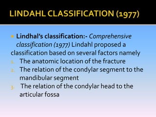
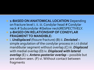


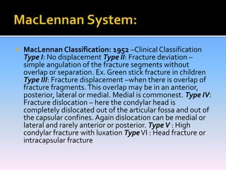


Condylar fractures can occur in different locations and with varying degrees of displacement. Treatment depends on factors like the patient's age, whether other fractures are present, and the level and displacement of the condylar fracture. Classification systems aim to describe the anatomic location and relationship of condylar fragments to help determine appropriate treatment, whether closed or open reduction is necessary. The goals of treatment are to relieve pain, achieve stable occlusion, restore jaw function, and avoid long-term complications.


