
Recommended
More Related Content
What's hot
What's hot (20)
Similar to ECG-Dr.Allam منقول.pdf
Similar to ECG-Dr.Allam منقول.pdf (20)
More from mernahazazah
More from mernahazazah (11)
Recently uploaded
Recently uploaded (20)
ECG-Dr.Allam منقول.pdf
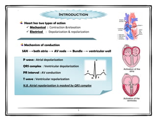
- 1. ` 1 P wave : Atrial depolarization QRS complex : Ventricular depolarization PR interval : AV conduction T wave : Ventricular repolarization N.B. Atrial repolarization is masked by QRS complex Heart has two types of action Mechanical : Contraction &relaxation Electrical : Depolarization & repolarization Mechanism of conduction SAN both atria AV node Bundle ventricular wall Introduction
- 2. ` 2 ECG LEADS 6 Limb Leads 6 Chest leads AV = augmented voltage AVR : inverted Blood supply of the heart Right coronary artery Left coronary artery Inferior wall Anterior descending artery Circumflex artery Anterior wall Lateral wall Topographism L II , L III , AVF V1,2,3,4 V5,V6
- 3. ` 3 Comment on ECG 1. Rhythm 2. Rate 3. Axis 4. P Wave 5. P-R Interval 6. QRS Complex 7. S-T Segment 8. T wave 9. Diagnosis 1 small square = 0.04 sec. 1 big square = 0. 2 sec. 1 sec.= 5 big square. 1 minute = 300 big square.
- 4. ` 4 1. Rhythm Regular Irregular 2. Rate Regular 300 R-R بين الكبيرة المربعات عدد Irregular 300 R-R بين الكبيرة المربعات عدد متوسط 3. Axis : LI AVF N.A.D. Rt.A.D. Lt.A.D.
- 5. ` 5 4. P Wave 5. P-R Interval وبداية بداية 6. QRS Complex ايه تمثل Atrial depolarization ازاى اعرفها 1st +ve wave before complex مكان احسن L11.V1 مقاستها less than (2.5 x 2.5 ) small ايه تمثل AVN conduction مكان احسن L11 مقاستها 3-5 small N.B. : If > 5 small square = H.B. ايه تمثل Ventricular depolarization ازاى اعرفها Complex مكان احسن Rt. Ventricle( V1,2 ) Lt .Ventricle ( V5,6 ) R : only +ve in complex عرضها = 2-3 small ارتفاعها = 1-5 big Q : 1st -ve in complex عرضها : small less than عمقها : less than 1/4 of next R
- 6. ` 6 7. S-T Segment وبداية نهاية 8. T wave NEVER ABSENT Tall T. (Himalaya T.) in hyperkalemia. ايه تمثل Ventricular repolarization Normally : Iso-electrical line as (P-R ) ايه تمثل Ventricular repolarization عرضها : Less than 6 small ارتفاعها : Less than 1/3 R ST elevation : Recent MI Pericarditis ( all leads ) Variant angina ST depression : Angina (Ischemia) Digitalis effect
- 7. ` 7 Abnormal ECG Bundle branch block (BBB) If QRS notched ( v1,2= Rt.BBB ) (V5,6= Lt.BBB) Atrial ++ Peaked > 2.5 = p. pulmonal P wave in LII , V1 Broad > 2.5 = p. mitral Ventricular++ Normal S>r in (V1,V2 ) & R>s in (V5,6 ) Lt. Ventricular ++ = Exaggeration of normal : deep S in v1 or 2 > 5 big square Tall R in V5 or 6> 5 big square R+S > 7 big square Strain ischemia in V5 & V6 Rt . ventricular ++ = Revisable of normal : Tall R in V1 or 2 Deep S in V5 or 6 Strain ischemia in V1 & V2
- 8. ` 8 Scheme for any segmented ECG 1- P wave in L II Peaked Rt.Atrim ++ Broad Lt.Atrium ++ 2- QRS V1,2 V5,6 a- Shape ( M notched) Rt.BBB Lt.BBB b- Direction R>s = Rt.v++ S>r = Rt.V++ c- Voltage S> 5 big square = Lt.V. ++ R>5 big square = Lt.V ++ 3- S.T. segment: All leads pericarditis Elevation ē topogrophism recent M.I All leads sagging digitalis effect Depression ē topographism Ischemia 4- Pathological Q: with topographism = old M.I 5- Tall T.: All leads = hyperkalemia
- 9. ` 9 Heart block 1st degree 2nd degree 3rd degree Prolonged P-R Mobitz 1 Mobitz 11 A.V. dissociation Progressive prolongation of P-R until drop of QRS Regular drop of QRS
- 10. ` 10
- 11. ` 11