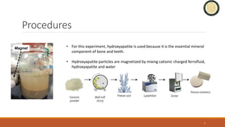
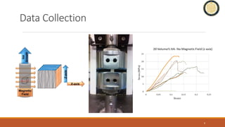
Magnetic Freeze Casting with Surface
Magnetized Hydroxyapatite for
Bioinspired Bone Implants
CINDY AYALA
BIOENGINEERING REU
DEPARTMENT OF MECHANICAL AND AEROSPACE ENGINEERING
ADVISOR: PROF. JOANNA MCKIT TRICK
GRAD STUDENT MENTOR: MICHAEL FRANK
Motivation
Osteoporosis
- causes erosion of trabecular (spongy) bone
- leads to decreased bone mass, especially in the elderly
- results in brittle bones that lead to unexpected failure
- titanium implants can lead to stress shielding
(reduction in bone density due to removal of stress from
bone by the implant)
- titanium implants often require adjustment surgeries
2
[1] http://www.medguidance.com/thread/What-Causes-Osteoporosis.html
Goal
Create a porous scaffold made of bone
mineral which mimics the structure of bone
Freeze Casting
3
Y
X
Z
Magnetic Freeze Casting
4
X-Axis
Y
X
Z
Procedures
• For this experiment, hydroxyapatite is used because it is the essential mineral
component of bone and teeth.
• Hydroxyapatite particles are magnetized by mixing cationic charged ferrofluid,
hydroxyapatite and water
5
Magnet
Data Collection
6
Results (10 Vol% Slurry, ≈ 85% Porosity)
7
SEM Imaging of Scaffolds
(10 Vol% Slurry, ≈ 85% Porosity, 50 mT Magnetic Field)
8
Magnetic Field (x-axis)
SEM Imaging of Scaffolds
(10 Vol% Slurry, ≈ 85% Porosity, 25 mT Magnetic Field)
9
Magnetic Field (x-axis)
SEM Imaging of Scaffolds
(10 Vol% Slurry, ≈ 85% Porosity, 25 mT Magnetic Field)
10
Magnetic Field (x-axis)
Results (20 Vol% Slurry, ≈75% Porosity)
11
Conclusion
• Hydroxyapatite particles (HA, 2 μm) are compared to
past work with alumina (195, 225, 350 nm).
• Bigger particle size (HA) needs lower magnetic field to
align more particle chains in scaffold center.
• Stiffness (Young’s Modulus) is enhanced when more
particle chains are aligned in scaffold center.
• 20 vol% HA (≈75% porosity) at 25 mT was best condition.
12
Scaffold
X-section
Scaffold
Center Cube
Particle
Chains
Goal
More particle chain alignment in
scaffold center rather than at poles
Future Work
13
• Take best condition (20 vol% HA, ≈75% porosity, 25 mT) and do further work to strengthen scaffold.
• Wrap scaffold cylinder with a biodegradable thermopolymer (polylactic acid, PLA) to resemble impact resistant
porcupine quill that is a keratin foam wrapped with a keratin cortex sheath.
• Infiltrate scaffold pores with a biodegradable polymer (polyethylene glycol diacrylated, PEGDA), photoinitiator
and phosphate binding element (calcium acetate) and then UV cure to crosslink PEGDA within HA scaffold.
• Compare overall mechanical properties of reinforced HA scaffold implant in axial and radial (Brazilian Test)
compression to assess viability of structural design for spongy bone biomedical implants.
Acknowledgements
•Professor McKittrick for welcoming me into her team
•PhD student Michael Frank for mentoring me through this project
•Fellow undergrads Sze Hei Siu, Louis Guibert, and Joyce Mok for assisting me in my research
14

Magnetic Freeze Casting Presentation

  • 1.
    Magnetic Freeze Castingwith Surface Magnetized Hydroxyapatite for Bioinspired Bone Implants CINDY AYALA BIOENGINEERING REU DEPARTMENT OF MECHANICAL AND AEROSPACE ENGINEERING ADVISOR: PROF. JOANNA MCKIT TRICK GRAD STUDENT MENTOR: MICHAEL FRANK
  • 2.
    Motivation Osteoporosis - causes erosionof trabecular (spongy) bone - leads to decreased bone mass, especially in the elderly - results in brittle bones that lead to unexpected failure - titanium implants can lead to stress shielding (reduction in bone density due to removal of stress from bone by the implant) - titanium implants often require adjustment surgeries 2 [1] http://www.medguidance.com/thread/What-Causes-Osteoporosis.html Goal Create a porous scaffold made of bone mineral which mimics the structure of bone
  • 3.
  • 4.
  • 5.
    Procedures • For thisexperiment, hydroxyapatite is used because it is the essential mineral component of bone and teeth. • Hydroxyapatite particles are magnetized by mixing cationic charged ferrofluid, hydroxyapatite and water 5 Magnet
  • 6.
  • 7.
    Results (10 Vol%Slurry, ≈ 85% Porosity) 7
  • 8.
    SEM Imaging ofScaffolds (10 Vol% Slurry, ≈ 85% Porosity, 50 mT Magnetic Field) 8 Magnetic Field (x-axis)
  • 9.
    SEM Imaging ofScaffolds (10 Vol% Slurry, ≈ 85% Porosity, 25 mT Magnetic Field) 9 Magnetic Field (x-axis)
  • 10.
    SEM Imaging ofScaffolds (10 Vol% Slurry, ≈ 85% Porosity, 25 mT Magnetic Field) 10 Magnetic Field (x-axis)
  • 11.
    Results (20 Vol%Slurry, ≈75% Porosity) 11
  • 12.
    Conclusion • Hydroxyapatite particles(HA, 2 μm) are compared to past work with alumina (195, 225, 350 nm). • Bigger particle size (HA) needs lower magnetic field to align more particle chains in scaffold center. • Stiffness (Young’s Modulus) is enhanced when more particle chains are aligned in scaffold center. • 20 vol% HA (≈75% porosity) at 25 mT was best condition. 12 Scaffold X-section Scaffold Center Cube Particle Chains Goal More particle chain alignment in scaffold center rather than at poles
  • 13.
    Future Work 13 • Takebest condition (20 vol% HA, ≈75% porosity, 25 mT) and do further work to strengthen scaffold. • Wrap scaffold cylinder with a biodegradable thermopolymer (polylactic acid, PLA) to resemble impact resistant porcupine quill that is a keratin foam wrapped with a keratin cortex sheath. • Infiltrate scaffold pores with a biodegradable polymer (polyethylene glycol diacrylated, PEGDA), photoinitiator and phosphate binding element (calcium acetate) and then UV cure to crosslink PEGDA within HA scaffold. • Compare overall mechanical properties of reinforced HA scaffold implant in axial and radial (Brazilian Test) compression to assess viability of structural design for spongy bone biomedical implants.
  • 14.
    Acknowledgements •Professor McKittrick forwelcoming me into her team •PhD student Michael Frank for mentoring me through this project •Fellow undergrads Sze Hei Siu, Louis Guibert, and Joyce Mok for assisting me in my research 14

Editor's Notes

  • #6 Superparamagnetic magnetite nanoparticles (≈ 10 nm) within cationic charged ferrofluid were electrostatically absorbed onto the surface of hydroxyapatite particles (≈ 2 µm) by mixing in water. Slurries of 10 and 20 vol % magnetized hydroxyapatite were then subjected to a low magnetic field (25 or 50 mT) during magnetic freeze casting. Subsequent freeze drying and sintering steps produced scaffolds with pores where ice crystals had previously grown in a uniform direction