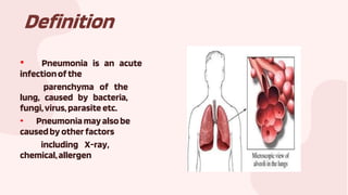
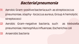
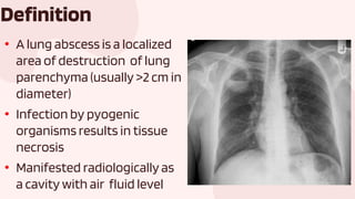
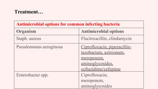
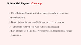
Dimas Fujiansyah- 2108260090
LUNGABSCESS
Defenitionand
Classification
Pneumonia
EtiologyandRisk
factorpneumonia
Pathophysiology
pneumonia
Defenitionand
classificationlungs
abscess
01 02
04 05
Tableofcontents
03
Clinical
Manifestatiom
pneumonia
Riskfactorand
etiologylung
abscess
Clinical
Manifestationlung
abscess
CMDlungAbscess Managementlung
abscess
06 07
09 10
Tableofcontents
08
Pathophysiology
lungabscess
Riskfactorand
etiologylung
abscess
Clinical
Manifestationlung
abscess
11 12
Tableofcontents
Defenitionand
Classification
Pneumonia
01
Definition
• Pneumonia is an acute
infectionofthe
parenchyma of the
lung, caused by bacteria,
fungi,virus,parasite etc.
• Pneumoniamayalsobe
causedby otherfactors
including X-ray,
chemical,allergen
Classification
• Classification of anatomy
• Classification of pathogen
• Classification of acquired environment
Ⅰ.Classificationbypathogen
Pathogen
classification is the
most useful to treat
the patients by
choosing effective
antimicrobial agents
Bacterialpneumonia
(1) Aerobic Gram-positive bacteria,such as streptococcus
pneumoniae, staphy- lococcus aureus, Group A hemolytic
streptococci
(2) Aerobic Gram-negative bacteria, such as klebsiella
pneumoniae, Hemophilus influenzae, Escherichia coli
(3) Anaerobicbacteria
Atypical pneumonia
Including Legionnaies pneumonia ,
Mycoplasmal pneumonia ,chlamydia pneumonia.
Fungal pneumonia
Fungal pneumonia is commonly caused by
candida and aspergilosis
Viral pneumonia
Viral pneumonia may be caused by adenoviruses, respiratory
syncytial virus , influenza, cytomegalovirus, herpes simplex
Ⅱ.Classification by anatomy
1. Lobar: Involvement of an entire lobe
2. Lobular: Involvement of parts of the lobe only, segmental
or of alveoli contiguous to bronchi (bronchopneumonia).
3. Interstitial
Lobarpneumonia
Lobularpneumonia
Classificationbyacquired
environment
◆Community acquired pneumonia ,CAP
◆Hospital acquired pneumonia ,HAP ,
NP
◆Nursing home acquired pneumonia, NHAP
◆Immunocompromised host
pneumonia, ICAP
EtiologyandRisk
factorpneumonia
02
Overview
◆ Pneumonia can result from a variety of causes,
including infection with bacteria, viruses, fungi,
parasites, and chemical or physical injury to the
lungs
◆ Source of Infection
◆ Aerosol
◆ Aspiration of amniotic fluid
◆ Blood-borne infection across the placenta
Common CausesbyAgeGroup
RiskFactors
Overview
◆ The risk factors for pneumonia include:
smoking, age>65, immuno-suppression,
exposure to chemicals, and underlying lung
disease.
Risk Factors
◆ AirwayObstruction
◆ When part of the airway (bronchi)leading to the alveoli is obstructed, lead to
infection of the fluid resulting in community-acquired pneumonia (CAP)
◆ Another cause of obstruction is lungcancer
◆ LungDisease
◆ Smoking, and diseases such as emphysema, result in more frequent and severe
bouts of CAP
◆ ImmuneCompromise
◆ more likely to get CAP
◆ active malignancy,immuno-suppression, neurological disease, congestive
heart failure, coronaryartery disease, and diabetes mellitus
Clinical
Manifestatiom
pneumonia
03
Clinicalmanifestations(1)
• Many patients have had an upperrespiratoryinfectionfor
several days before the onset of pneumonia
• Onset usually is sudden, half caseswith a shaking
chill
• The temperature rises during the first few hours to 39-
40℃
Clinicalmanifestations(2)
• Typically, patients have the symptoms of high
fever , shaking chill, sharp chest pain, cough,
dyspnea and blood-flecked sputum.
• But in some cases, especially those at age
extremes symptoms may be more insidious.
• The pulse accelerates
• Sharp pain in the involved hemi thorax
• The cough is initially dry with pinkish or
blood-flecked sputum
• Gastrointestinal symptoms such as,
anorexia, nausea, vomiting abdominal
pain, diarrhea may be mistaken as acute
abdominal inflammation
Clinicalmanifestations(3)
Pathophysiology
pneumonia
04
Defenitionand
classificationlungs
abscess
05
• A lung abscess is a localized
area of destruction of lung
parenchyma (usually >2 cm in
diameter)
• Infection by pyogenic
organisms results in tissue
necrosis
• Manifested radiologically as
a cavity with air fluid level
Definition
Classification
Lung abscess may be single or multiple and they
frequently containair-fluid levels
Lung abscesses can be classified based on the
duration & the likely etiology
Acute abscess
Chronic abscess
Acute
patient presents with symptoms of <2weeks
duration
more likely to have an infection caused by a
virulent aerobic bacterial agent (e.g. S. aureus)
Acute Lung Abscess
CXR of a patient who had foul-smelling & bad tasting sputum, an almost
diagnostic feature of anaerobic lung abscess
Classification…
Clinically useful during initial evaluation
Chronic:
A chronic lung abscess is defined by symptoms lasting for > 4 to 6 weeks.
Patients more like to have an underlying neoplasm or infection with a less
virulentanaerobicagent
Primary abscess
caused by aspirationor pneumonia in the healthy
host
Mostly result from necrosis in an existing
parenchymal process, usually untreated or
aspiration pneumonia
Classification…
Secondary abscess is caused by
Pre-existing condition eg bronchiectasis
Bronchial obstruction (eg- aspirated foreign body)
An immuno-compromised state
Spread from an extra-pulmonary site
Abscess that complicates either a septic vascular
embolus (eg- right sided endocarditis)
Etiologyandrisk
factorlungabscess
06
Causes of Lung abscess (A) Aspiration
A) Aspiration of infected material containing
oropharyngeal flora (commonest cause)
Organisms are anaerobic and
aerobic May be due to
 Dental/ periodontal sepsis esp following tooth
extraction, tonsillectomy and nasal operation
 Paranasal sinus infection
Causes…
B) Necrotizing Pneumonia / Inadequately Treated Pneumonia
 Aerobic bacteria (eg- Staphylococcus aureus, Strepto. Pneumoniae,
Streptococcus milleri/intermedius, Klebsiella pneumoniae,
Pseudomonas aeruginosa )
 Anaerobic bacteria
 Others:
 Mycobacteria
 Fungal
 Parasites
Causes…
C) Mechanical Bronchial obstruction by
 Tumour (Bronchial carcinoma/ Adenoma)
 Foreign body
 Enlarged lymph nodes
 Congenital abnormality – bronchial stenosis
D) Pre-existing lung disease
 Bronchiectasis
 Cystic fibrosis
Causes…
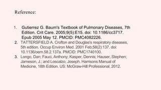
E)Haematogenous spread from a distal site [from
other infection as septic emboli]
 Urinary tract infection
 Abdominal sepsis
 Pelvic sepsis
 Infective endocarditis (right-sided)
 Intravenous drug abuse
 Infected IV cannulae
 Septic thrombophlebitis
 Salpingitis
 Appendicitis
 Pyaemia/ Septicaemia
Causes…
F) Extension from extra-pulmonary abscess/( transdiaphragmatic
spread)
 liver abscess
 subphrenic abscess
 Mediastinal abscess
G) Trauma/ Post traumatic
 Infected pulmonary haematoma
 Contaminated foreign body
H) Immunodeficiency
 Primary or
 Acquired
RISK FACTORS FOR
GRAM NEGATIVE
COLONIZATION
1. Malnutrition
2. Severe illness
3. Coma
4. Intubation
5. Diabetes
6. Prior surgery
7. Lung disease
1. Lung disease
2. Renal failure
3. Prior antibiotic uses
4. Hypotension
5. Cigarette smoking
6. Prolonged
hospitalization
ClinicalManifestation
lungabscess
07
Symptoms …
Patients present with
Severe cough with
Profuse foul smelling sputum, may be foetid
There may be large amounts of purulent sputum once a
bronchial communication has been established
Putrid sputum is a highly specific symptoms
pathognomonic for anaerobic infection
although present in only 50-60% of patients
that is
Haemoptysis (25% of patients) – not uncommon and may be life-
threatening
Symptoms …
Chest pain (pleuritic or deep-seated aching discomfort) – 60% of patients
Fever – usually high with chill & rigor, profuse night sweating
Constitutional upset like- malaise, weakness
Weight loss (60% of patients) – with an average loss of between 15 & 20
lbs
Anorexia
Symptoms of associated disease process eg-
 Bronchial obstruction due to lung cancer
 Oesophageal obstruction due to achalasia
 Right-sided endocarditis
Dyspnoea
Symptoms …
In most patients, presentation is insidious with symptoms lasting at
least 2 weeks before presentation
History
Includes risk factors for aspiration, eg-
 Alcoholism
 Drug overdose
 Seizures
 Head injury
 Stroke
Absence of such risk factors should prompt a search for a diagnosis
other than primary lung abscess
Signs
There is no signs specific for lung abscess
Patient is toxic with high temperature & Halitosis
Clubbing may develop within few weeks if treatment is
inadequate
usually in 10% cases after 3 weeks
Signs…
On chest exam
 Evidence of consolidation
Dullness to percussion and diminished breath sounds, if the abscess
is large and situated near the surface of the lung
The ‘amorphic’ or ‘cavernous’ breath sound traditionally associated
with lung cavities are rarely elicited in modern practice
Features Of Severe Aspiration Pneumonia
Chest radiographic findings
50% increase in the infiltrate in
48 hours
Respiratory rate >30 breaths/min SIRS (systemic inflammatory
Presence of shock
Urine output <<20 mL/h
Bilateral multilobar involvement Severe lung injury (PaO2/FIO2
response syndrome) or need for
vasopressors to support blood
pressure
ratio <<250 mm Hg)
Acute renal failure requiring
dialysis
Pathophysiology
lungabscess
08
CMDlungAbscess
09
Investigations
1.CBC
2.X-ray chest P/A view & lateral view
3.Sputum examination :
Gram staining
C/S (aerobic & anaerobic)
AFB, fungus & malignant cells
4.FOB
5.CT scan of chest in some cases
6.Blood sugar
Investigations
CBC
 Neutrophilic Leukocytosis
 WBC count may exceed 20,000
 Elevated ESR
 Anaemia of chronic inflammation
Imaging Studies…
X-ray chest
Radiographic abnormality may start with
a pneumonic infiltrate
followed by the development of one or
more spherical areas of more
homogeneous density in which air-fluid
levels often arise
 indicating the formation of a bronchial
communication
Abscess cavities/multilocular
Managementlung
abscess
10
Treatment
Principles:
Sputum is sent for C/S
& broad-spectrum antibiotic should be started
Postural drainage & chest physiotherapy
Surgery
Treatment of the cause if any
Treatment…
Antimicrobials
Currently the mainstay of therapy is antimicrobial therapy
Antibiotics should be given according to the culture & sensitivity
for prolonged period
Commonly sputum is sent for C/S and a broad-spectrum antibiotic
should be started
Treatment…
The majority of patients are treated empirically
Most lung abscess pathogens are sensitive to conventional
antimicrobial therapy
Majority of lung abscesses are related to aspiration and are caused by
anaerobes
About 90% of patients with anaerobic lung abscess responds to
medical treatment
Treatment…
Clindamycin associated with fewer treatment failure & a shorter
time to symptom resolution than penicillin
May be preferable to other agents. Dose is 600 mg IV every 6-8
hourly
Switching to oral therapy at a dose of 300 mg every 6-8 hourly
when the patient improves
Treatment…
Antibiotic Regimen For Aspiration Pneumonia
 Clindamycin + fluoroquinolone
Clindamycin + aminoglycosides
Clindamycin + third/fourth generation cepalosporin
Imipenem/meropenem
Treatment…
Antimicrobial options for common infecting bacteria
Organism Antimicrobial options
Staph. aureus Flucloxacillin, clindamycin
Pseudomonas aeruginosa Ciprofloxacin, piperacillin-
tazobactam, aztreonam,
meropenem,
aminoglycosides,
ceftazidime/cefepime
Enterobacter spp. Ciprofloxacin,
meropenem,
aminoglycosides
Defferential
diagnosis
11
Differential diagnosis/Clinically
Consolidation (during resolution stage), usually no clubbing
Bronchiectasis
Bronchial carcinoma, usually Squamous cell carcinoma
 Pulmonary tuberculosis (without causing abscess)
Rare infections, including – Actinomycosis, Nocardiasis, Fungal
pneumonia
Differential diagnosis (Contd)
In Lung abscess
Fever, systemic complaints
 purulent sputum
and WBC count >11x109/L more likely to be
found
 Response to antibiotic therapy
Differential diagnosis…/Radiologically
Necrosis in a lung tumour
Age more than 50 years
 No history
suggestive of
aspiration
Lesions need not be situated
in a typically dependent
segment of the lung
In CXR: an eccentric cavity
with thick irregular walls
Lung abscess Empyema
Fever, systemic complaints Purulent infection ,confined to pleural space
Purulent sputum Can developed as a complication, or be a cause, of a
lung abscess
WBC count >11x109/L If an empyema contains an air-fluid level,
then a broncho-pleural fistula is likely to be
present
Response to
antibiotic therapy
Often difficult to distinguish radiographically between
a localized empyema with a bronchopleural fistula and
a lung abscess.
In CT - wall is of varying
thickness with an
irregular itraluminal
margin and exterior
surface.
Seen on the lateral Chest X-ray as a D- shaped
opacity with the convexity projecting Anteriorly
from the Posterior Chest wall.
Differential diagnosis…/Radiologically
Complicationanf
prevention
12
Prevention
Prevention of aspiration is important to minimize the risk of lung abscess
 Vomiting patients should be placed on their sides
Improving oral hygiene and dental care in elderly and debilitated patients
 Positioning the supine patient at a 30° reclined angle minimizes the risk of
aspiration
Early intubation in patients who have diminished ability to protect the airway
from massive aspiration (cough, gag reflexes), should be considered
THANKYOU!
Reference:
1. Gutierrez G. Baum's Textbook of Pulmonary Diseases, 7th
Edition. Crit Care. 2005;9(5):E15. doi: 10.1186/cc3717.
Epub 2005 May 12. PMCID: PMC4082226.
2. TATTERSFIELD A. Crofton and Douglas's respiratory diseases,
5th edition. Occup Environ Med. 2001 Feb;58(2):137. doi:
10.1136/oem.58.2.137a. PMCID: PMC1740100.
3. Longo, Dan; Fauci, Anthony; Kasper, Dennis; Hauser, Stephen;
Jameson, J.; and Loscalzo, Joseph. Harrisons Manual of
Medicine, 18th Edition. US: McGraw-Hill Professional, 2012.

LUNG ABSCESS.pptx

  • 1.
  • 2.
  • 3.
  • 4.
  • 5.
  • 6.
    Definition • Pneumonia isan acute infectionofthe parenchyma of the lung, caused by bacteria, fungi,virus,parasite etc. • Pneumoniamayalsobe causedby otherfactors including X-ray, chemical,allergen
  • 7.
    Classification • Classification ofanatomy • Classification of pathogen • Classification of acquired environment
  • 8.
    Ⅰ.Classificationbypathogen Pathogen classification is the mostuseful to treat the patients by choosing effective antimicrobial agents
  • 9.
    Bacterialpneumonia (1) Aerobic Gram-positivebacteria,such as streptococcus pneumoniae, staphy- lococcus aureus, Group A hemolytic streptococci (2) Aerobic Gram-negative bacteria, such as klebsiella pneumoniae, Hemophilus influenzae, Escherichia coli (3) Anaerobicbacteria
  • 10.
    Atypical pneumonia Including Legionnaiespneumonia , Mycoplasmal pneumonia ,chlamydia pneumonia. Fungal pneumonia Fungal pneumonia is commonly caused by candida and aspergilosis
  • 11.
    Viral pneumonia Viral pneumoniamay be caused by adenoviruses, respiratory syncytial virus , influenza, cytomegalovirus, herpes simplex
  • 12.
    Ⅱ.Classification by anatomy 1.Lobar: Involvement of an entire lobe 2. Lobular: Involvement of parts of the lobe only, segmental or of alveoli contiguous to bronchi (bronchopneumonia). 3. Interstitial
  • 13.
  • 14.
  • 15.
    Classificationbyacquired environment ◆Community acquired pneumonia,CAP ◆Hospital acquired pneumonia ,HAP , NP ◆Nursing home acquired pneumonia, NHAP ◆Immunocompromised host pneumonia, ICAP
  • 16.
  • 17.
    Overview ◆ Pneumonia canresult from a variety of causes, including infection with bacteria, viruses, fungi, parasites, and chemical or physical injury to the lungs ◆ Source of Infection ◆ Aerosol ◆ Aspiration of amniotic fluid ◆ Blood-borne infection across the placenta
  • 18.
  • 19.
  • 20.
    Overview ◆ The riskfactors for pneumonia include: smoking, age>65, immuno-suppression, exposure to chemicals, and underlying lung disease.
  • 21.
    Risk Factors ◆ AirwayObstruction ◆When part of the airway (bronchi)leading to the alveoli is obstructed, lead to infection of the fluid resulting in community-acquired pneumonia (CAP) ◆ Another cause of obstruction is lungcancer ◆ LungDisease ◆ Smoking, and diseases such as emphysema, result in more frequent and severe bouts of CAP ◆ ImmuneCompromise ◆ more likely to get CAP ◆ active malignancy,immuno-suppression, neurological disease, congestive heart failure, coronaryartery disease, and diabetes mellitus
  • 22.
  • 23.
    Clinicalmanifestations(1) • Many patientshave had an upperrespiratoryinfectionfor several days before the onset of pneumonia • Onset usually is sudden, half caseswith a shaking chill • The temperature rises during the first few hours to 39- 40℃
  • 24.
    Clinicalmanifestations(2) • Typically, patientshave the symptoms of high fever , shaking chill, sharp chest pain, cough, dyspnea and blood-flecked sputum. • But in some cases, especially those at age extremes symptoms may be more insidious.
  • 25.
    • The pulseaccelerates • Sharp pain in the involved hemi thorax • The cough is initially dry with pinkish or blood-flecked sputum • Gastrointestinal symptoms such as, anorexia, nausea, vomiting abdominal pain, diarrhea may be mistaken as acute abdominal inflammation Clinicalmanifestations(3)
  • 26.
  • 28.
  • 29.
    • A lungabscess is a localized area of destruction of lung parenchyma (usually >2 cm in diameter) • Infection by pyogenic organisms results in tissue necrosis • Manifested radiologically as a cavity with air fluid level Definition
  • 30.
    Classification Lung abscess maybe single or multiple and they frequently containair-fluid levels Lung abscesses can be classified based on the duration & the likely etiology Acute abscess Chronic abscess
  • 31.
    Acute patient presents withsymptoms of <2weeks duration more likely to have an infection caused by a virulent aerobic bacterial agent (e.g. S. aureus)
  • 32.
    Acute Lung Abscess CXRof a patient who had foul-smelling & bad tasting sputum, an almost diagnostic feature of anaerobic lung abscess
  • 33.
    Classification… Clinically useful duringinitial evaluation Chronic: A chronic lung abscess is defined by symptoms lasting for > 4 to 6 weeks. Patients more like to have an underlying neoplasm or infection with a less virulentanaerobicagent
  • 34.
    Primary abscess caused byaspirationor pneumonia in the healthy host Mostly result from necrosis in an existing parenchymal process, usually untreated or aspiration pneumonia
  • 35.
    Classification… Secondary abscess iscaused by Pre-existing condition eg bronchiectasis Bronchial obstruction (eg- aspirated foreign body) An immuno-compromised state Spread from an extra-pulmonary site Abscess that complicates either a septic vascular embolus (eg- right sided endocarditis)
  • 36.
  • 37.
    Causes of Lungabscess (A) Aspiration A) Aspiration of infected material containing oropharyngeal flora (commonest cause) Organisms are anaerobic and aerobic May be due to  Dental/ periodontal sepsis esp following tooth extraction, tonsillectomy and nasal operation  Paranasal sinus infection
  • 38.
    Causes… B) Necrotizing Pneumonia/ Inadequately Treated Pneumonia  Aerobic bacteria (eg- Staphylococcus aureus, Strepto. Pneumoniae, Streptococcus milleri/intermedius, Klebsiella pneumoniae, Pseudomonas aeruginosa )  Anaerobic bacteria  Others:  Mycobacteria  Fungal  Parasites
  • 39.
    Causes… C) Mechanical Bronchialobstruction by  Tumour (Bronchial carcinoma/ Adenoma)  Foreign body  Enlarged lymph nodes  Congenital abnormality – bronchial stenosis D) Pre-existing lung disease  Bronchiectasis  Cystic fibrosis
  • 40.
    Causes… E)Haematogenous spread froma distal site [from other infection as septic emboli]  Urinary tract infection  Abdominal sepsis  Pelvic sepsis  Infective endocarditis (right-sided)  Intravenous drug abuse  Infected IV cannulae  Septic thrombophlebitis  Salpingitis  Appendicitis  Pyaemia/ Septicaemia
  • 41.
    Causes… F) Extension fromextra-pulmonary abscess/( transdiaphragmatic spread)  liver abscess  subphrenic abscess  Mediastinal abscess G) Trauma/ Post traumatic  Infected pulmonary haematoma  Contaminated foreign body H) Immunodeficiency  Primary or  Acquired
  • 42.
    RISK FACTORS FOR GRAMNEGATIVE COLONIZATION 1. Malnutrition 2. Severe illness 3. Coma 4. Intubation 5. Diabetes 6. Prior surgery 7. Lung disease 1. Lung disease 2. Renal failure 3. Prior antibiotic uses 4. Hypotension 5. Cigarette smoking 6. Prolonged hospitalization
  • 43.
  • 44.
    Symptoms … Patients presentwith Severe cough with Profuse foul smelling sputum, may be foetid There may be large amounts of purulent sputum once a bronchial communication has been established Putrid sputum is a highly specific symptoms pathognomonic for anaerobic infection although present in only 50-60% of patients that is Haemoptysis (25% of patients) – not uncommon and may be life- threatening
  • 45.
    Symptoms … Chest pain(pleuritic or deep-seated aching discomfort) – 60% of patients Fever – usually high with chill & rigor, profuse night sweating Constitutional upset like- malaise, weakness Weight loss (60% of patients) – with an average loss of between 15 & 20 lbs Anorexia Symptoms of associated disease process eg-  Bronchial obstruction due to lung cancer  Oesophageal obstruction due to achalasia  Right-sided endocarditis Dyspnoea
  • 46.
    Symptoms … In mostpatients, presentation is insidious with symptoms lasting at least 2 weeks before presentation History Includes risk factors for aspiration, eg-  Alcoholism  Drug overdose  Seizures  Head injury  Stroke Absence of such risk factors should prompt a search for a diagnosis other than primary lung abscess
  • 47.
    Signs There is nosigns specific for lung abscess Patient is toxic with high temperature & Halitosis Clubbing may develop within few weeks if treatment is inadequate usually in 10% cases after 3 weeks
  • 48.
    Signs… On chest exam Evidence of consolidation Dullness to percussion and diminished breath sounds, if the abscess is large and situated near the surface of the lung The ‘amorphic’ or ‘cavernous’ breath sound traditionally associated with lung cavities are rarely elicited in modern practice
  • 49.
    Features Of SevereAspiration Pneumonia Chest radiographic findings 50% increase in the infiltrate in 48 hours Respiratory rate >30 breaths/min SIRS (systemic inflammatory Presence of shock Urine output <<20 mL/h Bilateral multilobar involvement Severe lung injury (PaO2/FIO2 response syndrome) or need for vasopressors to support blood pressure ratio <<250 mm Hg) Acute renal failure requiring dialysis
  • 50.
  • 53.
  • 54.
    Investigations 1.CBC 2.X-ray chest P/Aview & lateral view 3.Sputum examination : Gram staining C/S (aerobic & anaerobic) AFB, fungus & malignant cells 4.FOB 5.CT scan of chest in some cases 6.Blood sugar
  • 55.
    Investigations CBC  Neutrophilic Leukocytosis WBC count may exceed 20,000  Elevated ESR  Anaemia of chronic inflammation
  • 56.
    Imaging Studies… X-ray chest Radiographicabnormality may start with a pneumonic infiltrate followed by the development of one or more spherical areas of more homogeneous density in which air-fluid levels often arise  indicating the formation of a bronchial communication
  • 57.
  • 58.
  • 59.
    Treatment Principles: Sputum is sentfor C/S & broad-spectrum antibiotic should be started Postural drainage & chest physiotherapy Surgery Treatment of the cause if any
  • 60.
    Treatment… Antimicrobials Currently the mainstayof therapy is antimicrobial therapy Antibiotics should be given according to the culture & sensitivity for prolonged period Commonly sputum is sent for C/S and a broad-spectrum antibiotic should be started
  • 61.
    Treatment… The majority ofpatients are treated empirically Most lung abscess pathogens are sensitive to conventional antimicrobial therapy Majority of lung abscesses are related to aspiration and are caused by anaerobes About 90% of patients with anaerobic lung abscess responds to medical treatment
  • 62.
    Treatment… Clindamycin associated withfewer treatment failure & a shorter time to symptom resolution than penicillin May be preferable to other agents. Dose is 600 mg IV every 6-8 hourly Switching to oral therapy at a dose of 300 mg every 6-8 hourly when the patient improves
  • 63.
    Treatment… Antibiotic Regimen ForAspiration Pneumonia  Clindamycin + fluoroquinolone Clindamycin + aminoglycosides Clindamycin + third/fourth generation cepalosporin Imipenem/meropenem
  • 64.
    Treatment… Antimicrobial options forcommon infecting bacteria Organism Antimicrobial options Staph. aureus Flucloxacillin, clindamycin Pseudomonas aeruginosa Ciprofloxacin, piperacillin- tazobactam, aztreonam, meropenem, aminoglycosides, ceftazidime/cefepime Enterobacter spp. Ciprofloxacin, meropenem, aminoglycosides
  • 65.
  • 66.
    Differential diagnosis/Clinically Consolidation (duringresolution stage), usually no clubbing Bronchiectasis Bronchial carcinoma, usually Squamous cell carcinoma  Pulmonary tuberculosis (without causing abscess) Rare infections, including – Actinomycosis, Nocardiasis, Fungal pneumonia
  • 67.
    Differential diagnosis (Contd) InLung abscess Fever, systemic complaints  purulent sputum and WBC count >11x109/L more likely to be found  Response to antibiotic therapy
  • 68.
    Differential diagnosis…/Radiologically Necrosis ina lung tumour Age more than 50 years  No history suggestive of aspiration Lesions need not be situated in a typically dependent segment of the lung In CXR: an eccentric cavity with thick irregular walls
  • 69.
    Lung abscess Empyema Fever,systemic complaints Purulent infection ,confined to pleural space Purulent sputum Can developed as a complication, or be a cause, of a lung abscess WBC count >11x109/L If an empyema contains an air-fluid level, then a broncho-pleural fistula is likely to be present Response to antibiotic therapy Often difficult to distinguish radiographically between a localized empyema with a bronchopleural fistula and a lung abscess. In CT - wall is of varying thickness with an irregular itraluminal margin and exterior surface. Seen on the lateral Chest X-ray as a D- shaped opacity with the convexity projecting Anteriorly from the Posterior Chest wall. Differential diagnosis…/Radiologically
  • 70.
  • 71.
    Prevention Prevention of aspirationis important to minimize the risk of lung abscess  Vomiting patients should be placed on their sides Improving oral hygiene and dental care in elderly and debilitated patients  Positioning the supine patient at a 30° reclined angle minimizes the risk of aspiration Early intubation in patients who have diminished ability to protect the airway from massive aspiration (cough, gag reflexes), should be considered
  • 72.
  • 73.
    Reference: 1. Gutierrez G.Baum's Textbook of Pulmonary Diseases, 7th Edition. Crit Care. 2005;9(5):E15. doi: 10.1186/cc3717. Epub 2005 May 12. PMCID: PMC4082226. 2. TATTERSFIELD A. Crofton and Douglas's respiratory diseases, 5th edition. Occup Environ Med. 2001 Feb;58(2):137. doi: 10.1136/oem.58.2.137a. PMCID: PMC1740100. 3. Longo, Dan; Fauci, Anthony; Kasper, Dennis; Hauser, Stephen; Jameson, J.; and Loscalzo, Joseph. Harrisons Manual of Medicine, 18th Edition. US: McGraw-Hill Professional, 2012.