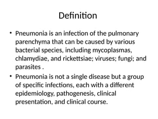
Definition
• Pneumonia isan infection of the pulmonary
parenchyma that can be caused by various
bacterial species, including mycoplasmas,
chlamydiae, and rickettsiae; viruses; fungi; and
parasites .
• Pneumonia is not a single disease but a group
of specific infections, each with a different
epidemiology, pathogenesis, clinical
presentation, and clinical course.
3.
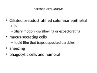
DEFENSE MECHANISMS
• Ciliatedpseudostratified columnar epithelial
cells
– ciliary motion –swallowing or expectorating
• mucus-secreting cells
– liquid film that traps deposited particles
• Sneezing
• phagocytic cells and humoral
4.
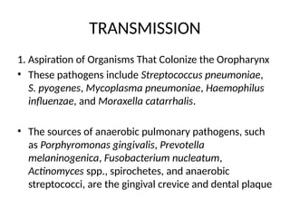
TRANSMISSION
1. Aspiration ofOrganisms That Colonize the Oropharynx
• These pathogens include Streptococcus pneumoniae,
S. pyogenes, Mycoplasma pneumoniae, Haemophilus
influenzae, and Moraxella catarrhalis.
• The sources of anaerobic pulmonary pathogens, such
as Porphyromonas gingivalis, Prevotella
melaninogenica, Fusobacterium nucleatum,
Actinomyces spp., spirochetes, and anaerobic
streptococci, are the gingival crevice and dental plaque
5.
Risk factors
• impairedlevel of consciousness ( alcoholics, drug
abusers, seizures, strokes, anaesthesia)
• Neurologic dysfunction of oropharynx
• swallowing disorders
• mechanical impediments – NGT, ETT)
• Impairment of the cough reflex
• mucociliary or alveolar macrophage dysfunction
6.
Transmission…
2. Inhalation ofInfectious Aerosols
• Particles <5 um in diameter (also called
airborne droplet nuclei) e.g tuberculosis,
influenza, legionellosis, psittacosis,
histoplasmosis, Q fever, and hantavirus
pulmonary syndrome (HPS
7.
Transmission
3. Hematogenous Disseminationfrom an
Extrapulmonary Site Infection, usually with
Staphylococcus aureus
• right- or left-sided bacterial endocarditis
• intravenous catheter infections.
• Fusobacterium infections of the retropharyngeal
tissues (Lemierre's syndrome i.e.,
retropharyngeal abscess and jugular venous
thrombophlebitis)
8.
Transmission
4. Direct Inoculationand Contiguous Spread
• tracheal intubation or stab wounds to the chest)
• contiguous spread from an adjacent site of
infection
• NB: Rheumatic fever has less possibilities of
causing pneumonia because there is no spread of
the bacteria from the infectious site as the
causation of the disease are the antibodies found
on the heart
9.
PATHOLOGY
• The pneumonicprocess may involve primarily the interstitium or the
alveoli.
• Lobar pneumonia - Involvement of an entire lobe caused by
S.pneumoniae, S.aureus, β haemolytic streptococcus, Pneumococcal
by gram negative aerobic bacteria like influenza, klebsiella,
pseudomonas and proteus. H.inlfuenza causes pnemonia below the
age of 3
• Bronchopneumonia - restricted to alveoli contiguous to bronchi,
infection of the terminal bronchioles that extend to the surrounding
alveoli resulting into patchy consolidation of te lung
• Necrotizing pneumonia (multiple small cavities, each <2 cm in
diameter,
• Lung abscess (one or more cavities >2 cm in diameter).
10.
• Pneumonia causedby aspiration of kerosene
is called lipid pneumonia
• Pneumonia caused by aspiration of gastric
contents is called chemical pneumonia
11.
Epidemiology
• The relativefrequency of various pulmonary
pathogens varies with the setting in which the
infection was acquired
– Community – community acquired pneumonia
– Nursing home
– Hospital – hospital acquired or nosocomial
infection
12.
CLINICAL MANIFESTATIONS
1. Community-AcquiredPneumonia
• Two syndromes: the typical presentation or the atypical
presentation.
• The "typical" pneumonia syndrome
– sudden onset of fever
– cough productive of purulent sputum
– shortness of breath
– pleuritic chest pain
– signs of pulmonary consolidation (dullness, increased fremitus,
egophony, bronchial breath sounds, and rales)
• Caused by the most common bacterial pathogen in community-
acquired pneumonia, S. pneumoniae
13.
CLINICAL MANIFESTATIONS
• The"atypical" pneumonia syndrome
– gradual onset, a dry cough, shortness of breath,
– a prominence of extrapulmonary symptoms (such
as headache, myalgias, fatigue, sore throat, nausea,
vomiting, and diarrhea)
– abnormalities on chest radiographs despite minimal
signs of pulmonary involvement (other than rales)
– Atypical pneumonia is classically produced by M.
pneumoniae
14.
Nosocomial Pneumonia
• Patientswith nosocomial pneumonia often pose
a diagnostic challenge.
• The differential diagnosis of acute respiratory
disease in critically ill, hospitalized patients is
diverse
• The usual criteria for nosocomial pneumonia,
which include new or progressive pulmonary
infiltrates, purulent tracheobronchial secretions,
fever, and leukocytosis, are frequently unreliable
15.
Aspiration Pneumonia andAnaerobic Lung
Abscess
• Aspiration of a sufficient volume of gastric acid
produces a chemical pneumonitis characterized by
– acute dyspnea and wheezing with hypoxemia
– infiltrates on chest radiographs in one or both lower
lobes.
• Although the aspiration of oral anaerobes can
initially lead to an infiltrative process, it ultimately
results in putrid sputum, tissue necrosis, and
pulmonary cavities.
16.
Aspiration pneumonia andAnaerobic Lung
Abscess
• In about three-quarters of cases, the clinical course
of an abscess of anaerobic polymicrobial etiology is
indolent OR SLOW and mimics that of pulmonary
tuberculosis,
• cough, shortness of breath
• Chills, fever, night sweats
• weight loss
• Pleuritic chest pain
• blood-streaked sputum lasting for several weeks or more
17.
DIAGNOSIS
• Radiography
– upto 30% of patients with PCP have false-negative
results
– can confirm the presence and location of the
pulmonary infiltrate
– assess the extent of the pulmonary infection
– detect pleural involvement, pulmonary cavitation,
or hilar lymphadenopathy
– gauge the response to antimicrobial therapy
18.
• Pneumocystic cariniicauses inhalation
pneumonia in children
• The pneumonia affects mainly
immunosupressed people eg HIV
19.
DIAGNOSIS
• Sputum Examination(Gram’s staining and
culture)
– Remains the mainstay of the evaluation of a patient
with acute bacterial pneumonia.
– Unfortunately, expectorated material is frequently
contaminated by potentially pathogenic bacteria that
colonize the upper respiratory tract without actually
causing disease.
– This contamination reduces the diagnostic specificity
of any lower respiratory tract specimen.
20.
DIAGNOSIS
Invasive Procedures
• TranstrachealAspiration (TTA)
– TTA is rarely performed today
• Percutaneous Transthoracic Lung Puncture
– This procedure employs a skinny (small-gauge)
needle that is advanced into the area of pulmonary
consolidation with computed tomographic
guidance
21.
DIAGNOSIS
Invasive Procedures
• FiberopticBronchoscopy
– Fiberoptic bronchoscopy is safe and relatively well tolerated
– Has become the standard invasive procedure used to obtain
lower respiratory tract secretions from seriously ill or
immunocompromised patients with complex or progressive
pneumonia.
– Samples are collected with
• a protected double-sheathed brush (PSB)
• bronchoalveolar lavage (BAL)
• transbronchial biopsy (TBB) at the site of pulmonary consolidation.
22.
DIAGNOSIS
• Open-Lung Biopsy
–when bronchoscopic results are unrevealing
• Other Diagnostic Tests
– Blood culture - at least two blood samples for culture should be
obtained from different venipuncture sites
– if empyema is a clinical consideration, diagnostic thoracentesis is
indicated.
– Positive blood or pleural fluid culture is generally considered diagnostic
of the etiology of pneumonia.
– Bacteremia and empyema each occur in fewer than 10 to 30% of
patients with pneumonia.
– Serologic studies (usually retrospective)
23.
TREATMENT
Community-Acquired Pneumonia: OutpatientManagement
• Treatment administered is frequently empirical (without ascertaining
the cause)
• The pathogen in such a situation is likely to be M. pneumoniae, S.
pneumoniae, or C. pneumoniae.
• In older patients with underlying chronic respiratory disease, L.
pneumophila, H. influenzae, or M. catarrhalis should also be considered.
• In patients at risk of aspiration, oral anaerobes may be involved.
• Few oral antimicrobial drugs have a reliable spectrum encompassing all
of these pathogens
• Whatever regimen is chosen, its antimicrobial activity should
encompass S. pneumoniae, the most common cause of pneumonia.
24.
TREATMENT
• Optimally, thechoice of antimicrobial drugs for
empirical therapy should be guided by local
resistance patterns, if known.
• Options for empirical antimicrobial therapy should
be modified in light of continually evolving
antimicrobial resistance patterns resulting from the
introduction of new resistant clones into the
community from other regions or the emergence of
resistant mutants under the selective pressure of
local patterns of antimicrobial use.
25.
TREATMENT
• The regimenshould be modified for patients
with particular epidemiologic factors or
comorbidities related to specific pathogens
e.g., structural lung disease or suspected
aspiration.
• Aspiration pneumonia can be treated with
amoxicillin/clavulanate, clindamycin, or
amoxicillin plus metronidazole because these
regimens are active against oral anaerobes.
26.
TREATMENT
Community-Acquired Pneumonia: InpatientManagement
• Patients who have community-acquired pneumonia and are
ill enough to be hospitalized
– must have a chest radiograph to establish the diagnosis of
pneumonia
– must undergo prompt microbiologic evaluation (including Gram's
staining and culture of sputum and culture of two blood samples
drawn by separate venipuncture)
– must receive empirical antimicrobial therapy based on Gram's
staining of sputum and knowledge of the current antimicrobial
sensitivities of the pulmonary pathogens in the local geographic
area
27.
TREATMENT
• Antimicrobial therapyshould be initiated
promptly (e.g., within 8 h of admission).
• Parenteral antimicrobial therapy in the
hospitalized patient is usually mandatory.
• A specific diagnosis should then be sought
aggressively so that optimal therapy can be
started promptly.
28.
TREATMENT
• Penicillin orampicillin remains the drug of choice
for infection due to penicillin-susceptible
pneumococci.
• Studies suggest that high-dose intravenous
penicillin G (e.g., 10 to 20 million units daily),
ampicillin (2 g every 6 h), ceftriaxone (1 or 2 g every
24 h), or cefotaxime (1 to 2 g every 6 h) constitutes
adequate therapy for pneumonia due to strains
exhibiting intermediate resistance to penicillin
29.
TREATMENT
• If theresult of Gram's staining of sputum is
not interpretable or not available, then treat
pt. with
– a b-lactam (e.g., ceftriaxone, cefotaxime) or a b-
lactam/b-lactamase inhibitor combination, with or
without a macrolide, or
– one of the fluoroquinolones alone
30.
TREATMENT
• Therapy canbe switched from intravenous to oral
agents within 3 days to complete a 7- to 10-day course
if the patient's clinical condition improves rapidly
• The presence of S. aureus or aerobic gram-negative
bacilli or the development of suppurative
complications requires a more prolonged course of
therapy.
• Pneumonia caused by Legionella, C. pneumoniae, or
Mycoplasma should be treated for 2 to 3 weeks unless
azithromycin is used.
31.
TREATMENT
• Anaerobic lungabscess should be treated with the regimens
suggested for aspiration pneumonia until a chest radiograph
(with radiography performed at 2-week intervals) is clear or
shows only a small stable scar.
• Therapy is prolonged for 6 weeks to prevent relapse, although
shorter courses are probably sufficient for many patients.
• Surgery is rarely required for lung abscess; indications for
surgery include massive hemoptysis and suspected neoplasm.
• Supportive measures include the administration of supplemental
oxygen and intravenous fluids, assistance in clearing secretions,
fiberoptic bronchoscopy, and (if necessary) ventilatory support
32.
TREATMENT
Institutionally Acquired Pneumonia
•Pneumonia acquired in institutions such as
nursing homes or hospitals is frequently caused
by enteric aerobic gram-negative bacilli, P.
aeruginosa, or S. aureus, with or without oral
anaerobes.
• Again, the selection of empirical antimicrobial
therapy should be guided by Gram's staining of
sputum
33.
PREVENTION
• The preventionof pneumonia involves either
– decreasing the likelihood of encountering the
pathogen or
– strengthening the host's response once the
pathogen is encountered.
34.
PREVENTION
– The firstapproach can include measures such as
• hand washing and glove use by persons who care for
patients infected with contact-transmitted pathogens
(e.g., aerobic gram-negative bacilli);
• use of face masks or negative-pressure isolation rooms
for patients with pneumonia due to pathogens spread
by the aerosol route (e.g., M. tuberculosis)
• prompt institution of effective chemotherapy for
patients with contagious illnesses
• correction of conditions that facilitate aspiration
35.
PREVENTION
• The secondapproach includes the use of
chemoprophylaxis or immunization for patients at risk.
• Chemoprophylaxis may be administered to patients who
have encountered or are likely to encounter the pathogen
before they become symptomatic
– amantadine during a community outbreak of influenza A
– isoniazid for tuberculosis
– TMP-SMZ (co-trimoxazole) for pneumocystosis
• Or to patients who are likely to have a recurrence following
recovery from a symptomatic episode
• TMP-SMZ for pneumocystosis in patients with HIV infection
36.
PREVENTION
• The preventionof nosocomial pneumonia requires
– good infection control practices
– judicious use of broad-spectrum antimicrobial agents
– maintenance of patients' gastric acidity a major factor
that prevents colonization of the gastrointestinal tract by
nosocomial gram-negative bacillary pathogens.
– To prevent stress ulceration, it is preferable to use
sucralfate, which maintains gastric acidity, rather than
H2-blocking agents.
37.
PREVENTION
• Vaccines areavailable for immunization against
– S. pneumoniae
– H. influenzae type b
– influenza viruses A and B
– measles virus
• Influenza vaccine is strongly recommended for individuals
> 55 years old and pneumococcal vaccine for those > 65
years old; these vaccines should be administered to
persons of any age who are at risk of adverse
consequences of influenza or pneumonia because of
underlying conditions. E.g HIV patients
Editor's Notes
#12 EGOPHONY is an increased resonance of voice sounds[1] heard when auscultating the lungs, often caused by lung consolidation and fibrosis. It is due to enhanced transmission of high-frequency noise across fluid, such as in abnormal lung tissue, with lower frequencies filtered out.
#22 Thoracentesis (pronounced /ˌθɔrəsɨnˈtiːsɨs/) (also known as thoracocentesis or pleural tap) is an invasive procedure to remove fluid or air from the pleural space for diagnostic or therapeutic purposes. A cannula, or hollow needle, is carefully introduced into the thorax, generally after administration of local anesthesia.


![CTEV [ clubfoot] DR ARUN LAL ,DR MOHAMED ASHRAF travancore medical college k...](https://cdn.slidesharecdn.com/ss_thumbnails/ctevclubfootdrarunlaldrmohamedashraftravancoremedicalcollegekollamkeralaindia-260208063247-18fc466c-thumbnail.jpg?width=640&height=640&fit=bounds)


![PERI-PROSTHETIC FRACTURE NAIL-PLATE CONSTRUCT [NPC].pptx](https://cdn.slidesharecdn.com/ss_thumbnails/drarunkumardrmohamedashrafperiprostheticfrasturenail-plateconstructnpc-260209164459-7e9d15a1-thumbnail.jpg?width=640&height=640&fit=bounds)