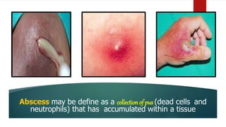
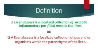
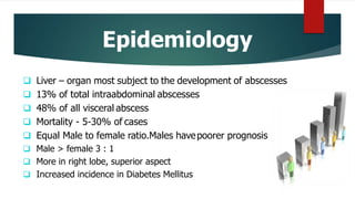
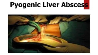
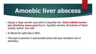
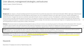
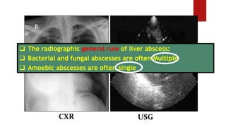
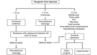
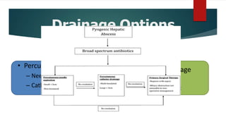
Liver Abscess
PRESENTED BY: ANJALI ARORA
M.SC. NURSING -1ST YEAR
COLLEGE OF NURSING
INSTITUTE OF LIVER AND BILIARY SCIENCES
Case -1
 A 29 year old Indian man reported a 4-day
history of RUQ abdominal pain. fever, chills and
fatigue.
 Laboratory tests
 Leukocytosis- 14040 cells/mm3
 Total bilirubin- 1.6 mg/dl,
 ALT-100 U/L, AST- 69 U/L.
 Chest X-ray - right hemidiaphragm.
 USG- 10 cm diameter lesion in the right lobe of
the liver.
 CT scan - 10 cm hypodense lesion
Case-2
 A 76 year old presented with a 5-day history of
spiking fever and chills with past medical
history of cholecystectomy.
 Laboratory tests
 C Reactive Protein 31 mg/dl
 S.creatinine-1.8 mg/dl
 Alkaline phosphatase (823 U/L)
 Total bilirubin- 2.12mg/dl,
 ALT-110 U/L, AST- 139 U/L.
 Chest X-ray - right hemidiaphragm.
 USG- 3 cm diameter lesion in the right lobe of
the liver.
 CT scan - 3cm hypodense lesion
Anatomy of liver
Introduction
Hepatic abscess (HA) is a localized process of
encapsulated suppurative material.
Although rare but it is associated with morbidity and
mortality figures of 2-12%.
Introduction
 Liver abscess usually develops in association with one of the following
three conditions:
• Bacterial cholangitis, which results from obstruction of the bile ducts
by stone or stricture
• Portal vein bacteraemia, which may develop following bowel
inflammation or organ perforation
• Amebiasis (infection with amoeba)
Abscess may be define as a collectionof pus (dead cells and
neutrophils) that has accumulated within a tissue
Definition
 Liver abscess is a localized collection of necrotic
inflammatory pus-filled mass in the liver.
OR
 A liver abscess is a localized collection of pus and or
organisms within the parenchyma of the liver.
Historical facts
 Liver abscess has long been recognized
since the age of Hippocrates (400 BC).
 In 1883, Koch described the amoebae as
a cause of liver abscess, and the first
published review was in 1936 by Bright.
 In 1938, Ochsner and Debakey
published the largest series of pyogenic
and amebic liver abscesses in the
literature.
Epidemiology
 Liver – organ most subject to the development of abscesses
 13% of total intraabdominal abscesses
 48% of all visceral abscess
 Mortality - 5-30% of cases
 Equal Male to female ratio.Males havepoorer prognosis
 Male > female 3 : 1
 More in right lobe, superior aspect
 Increased incidence in Diabetes Mellitus
Liver abscess
 A liver abscess is a collection of pus in the liver caused by bacteria, fungi, or
parasites. It may occur as a single lesion or as multiple lesions of different
sizes. The abscess may contain thick, bad smelling pus or reddish-brown
anchovy paste-like fluid with no odor.
 Occurs when bacteria/protozoa destroy hepatic tissue, produces a cavity which
fills up with infective organisms, liquefied cells & leucocytes. Necrotic tissue
then falls off the cavity from rest of the liver.
More common in Right lobe
WHY?
Liver Abscess
 Left lobe receives blood from
– Inferior mesenteric
– Splenic veins
 Right lobe receives blood from
– Superior mesenteric
– Portal veins
Streaming effect in portal circulation is causative.
Risk factors
 Age: Advanced age, particularly in people
older than 70 years.
 Health: Having a long-term disease,
(cancer, diabetes, tuberculosis) or
splenectomy, a weak immune system,
AIDS.
 Taking Drugs: Such as steroids,
chemotherapy.
 Lifestyle: Drinking too much alcohol, too
often. Living in over crowding area, poor
sanitation
 Nutrition: Being malnourished (having poor
nutrition).
 Activity: Traveling to places where amebiasis
is common. Eating foods and drinking liquids
that are sold in the street may further
increase risk.
 Liver abscess usually develops in
association with one of the
following three conditions:
• Bacterial cholangitis
• Portal vein bacteraemia
• Amebiasis.
Others:
1.Infected hepatic cyst
2.Metastatic liver tumours
3.Diverticulitis
Etiology
Pathophysiology
Whenever an infection develops, infecting organisms may reach
the liver.
Most bacteria are destroyed promptly, but occasionally some
gain a foothold.
However, Kupffer cells lining the hepatic sinusoids clear pathogens so efficiently that
infection rarely occurs but when pathogens exceed ,they destroy hepatic tissue.
Pathophysiology
The bacterial toxins destroy the neighbouring liver cells
Meanwhile, leukocytes migrate into the infected area and produce a Necrotic
filled cavity, with the infectious organism,
The result is an abscess cavity full of a liquid containing living and dead
leukocytes, liquefied liver cells, and bacteria.
liquefied liver cells and leukocytes, which walls off from the rest of the
liver forming a liverabscess
Classification
Based on cause
Based on the duration
Based on Associated
conditions
CLASSIFICATION
Classification
Based on cause
CLASSIFICATION
Pyogenic Liver Abscess
Amoebic Liver Abscess
Fungal Liver Abscess
Parasitic liver Abscess
TB related Liver Abscess-Rare
Classification
Based on the duration
CLASSIFICATION
Acute Liver abscess
Chronic Liver abscess
Classification
Based on Associated
conditions
CLASSIFICATION
Primary liver abscess
Secondary liver abscess
Classification
Based on cause
Based on the duration
Based on Associated
conditions
CLASSIFICATION
Pyogenic liver
abscess
Pyogenic Liver Abscess
 Pyogenic liver abscess is most often polymicrobial, accounts
for 80% of hepatic abscess cases in the United States.
 A pyogenic liver abscess is a type of liver abscess caused by
bacteria, can be single or multiple.
 The cluster sign is characteristic of the PHA, consisting of
several lesions grouping to form a single multiloculated cavity.
Pyogenic Liver Abscess
CLUSTER SIGN
Amoebic liver
abscess
Amoebic liver abscess
Amoebic liver abscess due to Entamoebahistolytica accounts for 10% of
cases.
 E. histolytica causes amoebic colitis and dysentery but liver abscess is the
most common extra-intestinal manifestation of infection
 Route of entry via oro-fecal route by ingestion of contaminated food or
water. Amoebae invade intestinal mucosa and can gain access to the portal
venous system.
Amoebic liver abscess
 Causes a large necrotic area which is liquefied into thick reddish-brown
pus (Anchovy sauce pus) due to liquefied necrosis, thrombosis of blood
vessels, lysisof liver cells
 It affects the right lobe in 80%.
 This type is common in overcrowded areas with poor sanitation and in
alcoholics.
Surgical piece of hepatectomy by PHA
Well circumscribed cavity, with uneven contours, with dark material
(“anchovy paste”) content.
Parasitic liver
abscess
Parasitic Liver abscess
1. Parasitic protozoa: Entamoeba histolytica (10%).
2. Parasitic helminths: Hepatic abscess is rarely associated with it.
 Mode of transmission:
 Ingestion of cysts.
Parasitic Liver abscess
Pathology: Two-stage life cycle.
 The trophozoite (ameba stage) is motile. The cyst stage is nonmotile.
 Trophozoites are found in the intestinal and extraintestinal lesions.
 Cysts predominate in the stools, with some trophozoites present.
Fungal liver abscess
 Fungal abscess, most often due to Candida species, accounts for less than 10%
of cases.
 Fungal abscesses is a less common type, primarily due to Candida albicans and
occur in individuals with :
 Prolonged exposure to antimicrobials
 Hematologic malignancies
 Solid-organ transplants
 Congenital and acquired immunodeficiency.
Classification
Based on cause
Based on the duration
Based on Associated
conditions
CLASSIFICATION
Liver Abscess
 Based on the duration of symptoms
 Acute (Less than a month)
 Chronic (Greater than a month)
Classification
Based on cause
Based on the duration
Based on Associated
conditions
CLASSIFICATION
Liver Abscess
 Based on Associated conditions
 Primary Liver Abscess: if patient was previously healthy
 Secondary Liver Abscess: When associated with any systemic diseases
or risk factors that could compromise the immune system such as HIV,
infection, Neoplasm etc.
Route of Infection
1) Biliary tract
2) Hematogenous (via hepatic artery)
3) Cryptogenic abscesses
4) Blunt or penetrating trauma
5) Direct infiltration and penetrating injuries tend
to cause a large abscess
Clinical Manifestations
Fever (either continuous or spiking),(PUO in AmoebicAbscess)
Chills
Right upper quadrant pain
Pleuritic chest pain
Anorexia
Malaise
Diarrhoea is present in 1/3 patients with amoebic liver abscess
Clay-colored stools
Clinical Manifestations
 Abdominal pain
 Weight loss
 Nausea, Vomiting
 Right shoulder pain
 Cough and Dyspnoea (due to hepatic friction rub associated with
diaphragmatic irritation or inflammation of Glisson capsule)
 Hepatomegaly
 Tenderness
 Rebound tenderness
 Jaundice (late)
https://youtu.be/LHqqvrm2j6g
Basilar rales (crackles)
https://youtu.be/LHqqvrm2j6g
Assessment
History Collection
Physical Examination
Diagnostic evaluation
 NON SPECIPIC
 Increase TLC - Leucocytosis
 Increase LFT’s
 SPECIFIC
 USG
 CT SCAN
 Image guided Aspiration
 Culture and sensitivity
 Fluorescent antibody test for Entamoeba(can be positive even after clinical
cure)
 ELISA should be performed–to detect E histolytica
 Indirect Haemagglutinin assays (IHA)
Imaging studies
Diagnostic evaluation
 CBC
 Increased WBC, usually NeutrophilicLeukocytosis.
 Raised erythrocyte sedimentation rate(ESR).
 Mild normochromic normocyticanaemia.
 Liver functionstudies
 Hypoalbuminemia
 Elevation of alkalinephosphatase
 Elevations of transaminase and bilirubin levels(variable)
Diagnostic evaluation
 Blood cultures are positive in roughly 50% of cases.
 Stool Analysis: Stools can contain cysts or trophozoites of E. histolytica.
 Serology should be carried out if E. histolyticais suspected.
 Culture of abscess fluid should be the goal in establishing
microbiologic diagnosis. Usually done through Percutaneous needle
aspiration (under CTor Ultrasound Guidance)
Diagnostic evaluation
1. ChestX-Ray: May show raised right hemi-diaphragm on.
2. Ultrasonography
a) Can show abscess and also allow guided percutaneous aspiration and drainage and biliary
tree examination. A Doppler ultrasound study may be done to check for blood flow in
your liver.
3. CTscanning
a) Can show the abscess, allow guided aspiration and drainage and showother intra-
abdominal abscessesora possiblecause such as diverticulardisease, appendicitis, etc. It is
good forthedetection of small abscesses.
4. MRI
Diagnostic evaluation
 Microscopic stool examinations for trophozoites and cysts are
performed in 3 to 6 stool samples for concentration and specificity and are
positive in 25% cases.
WHO recommends that intestinal amoebiasis should be
diagnosed with specific stool E. histolytica testing (such as
cultures, serology, antigen testing, and Enzyme immunoassay,
hemagglutination tests or PCR) rather than microscopy for ova
and cysts.
CXR USG
 The radiographic general rule of liver abscess:
 Bacterial and fungal abscesses are often Multiple
 Amoebic abscesses are often single
X-Ray features
 Gas within the abscess cavity,
 Gas in biliary tree (pneumobilia) or
 Gas beneath the diaphragm (elevating the diaphragm)
 right pleural effusion
USG features
 Intrahepatic abscesses larger than 2 cm
 Hypoechoic round or oval lesions (still with some internal
echoes however), and irregularly shaped well defined borders
 Limitation in abscesses located in the dome of the liver and
liquefied necrotic area does not enhance
USG features for Amoebic
abscess
 Round or oval shaped lesion
 Absence of prominent abscess wall
 Hypoechogenicity
 Contiguity with diaphragm
USG features for candida
abscess
 USG features
 Wheel within a wheel
 Bull’s eye
 Uniformly hypoechoic
 Echogenic
BULL’S EYE
USG of amebic abscess
Note: Peripheral location, rounded shape, poor rim with internal echoes
CT features
 CT scan is superior in detecting abscess(es) as small as 0.5 cm
and reveals the abscess as:
 Lobar involvement
 Solid appearance (mimicking a hepatic tumour)
 Contain gas in 20% of lesions
 Association with thrombophlebitis
(a) Contrast-enhanced CT scan demonstrates a large, lobulated,well-
defined cystic mass in the right hepatic lobe.
Double ring /double target sign
CT Scan
CT scan of amebic abscess
Note: The lesion is peripherally located and round. Rim is non enhancing but shows
peripheral edema (black arrows).
Note the extension into the intercostal space (white arrows).
Faint rim enhancement and perilesional edema
(CLUSTER SIGN)
CT SCAN (Candidiasis) MRI (Candidiasis)
Contrast-enhanced CT scan shows multiple hypo attenuating micro abscesses (<1 cm)
MRI
Imaging difference in amoebic and
bacterial liver abscess
Management
Principles of Management
• Drain the pus
• Institute appropriate antibiotics, and
• Deal with any underlying source of infection,
Percutaneous drainage combined with antibiotics has become the first line
and mainstay of treatment for most PLAs
Management
 Intervention in hepatic abscess consists of:
(1) Percutaneous drainage of the abscess with antimicrobial therapy,
(2) Surgical drainage of large abscess with post operative antimicrobial
therapy
(3) Antimicrobial therapy without drainage for a few months.
 Abscesses resulting from amoebic infestation (such as by Entamoeba
histolytica) call for treatment with metronidazole (Flagyl) or chloroquine
phosphate instead of broad-spectrum antibiotics.
Medical Management
 Antibiotic therapy
 Continuous supportive care
 Open surgical drainage may be required.
 Percutaneous drainage are ineffective.
Antibiotic therapy
• Antibiotic therapy should cover gram negative organisms and
anaerobes
• First line antibiotics are
– Penicillin's, aminoglycosides and metronidazole or
– Cephalosporin and metronidazole
• Can be changed after Culture report
• IV antibiotictherapy should be
continued for at least 8 weeks
• Some studies suggest antibiotics should be administered parenterally
for 2 weeks
• Then appropriate oral agents may be used for a further 6 weeks
Empirical antibiotic Regimen
Management
 Pain Management: Alleviation or reduction in pain
 Nutrition Management: Assisting with orproviding a balanced dietary
intake of foodsand fluids.
 Infection Protection: Infection Control, Prevention and early detection of
infection in a patient at risk.
Pyogenic liver abscess
 Broad spectrum antibioticsshould be started before waiting for
culture results.
 Usually start treatment with tri-therapy included the use of
penicillin, amino-glycoside and metronidazole.
 A third-generation cephalosporin can be considered in the
elderly or if renal function is impaired.
 Antibiotic therapy can be modified once culture results are
available.
 Treatment may be needed for up to 12 weeks and should be guided
by the clinical picture and radiological monitoring.
Amoebic liver abscess
Metronidazole is the treatment of choice. Most patients show a
response to treatment within 72-96 hours.
Diloxanide furoate should be prescribed for 10 days to eliminate
intestinal amoebae after the abscess has been successfully treated.
Antifungal agents such as amphotericin B are used if fungal
abscess is suspected.
Algorithm
INTESTINAL OBSTRUCTION
Drainage Options
• Percutaneous
– Needle aspiration
– Catheter drainage
• Surgical drainage
– Open
– Laparoscopic
Percutaneous needle
aspiration
• Under CT or USG guidance, needle aspiration of cavity material can
be performed.
• Needle aspiration enables
– rapid recovery of material for microbiologic and pathologic
evaluation.
• Large percentage requires second or third aspirations to
achieve success
Percutaneous catheter
drainage
• Percutaneous drainage has become the standard of
care.
• Should be the first intervention considered for
– Small cysts.
– The pus is too thick to be aspirated
– The wall is thick and non-collapsible
– The PLA is multi-loculated
Advantages
 reduced costs, recovery time
 it eliminates the need for general anesthesia
 This also allows for gradual, controlled drainage
Percutaneous catheter
drainage
• A catheter is placed under ultrasonographic or CT guidance via
the Seldinger or trocar techniques.
• The catheter is flushed daily until output is less than 10
mL/day or cavity collapse is documented by serial CT.
Surgical drainage
Indications
– Failure of non operative treatment
– Intraperitoneal rupture
– the presence of a complicated, multiloculated, thick-walled
abscess with viscous pus
– treatment of underlying intra-abdominal processes,
• peritonitis;
• existence of a known abdominal surgical pathology (eg, diverticular
abscess)
Approaches
(Open)
• A transperitoneal approach
– allows for abscess drainage and
– abdominal exploration to identify previously undetected abscesses and
the location of an etiologic source
• Transpleural approach
– For high posterior lesions,
– easier access to the abscess,
– the identification of multiple lesions or a concurrent intra-abdominal
pathology is lost
Laparoscopic approach
• Used in selected cases
• Experienced and well equipped setups
Surgical Management
Surgical therapy is necessary for multiple macroscopic or multiloculated
abscesses, or those in the left lobe, after percutaneous drainage failure.
 Hepatectomy (Hemi/Segmental Resection)
Complication of Liver
abscess
 Rupture and peritonitis
 Rupture into lung
 Bronchopleural fistula
 Subphrenic abscess
 Retroperitoneal abscess
 Cardiac tamponade
 Septicaemia
 Encephalopathy
 liver failure
 Liver cysts
Nursing Management
Assessment
 History
 Physical examination
Assess any
pre-existing
illness
Nursing Diagnosis
 Ineffective breathing pattern related to restriction of thoracic excursion
as evidenced by dyspnoea.
 Acute Pain related to tissue as evidenced by facial expressions and
verbalization.
 Impaired thermoregulatory pattern related to inflammation as
evidenced by Fever.
Nursing Diagnosis
 Impaired fluid and electrolytes related to in-situ drainage as evidenced by
dehydration
 Impaired skin integrity related to pruritus from jaundice and edema.
 Activity intolerance related to fatigue, lethargy, and malaise.
Nursing Diagnosis
 Imbalanced nutrition: less than body requirements related to anorexia
as evidenced by less intake of food.
 Risk for infection related to open wound
 Risk for injury related to altered clotting mechanisms and altered level of
consciousness.
Planning
 Have adequate relief from the underlying health problem
 Have satisfactory pain management
 Cope with the body image changes
 Make satisfying lifestyle adjustment
Nursing Considerations
Intervention
 Health Promotion
 Acute Care
 Pre-operative care
 Post operative care
 Ambulatory care
 Special considerations
Health Promotion
 Prompt treatment of abdominal and other infections.
 Educating and exercising proper sanitary measures
 Avoiding fecal contamination of food and water
 Boiling is the only effective means of eradicating cyst in water.
 Travelers should avoid drinking local water including ice cubes frequently
used for cocktails
 Eat a variety of healthy foods.
 Do not drink alcohol.
Nursing Management
 The nursing management depends on the patient's physical status and
the medical management that is indicated.
 For patients who undergo evacuation and drainage of an abscess,
monitoring of the drainage and skin care are imperative.
 Vital signs are monitored to detect changes in the patient's physical
status. Deterioration in vital signs or the onset of new symptoms such
as increasing pain, which may indicate rupture or extension of the
abscess, is reported promptly.
 The nurse administers IV antibiotic therapy as prescribed.
Nursing Management
 The white blood cell count and other laboratory test results are monitored closely
for changes consistent with worsening infection.
 The nurse prepares the patient for discharge by providing instruction about symptom
management, signs and symptoms that should be reported to the physician, management of
drainage, and the importance of taking antibiotics as prescribed.
 Encourage movement, coughing, and deep breathing to prevent or limit pulmonary
complications related to hepatic abscess.
 Increase the client's fluid in take and provide skin care in the event of hyperpyrexia.
Dispose of faeces carefully, and wash your hands to prevent transmission of amoebic
infestations.
Video
 https://media.springernature.com/original/springer-static/image/chp%3A10.1007%2F978-1-4614-6123-
4_22/MediaObjects/217224_1_En_22_Fig2_HTML.gif
 https://www.youtube.com/watch?v=Zcbra9s2Mpk
 https://www.youtube.com/watch?v=fWSxYxDL_qI
Evaluation
 Accept and Integrate changes into lifestyle
 Have no evidence of open skin breakdown
 Have reduction or absence of pain
 Become mobile within limitations.
Documentation Guidelines
 Condition of the surgical site
 Control of initial postoperative pain by Pain Medication
 Presence of any complication
Discharge guidelines
 The clinical and radiographic progress of the patient should guide the
length of therapy.
 Intravenous medication should be administered for 14 days, and then
replaced with oral medications to complete a 4-6 week course following
adequate drainage.
 Multiple abscesses require up to 12 weeks of therapy.
 A 10-day course of Diloxanide furoate should be given at the completion of
metronidazole to destroy any amoebae in the gut.
Follow up  Patients prolonged parenteral medications may
continue after discharge.
 Monitoring of vital organ functions and
complete blood count will be needed.
 Imaging studies
Long-Term Monitoring
 Weekly serial computed tomography (CT) or
ultrasound examinations to document adequate
drainage
 Maintain drains until the output is less than 10
mL/day
 Monitor fever curves.
 Persistent fever after 2 weeks of therapy may
indicate the need for more aggressive drainage.
Prognosis
 With better diagnostic techniques and early treatment Prognosis is good as
abscess resolves completely within 8 months to 2 years.
 Co-morbidities could alter the prognosis.
 Recurrence is common if pathogen(s)-causing abscess are not completely
eradicated.
 With treatment, (despite co-morbidities) mortality is less than 15%.
But without treatment, the mortality rate approaches 100%.
 The most common causes of death include sepsis, multi-organ failure and
hepatic failure. Also mortality has increased significantly with laparotomy
Summary
Conclusion
 Liver abscesses are of significant relevance, which is related to the high
morbidity and mortality figures which they can cause if not detected
and treated in time.
 If left untreated, these lesions are invariably fatal.
References
Abbas, M. T.; Khan, F. Y.; Muhsin, S. A.; Al-Dehwe, B.; Abukamar, M. & Elzouki, A. N. Epidemiology, clinical
features and outcome of liver abscess: a single reference center experience in Qatar. Oman Med. J.,
29(4):260-3, 2014.
Akhondi, H. & Sabih, D. E. Liver Abscess. StatPearls website. Treasure Island (FL), StatPearls Publishing,
2019.
Alam, F.; Salam, M. A.; Hassan, P.; Mahmood, I.; Kabir, M. & Haque, R. Amebic liver abscess in Northern
Region of Bangladesh: sociodemographic determinants and clinical outcomes. BMC Res. Notes, 7:625, 2014.
Alghofaily, K. A.; Saeedan, M. B.; Aljohani, I. M.; Alrasheed, M.; McWilliams, S.; Aldosary, A. & Neimatallah,
M. Hepatic hydatid disease complications: review of imaging findings and clinical implications. Abdom.
Radiol. (N. Y.), 42(1):199-210, 2017.
Anesi, J. A.; & Gluckman, S. Amebic liver abscess. Clin. Liver Dis. (Hoboken), 6(2):41-3, 2015. [ Links ]
Barosa, R.; Pinto, J.; Caldeira, A. & Pereira, E. Modern role of clinical ultrasound in liver abscess and
echinococcosis. J. Med. Ultrason. (2001), 44(3):239-45, 2017.
Thank you

Liver abscess

  • 1.
    Liver Abscess PRESENTED BY:ANJALI ARORA M.SC. NURSING -1ST YEAR COLLEGE OF NURSING INSTITUTE OF LIVER AND BILIARY SCIENCES
  • 2.
    Case -1  A29 year old Indian man reported a 4-day history of RUQ abdominal pain. fever, chills and fatigue.  Laboratory tests  Leukocytosis- 14040 cells/mm3  Total bilirubin- 1.6 mg/dl,  ALT-100 U/L, AST- 69 U/L.  Chest X-ray - right hemidiaphragm.  USG- 10 cm diameter lesion in the right lobe of the liver.  CT scan - 10 cm hypodense lesion
  • 3.
    Case-2  A 76year old presented with a 5-day history of spiking fever and chills with past medical history of cholecystectomy.  Laboratory tests  C Reactive Protein 31 mg/dl  S.creatinine-1.8 mg/dl  Alkaline phosphatase (823 U/L)  Total bilirubin- 2.12mg/dl,  ALT-110 U/L, AST- 139 U/L.  Chest X-ray - right hemidiaphragm.  USG- 3 cm diameter lesion in the right lobe of the liver.  CT scan - 3cm hypodense lesion
  • 4.
  • 7.
    Introduction Hepatic abscess (HA)is a localized process of encapsulated suppurative material. Although rare but it is associated with morbidity and mortality figures of 2-12%.
  • 8.
    Introduction  Liver abscessusually develops in association with one of the following three conditions: • Bacterial cholangitis, which results from obstruction of the bile ducts by stone or stricture • Portal vein bacteraemia, which may develop following bowel inflammation or organ perforation • Amebiasis (infection with amoeba)
  • 9.
    Abscess may bedefine as a collectionof pus (dead cells and neutrophils) that has accumulated within a tissue
  • 10.
    Definition  Liver abscessis a localized collection of necrotic inflammatory pus-filled mass in the liver. OR  A liver abscess is a localized collection of pus and or organisms within the parenchyma of the liver.
  • 11.
    Historical facts  Liverabscess has long been recognized since the age of Hippocrates (400 BC).  In 1883, Koch described the amoebae as a cause of liver abscess, and the first published review was in 1936 by Bright.  In 1938, Ochsner and Debakey published the largest series of pyogenic and amebic liver abscesses in the literature.
  • 12.
    Epidemiology  Liver –organ most subject to the development of abscesses  13% of total intraabdominal abscesses  48% of all visceral abscess  Mortality - 5-30% of cases  Equal Male to female ratio.Males havepoorer prognosis  Male > female 3 : 1  More in right lobe, superior aspect  Increased incidence in Diabetes Mellitus
  • 13.
    Liver abscess  Aliver abscess is a collection of pus in the liver caused by bacteria, fungi, or parasites. It may occur as a single lesion or as multiple lesions of different sizes. The abscess may contain thick, bad smelling pus or reddish-brown anchovy paste-like fluid with no odor.  Occurs when bacteria/protozoa destroy hepatic tissue, produces a cavity which fills up with infective organisms, liquefied cells & leucocytes. Necrotic tissue then falls off the cavity from rest of the liver.
  • 14.
    More common inRight lobe WHY?
  • 15.
    Liver Abscess  Leftlobe receives blood from – Inferior mesenteric – Splenic veins  Right lobe receives blood from – Superior mesenteric – Portal veins Streaming effect in portal circulation is causative.
  • 20.
    Risk factors  Age:Advanced age, particularly in people older than 70 years.  Health: Having a long-term disease, (cancer, diabetes, tuberculosis) or splenectomy, a weak immune system, AIDS.  Taking Drugs: Such as steroids, chemotherapy.  Lifestyle: Drinking too much alcohol, too often. Living in over crowding area, poor sanitation  Nutrition: Being malnourished (having poor nutrition).  Activity: Traveling to places where amebiasis is common. Eating foods and drinking liquids that are sold in the street may further increase risk.
  • 21.
     Liver abscessusually develops in association with one of the following three conditions: • Bacterial cholangitis • Portal vein bacteraemia • Amebiasis. Others: 1.Infected hepatic cyst 2.Metastatic liver tumours 3.Diverticulitis Etiology
  • 22.
    Pathophysiology Whenever an infectiondevelops, infecting organisms may reach the liver. Most bacteria are destroyed promptly, but occasionally some gain a foothold. However, Kupffer cells lining the hepatic sinusoids clear pathogens so efficiently that infection rarely occurs but when pathogens exceed ,they destroy hepatic tissue.
  • 23.
    Pathophysiology The bacterial toxinsdestroy the neighbouring liver cells Meanwhile, leukocytes migrate into the infected area and produce a Necrotic filled cavity, with the infectious organism, The result is an abscess cavity full of a liquid containing living and dead leukocytes, liquefied liver cells, and bacteria. liquefied liver cells and leukocytes, which walls off from the rest of the liver forming a liverabscess
  • 24.
    Classification Based on cause Basedon the duration Based on Associated conditions CLASSIFICATION
  • 25.
    Classification Based on cause CLASSIFICATION PyogenicLiver Abscess Amoebic Liver Abscess Fungal Liver Abscess Parasitic liver Abscess TB related Liver Abscess-Rare
  • 26.
    Classification Based on theduration CLASSIFICATION Acute Liver abscess Chronic Liver abscess
  • 27.
  • 28.
    Classification Based on cause Basedon the duration Based on Associated conditions CLASSIFICATION
  • 29.
  • 30.
    Pyogenic Liver Abscess Pyogenic liver abscess is most often polymicrobial, accounts for 80% of hepatic abscess cases in the United States.  A pyogenic liver abscess is a type of liver abscess caused by bacteria, can be single or multiple.  The cluster sign is characteristic of the PHA, consisting of several lesions grouping to form a single multiloculated cavity.
  • 31.
  • 32.
  • 33.
  • 34.
    Amoebic liver abscess Amoebicliver abscess due to Entamoebahistolytica accounts for 10% of cases.  E. histolytica causes amoebic colitis and dysentery but liver abscess is the most common extra-intestinal manifestation of infection  Route of entry via oro-fecal route by ingestion of contaminated food or water. Amoebae invade intestinal mucosa and can gain access to the portal venous system.
  • 35.
    Amoebic liver abscess Causes a large necrotic area which is liquefied into thick reddish-brown pus (Anchovy sauce pus) due to liquefied necrosis, thrombosis of blood vessels, lysisof liver cells  It affects the right lobe in 80%.  This type is common in overcrowded areas with poor sanitation and in alcoholics.
  • 37.
    Surgical piece ofhepatectomy by PHA Well circumscribed cavity, with uneven contours, with dark material (“anchovy paste”) content.
  • 38.
  • 39.
    Parasitic Liver abscess 1.Parasitic protozoa: Entamoeba histolytica (10%). 2. Parasitic helminths: Hepatic abscess is rarely associated with it.  Mode of transmission:  Ingestion of cysts.
  • 40.
    Parasitic Liver abscess Pathology:Two-stage life cycle.  The trophozoite (ameba stage) is motile. The cyst stage is nonmotile.  Trophozoites are found in the intestinal and extraintestinal lesions.  Cysts predominate in the stools, with some trophozoites present.
  • 42.
  • 43.
    Fungal liver abscess Fungal abscess, most often due to Candida species, accounts for less than 10% of cases.  Fungal abscesses is a less common type, primarily due to Candida albicans and occur in individuals with :  Prolonged exposure to antimicrobials  Hematologic malignancies  Solid-organ transplants  Congenital and acquired immunodeficiency.
  • 44.
    Classification Based on cause Basedon the duration Based on Associated conditions CLASSIFICATION
  • 45.
    Liver Abscess  Basedon the duration of symptoms  Acute (Less than a month)  Chronic (Greater than a month)
  • 46.
    Classification Based on cause Basedon the duration Based on Associated conditions CLASSIFICATION
  • 47.
    Liver Abscess  Basedon Associated conditions  Primary Liver Abscess: if patient was previously healthy  Secondary Liver Abscess: When associated with any systemic diseases or risk factors that could compromise the immune system such as HIV, infection, Neoplasm etc.
  • 48.
    Route of Infection 1)Biliary tract 2) Hematogenous (via hepatic artery) 3) Cryptogenic abscesses 4) Blunt or penetrating trauma 5) Direct infiltration and penetrating injuries tend to cause a large abscess
  • 50.
    Clinical Manifestations Fever (eithercontinuous or spiking),(PUO in AmoebicAbscess) Chills Right upper quadrant pain Pleuritic chest pain Anorexia Malaise Diarrhoea is present in 1/3 patients with amoebic liver abscess Clay-colored stools
  • 51.
    Clinical Manifestations  Abdominalpain  Weight loss  Nausea, Vomiting  Right shoulder pain  Cough and Dyspnoea (due to hepatic friction rub associated with diaphragmatic irritation or inflammation of Glisson capsule)  Hepatomegaly  Tenderness  Rebound tenderness  Jaundice (late)
  • 52.
  • 53.
  • 55.
  • 56.
    Diagnostic evaluation  NONSPECIPIC  Increase TLC - Leucocytosis  Increase LFT’s  SPECIFIC  USG  CT SCAN  Image guided Aspiration  Culture and sensitivity  Fluorescent antibody test for Entamoeba(can be positive even after clinical cure)  ELISA should be performed–to detect E histolytica  Indirect Haemagglutinin assays (IHA)
  • 57.
  • 58.
    Diagnostic evaluation  CBC Increased WBC, usually NeutrophilicLeukocytosis.  Raised erythrocyte sedimentation rate(ESR).  Mild normochromic normocyticanaemia.  Liver functionstudies  Hypoalbuminemia  Elevation of alkalinephosphatase  Elevations of transaminase and bilirubin levels(variable)
  • 59.
    Diagnostic evaluation  Bloodcultures are positive in roughly 50% of cases.  Stool Analysis: Stools can contain cysts or trophozoites of E. histolytica.  Serology should be carried out if E. histolyticais suspected.  Culture of abscess fluid should be the goal in establishing microbiologic diagnosis. Usually done through Percutaneous needle aspiration (under CTor Ultrasound Guidance)
  • 60.
    Diagnostic evaluation 1. ChestX-Ray:May show raised right hemi-diaphragm on. 2. Ultrasonography a) Can show abscess and also allow guided percutaneous aspiration and drainage and biliary tree examination. A Doppler ultrasound study may be done to check for blood flow in your liver. 3. CTscanning a) Can show the abscess, allow guided aspiration and drainage and showother intra- abdominal abscessesora possiblecause such as diverticulardisease, appendicitis, etc. It is good forthedetection of small abscesses. 4. MRI
  • 61.
    Diagnostic evaluation  Microscopicstool examinations for trophozoites and cysts are performed in 3 to 6 stool samples for concentration and specificity and are positive in 25% cases. WHO recommends that intestinal amoebiasis should be diagnosed with specific stool E. histolytica testing (such as cultures, serology, antigen testing, and Enzyme immunoassay, hemagglutination tests or PCR) rather than microscopy for ova and cysts.
  • 62.
    CXR USG  Theradiographic general rule of liver abscess:  Bacterial and fungal abscesses are often Multiple  Amoebic abscesses are often single
  • 63.
    X-Ray features  Gaswithin the abscess cavity,  Gas in biliary tree (pneumobilia) or  Gas beneath the diaphragm (elevating the diaphragm)  right pleural effusion
  • 64.
    USG features  Intrahepaticabscesses larger than 2 cm  Hypoechoic round or oval lesions (still with some internal echoes however), and irregularly shaped well defined borders  Limitation in abscesses located in the dome of the liver and liquefied necrotic area does not enhance
  • 65.
    USG features forAmoebic abscess  Round or oval shaped lesion  Absence of prominent abscess wall  Hypoechogenicity  Contiguity with diaphragm
  • 66.
    USG features forcandida abscess  USG features  Wheel within a wheel  Bull’s eye  Uniformly hypoechoic  Echogenic BULL’S EYE
  • 67.
    USG of amebicabscess Note: Peripheral location, rounded shape, poor rim with internal echoes
  • 68.
    CT features  CTscan is superior in detecting abscess(es) as small as 0.5 cm and reveals the abscess as:  Lobar involvement  Solid appearance (mimicking a hepatic tumour)  Contain gas in 20% of lesions  Association with thrombophlebitis (a) Contrast-enhanced CT scan demonstrates a large, lobulated,well- defined cystic mass in the right hepatic lobe. Double ring /double target sign
  • 69.
  • 70.
    CT scan ofamebic abscess Note: The lesion is peripherally located and round. Rim is non enhancing but shows peripheral edema (black arrows). Note the extension into the intercostal space (white arrows).
  • 71.
    Faint rim enhancementand perilesional edema (CLUSTER SIGN)
  • 72.
    CT SCAN (Candidiasis)MRI (Candidiasis) Contrast-enhanced CT scan shows multiple hypo attenuating micro abscesses (<1 cm)
  • 73.
  • 74.
    Imaging difference inamoebic and bacterial liver abscess
  • 76.
  • 77.
    Principles of Management •Drain the pus • Institute appropriate antibiotics, and • Deal with any underlying source of infection, Percutaneous drainage combined with antibiotics has become the first line and mainstay of treatment for most PLAs
  • 78.
    Management  Intervention inhepatic abscess consists of: (1) Percutaneous drainage of the abscess with antimicrobial therapy, (2) Surgical drainage of large abscess with post operative antimicrobial therapy (3) Antimicrobial therapy without drainage for a few months.  Abscesses resulting from amoebic infestation (such as by Entamoeba histolytica) call for treatment with metronidazole (Flagyl) or chloroquine phosphate instead of broad-spectrum antibiotics.
  • 79.
    Medical Management  Antibiotictherapy  Continuous supportive care  Open surgical drainage may be required.  Percutaneous drainage are ineffective.
  • 80.
    Antibiotic therapy • Antibiotictherapy should cover gram negative organisms and anaerobes • First line antibiotics are – Penicillin's, aminoglycosides and metronidazole or – Cephalosporin and metronidazole • Can be changed after Culture report
  • 81.
    • IV antibiotictherapyshould be continued for at least 8 weeks • Some studies suggest antibiotics should be administered parenterally for 2 weeks • Then appropriate oral agents may be used for a further 6 weeks
  • 82.
  • 83.
    Management  Pain Management:Alleviation or reduction in pain  Nutrition Management: Assisting with orproviding a balanced dietary intake of foodsand fluids.  Infection Protection: Infection Control, Prevention and early detection of infection in a patient at risk.
  • 84.
    Pyogenic liver abscess Broad spectrum antibioticsshould be started before waiting for culture results.  Usually start treatment with tri-therapy included the use of penicillin, amino-glycoside and metronidazole.  A third-generation cephalosporin can be considered in the elderly or if renal function is impaired.  Antibiotic therapy can be modified once culture results are available.  Treatment may be needed for up to 12 weeks and should be guided by the clinical picture and radiological monitoring.
  • 85.
    Amoebic liver abscess Metronidazoleis the treatment of choice. Most patients show a response to treatment within 72-96 hours. Diloxanide furoate should be prescribed for 10 days to eliminate intestinal amoebae after the abscess has been successfully treated. Antifungal agents such as amphotericin B are used if fungal abscess is suspected.
  • 86.
  • 90.
    Drainage Options • Percutaneous –Needle aspiration – Catheter drainage • Surgical drainage – Open – Laparoscopic
  • 91.
    Percutaneous needle aspiration • UnderCT or USG guidance, needle aspiration of cavity material can be performed. • Needle aspiration enables – rapid recovery of material for microbiologic and pathologic evaluation. • Large percentage requires second or third aspirations to achieve success
  • 92.
    Percutaneous catheter drainage • Percutaneousdrainage has become the standard of care. • Should be the first intervention considered for – Small cysts. – The pus is too thick to be aspirated – The wall is thick and non-collapsible – The PLA is multi-loculated
  • 93.
    Advantages  reduced costs,recovery time  it eliminates the need for general anesthesia  This also allows for gradual, controlled drainage
  • 94.
    Percutaneous catheter drainage • Acatheter is placed under ultrasonographic or CT guidance via the Seldinger or trocar techniques. • The catheter is flushed daily until output is less than 10 mL/day or cavity collapse is documented by serial CT.
  • 95.
    Surgical drainage Indications – Failureof non operative treatment – Intraperitoneal rupture – the presence of a complicated, multiloculated, thick-walled abscess with viscous pus – treatment of underlying intra-abdominal processes, • peritonitis; • existence of a known abdominal surgical pathology (eg, diverticular abscess)
  • 96.
    Approaches (Open) • A transperitonealapproach – allows for abscess drainage and – abdominal exploration to identify previously undetected abscesses and the location of an etiologic source • Transpleural approach – For high posterior lesions, – easier access to the abscess, – the identification of multiple lesions or a concurrent intra-abdominal pathology is lost
  • 97.
    Laparoscopic approach • Usedin selected cases • Experienced and well equipped setups
  • 98.
    Surgical Management Surgical therapyis necessary for multiple macroscopic or multiloculated abscesses, or those in the left lobe, after percutaneous drainage failure.  Hepatectomy (Hemi/Segmental Resection)
  • 99.
    Complication of Liver abscess Rupture and peritonitis  Rupture into lung  Bronchopleural fistula  Subphrenic abscess  Retroperitoneal abscess  Cardiac tamponade  Septicaemia  Encephalopathy  liver failure  Liver cysts
  • 100.
  • 101.
    Assessment  History  Physicalexamination Assess any pre-existing illness
  • 102.
    Nursing Diagnosis  Ineffectivebreathing pattern related to restriction of thoracic excursion as evidenced by dyspnoea.  Acute Pain related to tissue as evidenced by facial expressions and verbalization.  Impaired thermoregulatory pattern related to inflammation as evidenced by Fever.
  • 103.
    Nursing Diagnosis  Impairedfluid and electrolytes related to in-situ drainage as evidenced by dehydration  Impaired skin integrity related to pruritus from jaundice and edema.  Activity intolerance related to fatigue, lethargy, and malaise.
  • 104.
    Nursing Diagnosis  Imbalancednutrition: less than body requirements related to anorexia as evidenced by less intake of food.  Risk for infection related to open wound  Risk for injury related to altered clotting mechanisms and altered level of consciousness.
  • 105.
    Planning  Have adequaterelief from the underlying health problem  Have satisfactory pain management  Cope with the body image changes  Make satisfying lifestyle adjustment
  • 106.
  • 107.
    Intervention  Health Promotion Acute Care  Pre-operative care  Post operative care  Ambulatory care  Special considerations
  • 108.
    Health Promotion  Prompttreatment of abdominal and other infections.  Educating and exercising proper sanitary measures  Avoiding fecal contamination of food and water  Boiling is the only effective means of eradicating cyst in water.  Travelers should avoid drinking local water including ice cubes frequently used for cocktails  Eat a variety of healthy foods.  Do not drink alcohol.
  • 109.
    Nursing Management  Thenursing management depends on the patient's physical status and the medical management that is indicated.  For patients who undergo evacuation and drainage of an abscess, monitoring of the drainage and skin care are imperative.  Vital signs are monitored to detect changes in the patient's physical status. Deterioration in vital signs or the onset of new symptoms such as increasing pain, which may indicate rupture or extension of the abscess, is reported promptly.  The nurse administers IV antibiotic therapy as prescribed.
  • 110.
    Nursing Management  Thewhite blood cell count and other laboratory test results are monitored closely for changes consistent with worsening infection.  The nurse prepares the patient for discharge by providing instruction about symptom management, signs and symptoms that should be reported to the physician, management of drainage, and the importance of taking antibiotics as prescribed.  Encourage movement, coughing, and deep breathing to prevent or limit pulmonary complications related to hepatic abscess.  Increase the client's fluid in take and provide skin care in the event of hyperpyrexia. Dispose of faeces carefully, and wash your hands to prevent transmission of amoebic infestations.
  • 111.
  • 112.
    Evaluation  Accept andIntegrate changes into lifestyle  Have no evidence of open skin breakdown  Have reduction or absence of pain  Become mobile within limitations.
  • 113.
    Documentation Guidelines  Conditionof the surgical site  Control of initial postoperative pain by Pain Medication  Presence of any complication
  • 114.
    Discharge guidelines  Theclinical and radiographic progress of the patient should guide the length of therapy.  Intravenous medication should be administered for 14 days, and then replaced with oral medications to complete a 4-6 week course following adequate drainage.  Multiple abscesses require up to 12 weeks of therapy.  A 10-day course of Diloxanide furoate should be given at the completion of metronidazole to destroy any amoebae in the gut.
  • 115.
    Follow up Patients prolonged parenteral medications may continue after discharge.  Monitoring of vital organ functions and complete blood count will be needed.  Imaging studies Long-Term Monitoring  Weekly serial computed tomography (CT) or ultrasound examinations to document adequate drainage  Maintain drains until the output is less than 10 mL/day  Monitor fever curves.  Persistent fever after 2 weeks of therapy may indicate the need for more aggressive drainage.
  • 116.
    Prognosis  With betterdiagnostic techniques and early treatment Prognosis is good as abscess resolves completely within 8 months to 2 years.  Co-morbidities could alter the prognosis.  Recurrence is common if pathogen(s)-causing abscess are not completely eradicated.  With treatment, (despite co-morbidities) mortality is less than 15%. But without treatment, the mortality rate approaches 100%.  The most common causes of death include sepsis, multi-organ failure and hepatic failure. Also mortality has increased significantly with laparotomy
  • 117.
  • 118.
    Conclusion  Liver abscessesare of significant relevance, which is related to the high morbidity and mortality figures which they can cause if not detected and treated in time.  If left untreated, these lesions are invariably fatal.
  • 119.
    References Abbas, M. T.;Khan, F. Y.; Muhsin, S. A.; Al-Dehwe, B.; Abukamar, M. & Elzouki, A. N. Epidemiology, clinical features and outcome of liver abscess: a single reference center experience in Qatar. Oman Med. J., 29(4):260-3, 2014. Akhondi, H. & Sabih, D. E. Liver Abscess. StatPearls website. Treasure Island (FL), StatPearls Publishing, 2019. Alam, F.; Salam, M. A.; Hassan, P.; Mahmood, I.; Kabir, M. & Haque, R. Amebic liver abscess in Northern Region of Bangladesh: sociodemographic determinants and clinical outcomes. BMC Res. Notes, 7:625, 2014. Alghofaily, K. A.; Saeedan, M. B.; Aljohani, I. M.; Alrasheed, M.; McWilliams, S.; Aldosary, A. & Neimatallah, M. Hepatic hydatid disease complications: review of imaging findings and clinical implications. Abdom. Radiol. (N. Y.), 42(1):199-210, 2017. Anesi, J. A.; & Gluckman, S. Amebic liver abscess. Clin. Liver Dis. (Hoboken), 6(2):41-3, 2015. [ Links ] Barosa, R.; Pinto, J.; Caldeira, A. & Pereira, E. Modern role of clinical ultrasound in liver abscess and echinococcosis. J. Med. Ultrason. (2001), 44(3):239-45, 2017.
  • 120.