This document presents an algorithm to automatically detect glaucoma from ultrasound images of the eye. Glaucoma occurs when fluid pressure inside the eye increases, damaging the optic nerve. Current detection methods like tonometry and ophthalmoscopy are manual and inaccurate. The proposed algorithm first enhances low-resolution ultrasound images using contrast improvement and speckle noise reduction. It then locates the anterior chamber and calculates the angle between the iris and cornea, which is used to diagnose glaucoma. Testing on sample images found the algorithm identified clinical parameters accurately in 97% of cases, outperforming manual analysis. This automatic detection method could improve efficiency and accuracy of glaucoma screening.

![K.Chiranjeevi et al. / (IJAEST) INTERNATIONAL JOURNAL OF ADVANCED ENGINEERING SCIENCES AND TECHNOLOGIES
Vol No. 6, Issue No. 1, 077 - 080
TABLE :4 Comparison of Results with Current Techniques for Image 4 patient’s ultrasound image, leading hopefully to an increase in
efficiency and a reduction of cost.
ACKNOWLEDGMENT
The Authors wish to thank Guru Kamesh Reddy,
JTO,AP,India. For his sugessions which have the improved
the presentation of the material in this paper.
REFERENCES
[1] R. Youmaran, P. Dicorato, R. Munger, T.Hall, A. Adler -
Automatic Detection of Features in Ultrasound Images of the Eye,
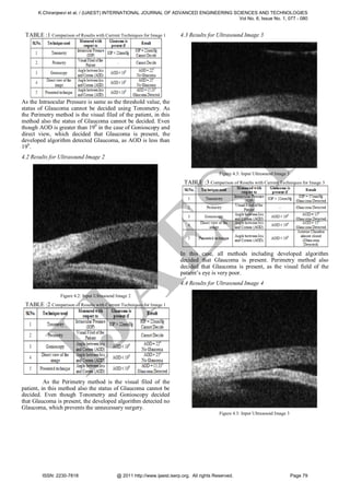
In this case, all methods including developed IMTC 2005 – Instrumentation and Measurement Technology
algorithm decided that Glaucoma is present. Conference, Ottawa, Canada, 17-19 May 2005.
[2] Xiaoyang Song, Keou Song, Yazhu Chen - A Computer-based
In ultrasound imaging, speckle noise severely Diagnosis System for Early Glaucoma Screening, Proceedings of
degrades the visual quality of the image. In order to achieve the 2005 IEEE Engineering in Medicine and Biology 27th Annual
high accuracy when extracting features, speckle must be Conference Shanghai, China, September 1-4, 2005.
filtered without destroying any important characteristics in the [3] Rafael C. Gonzalez, Richard E. Woods, ― Digital Image
image. In the developed algorithm, speckle noise was reduced Processing‖, Second Edition, Pearson Education Asia Publications.
T
using a multi-scale algorithm. It is worthwhile to investigate a [4] Rafael C. Gonzalez, Richard E. Woods, Steven L. Eddins, ― Digital
different speckle reduction technique that do not depend on Image Processing using MATLAB®‖, Pearson Education Asia
the selection of the window size and that can be used on the Publications.
ultrasound images of the eye before edge enhancement. One [5] Glaucoma Research Foundation - funding innovative research to
find a cure for Glaucoma.251 Post Street, Suite 600, San
easy way to reduce speckle is to average multiple uncorrelated Francisco, CA.
images of the same object obtained from different spatial
positions. However, this procedure is computationally costly
ES
and will increase the processing time of the algorithm. It
seems to design an algorithm for fine enhancement, which
does not require the selection of a fixed window size and to
reduce speckle noise based on each pixel surrounding area.
K.Chiranjeevi received B.E Electronics and
Communication Engineering from
from JNTU Kakinada, He is working as Asst.
Professor in GMR Institute of Technology. His
J.N.T.U.
Anantapur, and M.Tech Instrumentation and Control
research interests are in Signal Processing and Image
However, for images with very poor resolution, more iteration Processing.
can be applied until all pixels lying in the same local
neighborhood have similar intensity values close to the initial
spike value. If this technique shows improvement in speckle
T.Prabhakar received M.Tech degree from Jawarlal
reduction and does not destroy edges in the original image, the
Nehru Technological University Kakinada, Andhra
enhancement process in the algorithm will require less Pradesh, India. B.Tech degree in Electronics and
iteration, resulting in a considerable reduction of the Communication Engineering from SIR C.R.Reddy
processing time.
A
College of Engineering, Eluru, Andhra Pradesh,
India. He is joined as Lecturer in the Department. Of
Electronics and Communication Engineering at
V. CONCLUSION GMR Institute of Technology, Rajam, Srikakulam
This thesis has developed an algorithm to District, Andhra Pradesh, India in 2002. Prior to join in this Institute he
worked as a Service Engineer in Machine Diagnostics and Deployed to work
automatically identify clinical features in ultrasound images of at National Remote Sensing agency, Department. Of. Space, Hyderabad for 1
the eye. The algorithm computes the AOD 500 used to year 1 month and Trainee Programmer in Indo Tech Computers, for 8 months
measure the presence and severity of glaucoma. Overall, the in Hyderabad. He is presently working as Senior. Assistant Professor in the
IJ
algorithm predictions are very advantageous compared to the Department. Of Electronics and Communication Engineering at GMR
technologist’s observation. In the processed images, features Institute of Technology. Having Total experience is 12 years out of which 10
years in Teaching (GMRIT) and 2 Years in Industry. His research interests are
were correctly identified in 97% of the cases. 3% of images Communication, Signal Processing and Image Processing. He has published
presented inaccurate approximation of the clinical parameters. 10 Technical papers in various International journals and conferences. He is a
The difficulties encountered in measuring clinical parameters, life member of ISTE Since 2002.
which are associated with the speckle noise, poor contrast,
poor resolution, and weak edge delineation present in the
processed ultrasound images, are accurately eliminated.
However, the designed algorithm failed for a few of images,
where more noise is present. The algorithm was designed with
a goal of robustness through the use of enhancement process
on the original image, and by validation of the proper
segmentation of the anterior chamber at each step. Overall, the
benefit of this work is the ability of algorithm to reduce the
processing time and improve processing consistency for each
ISSN: 2230-7818 @ 2011 http://www.ijaest.iserp.org. All rights Reserved. Page 80](https://image.slidesharecdn.com/leeyoutaidannyfyp-130314030258-phpapp01/85/Lee-youtaidanny-_fyp-4-320.jpg)


