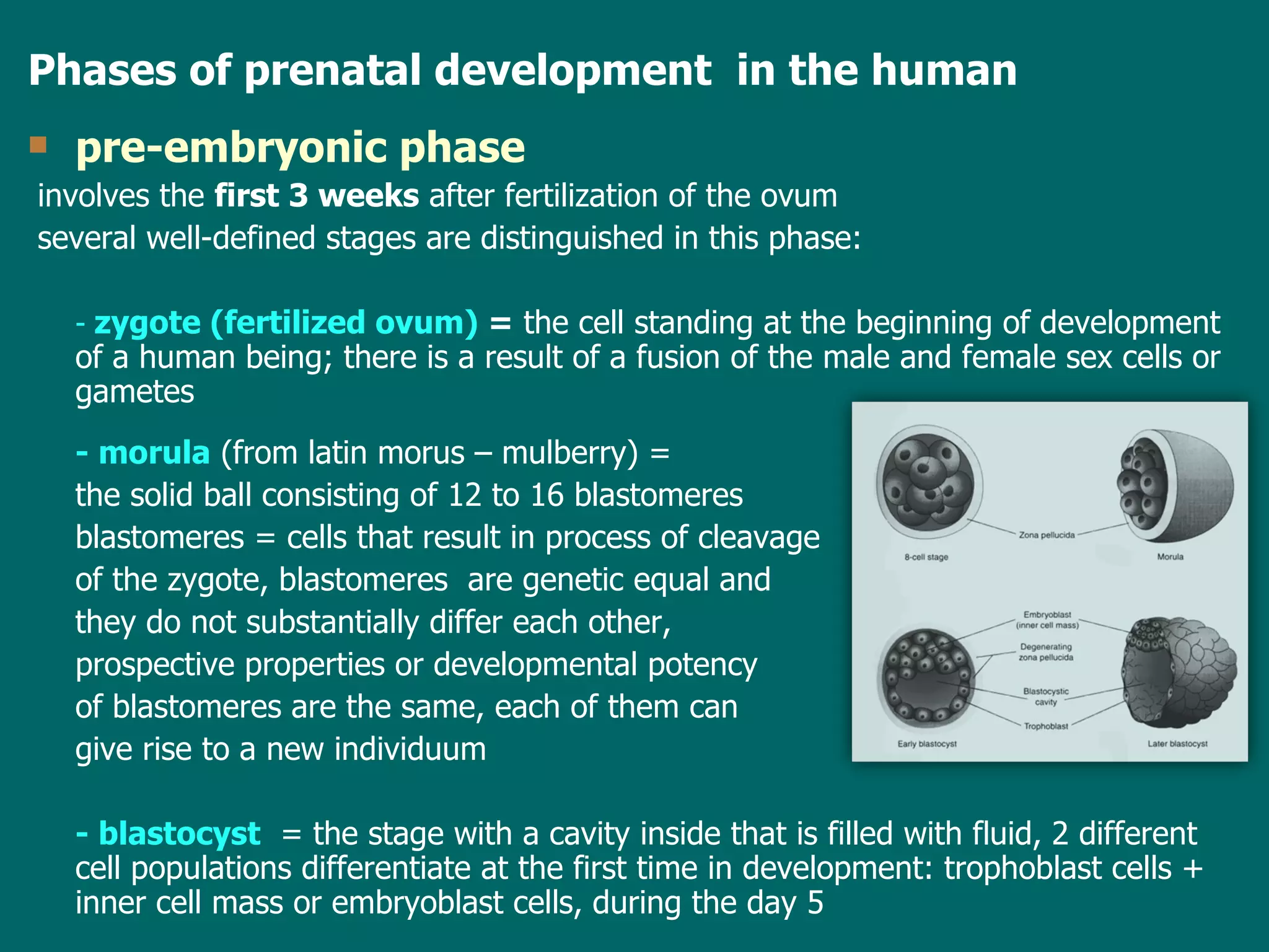
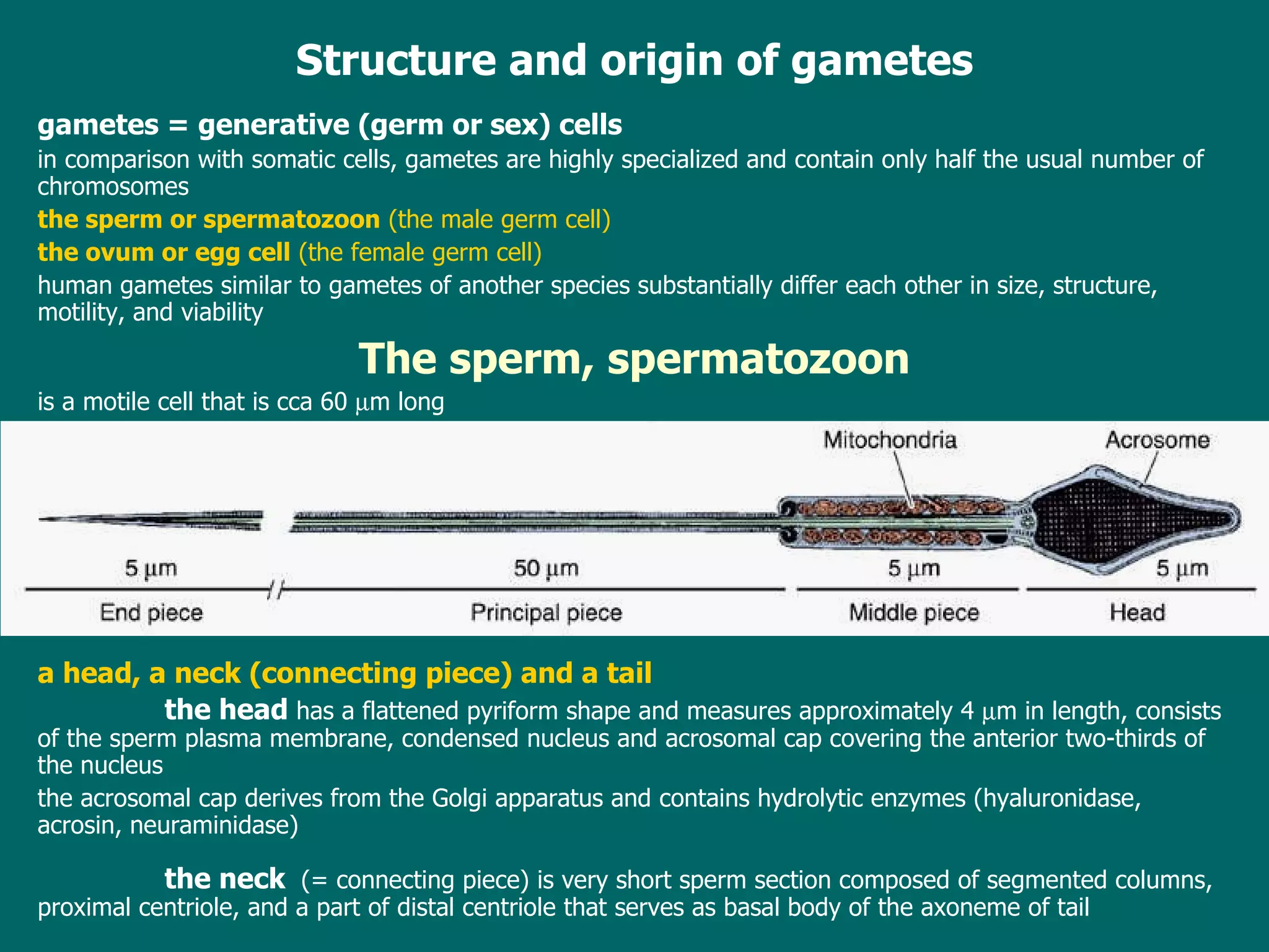
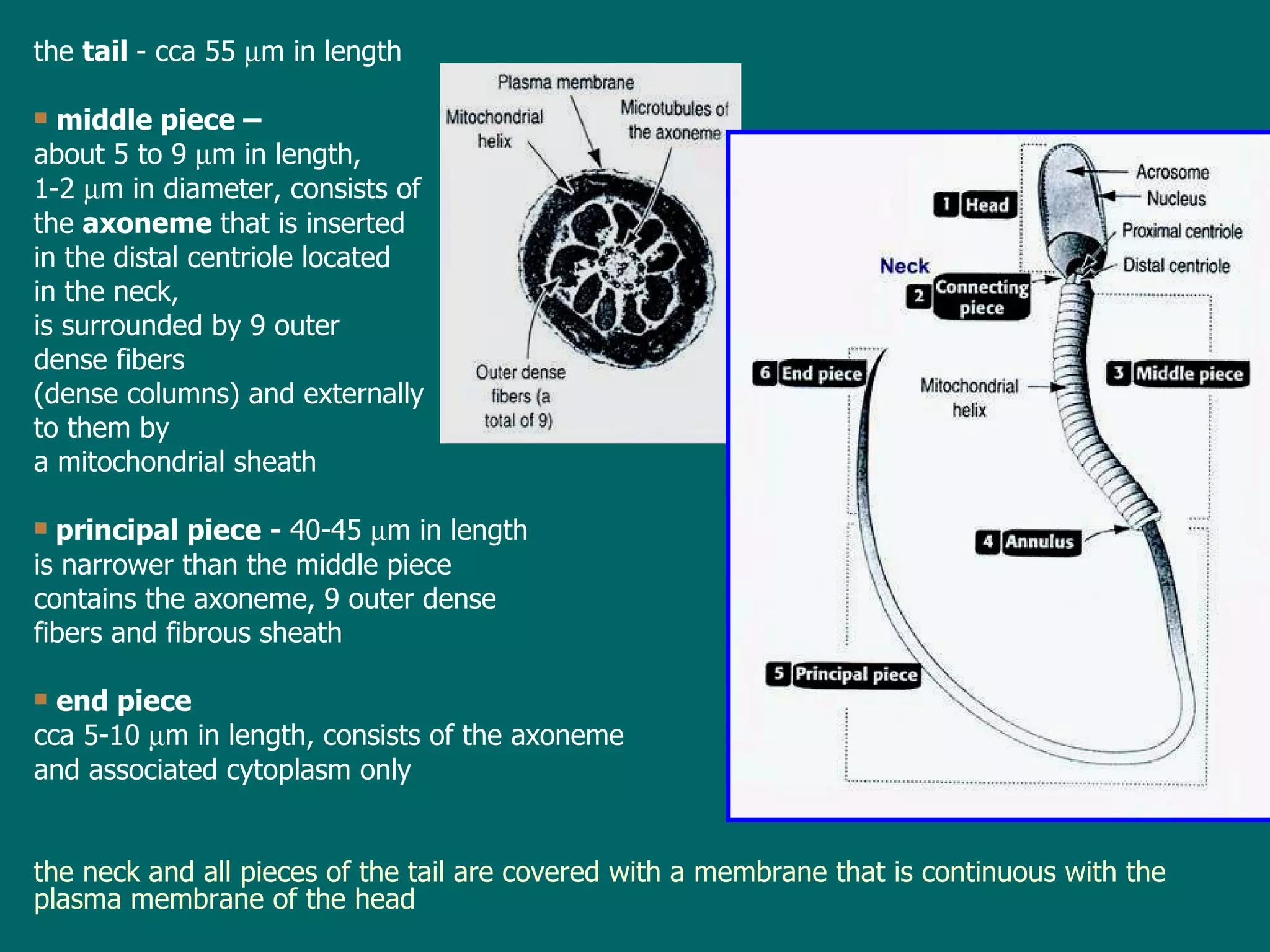
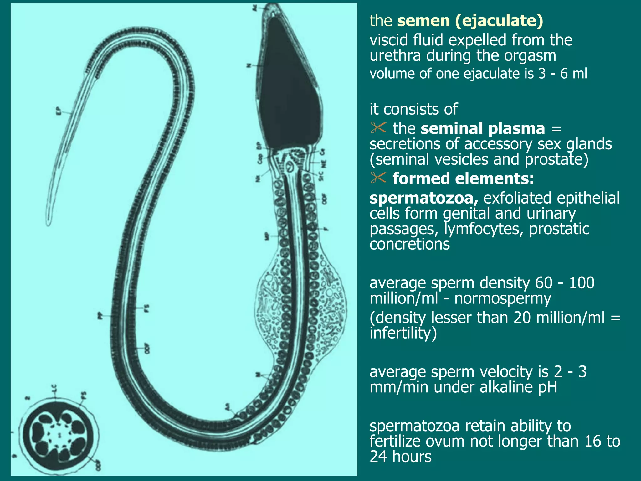
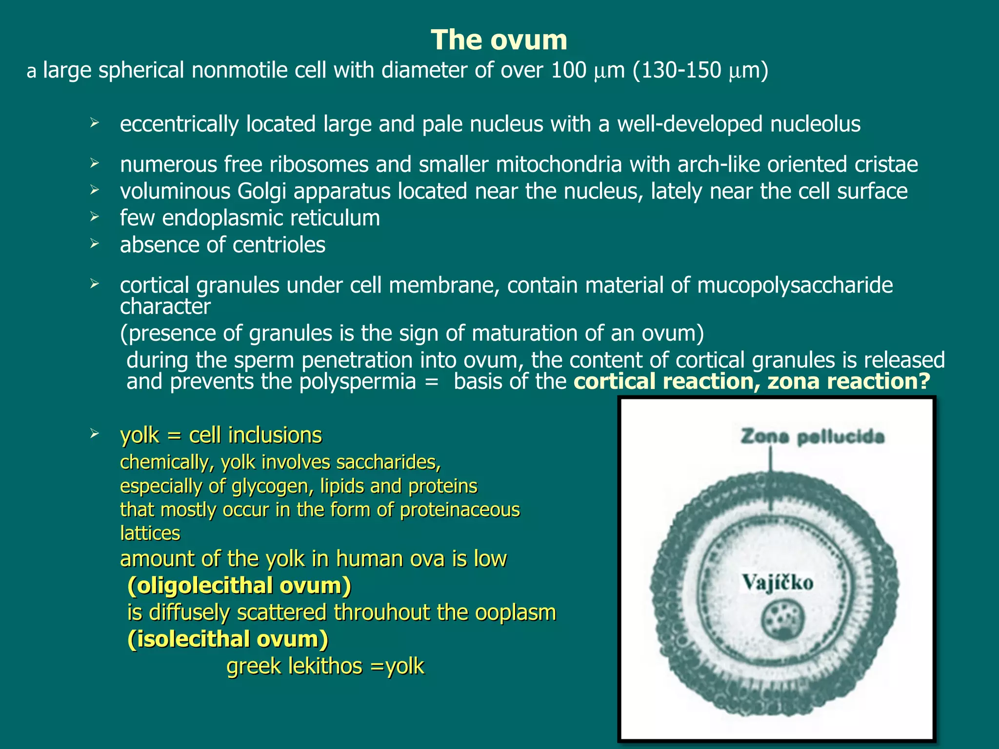
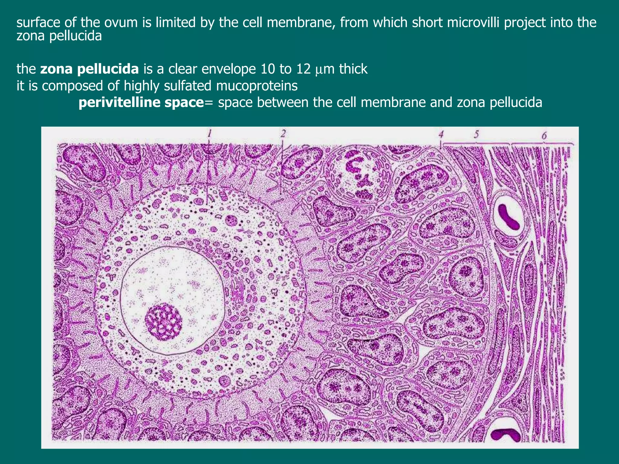
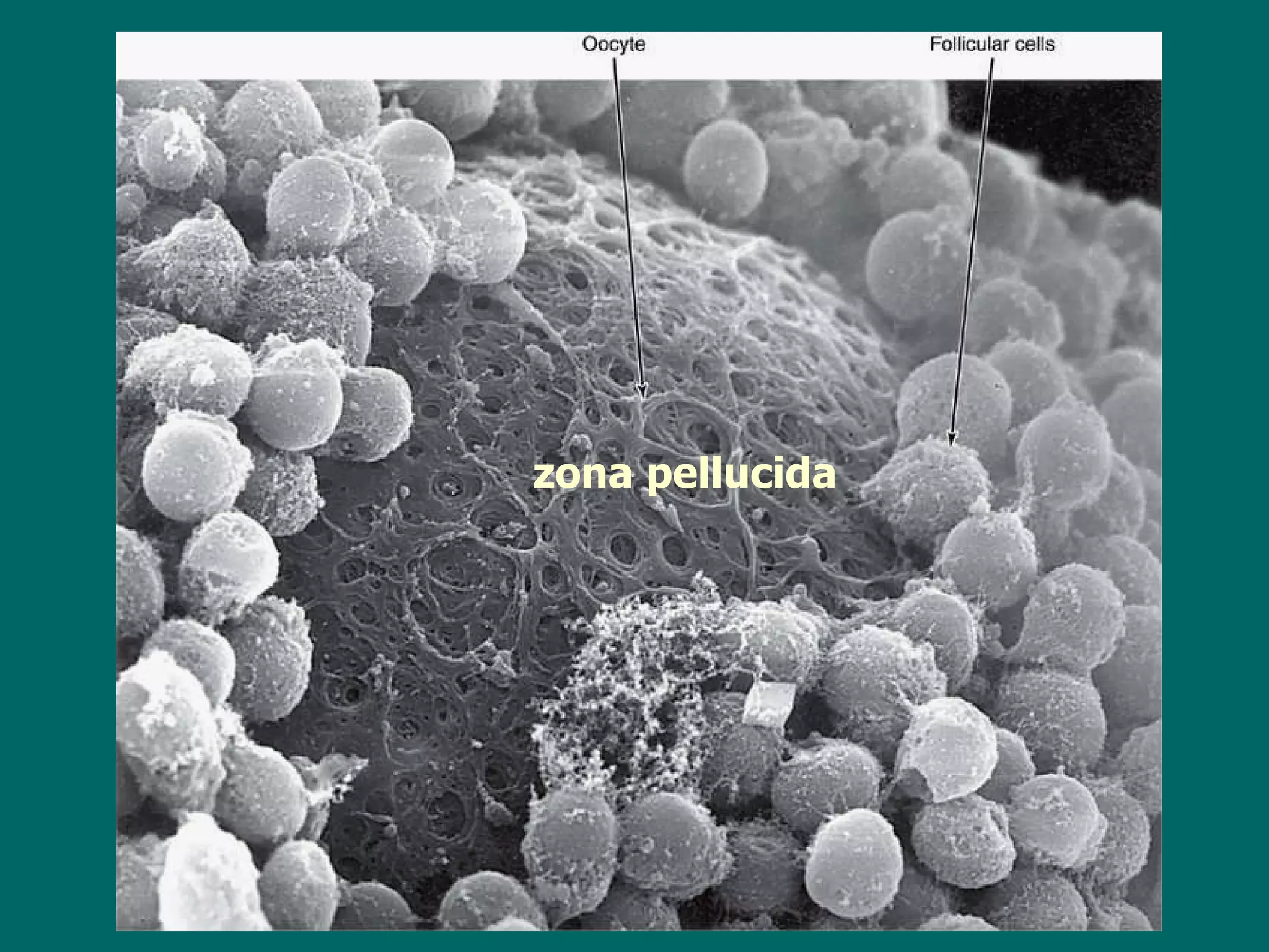
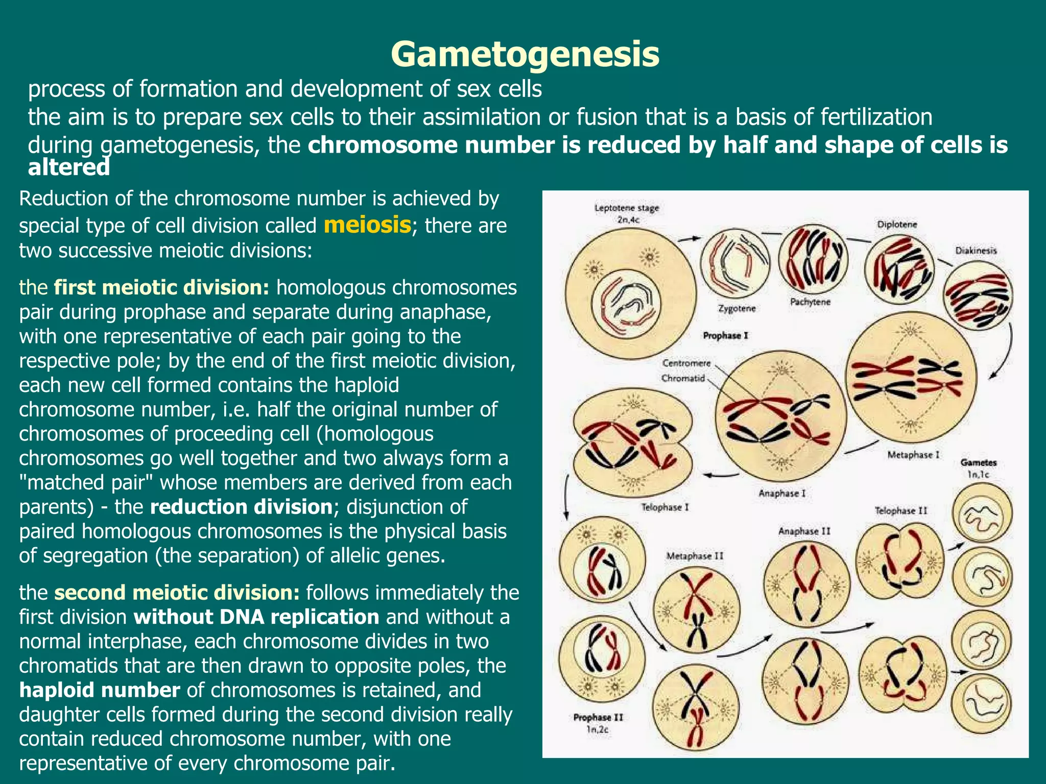
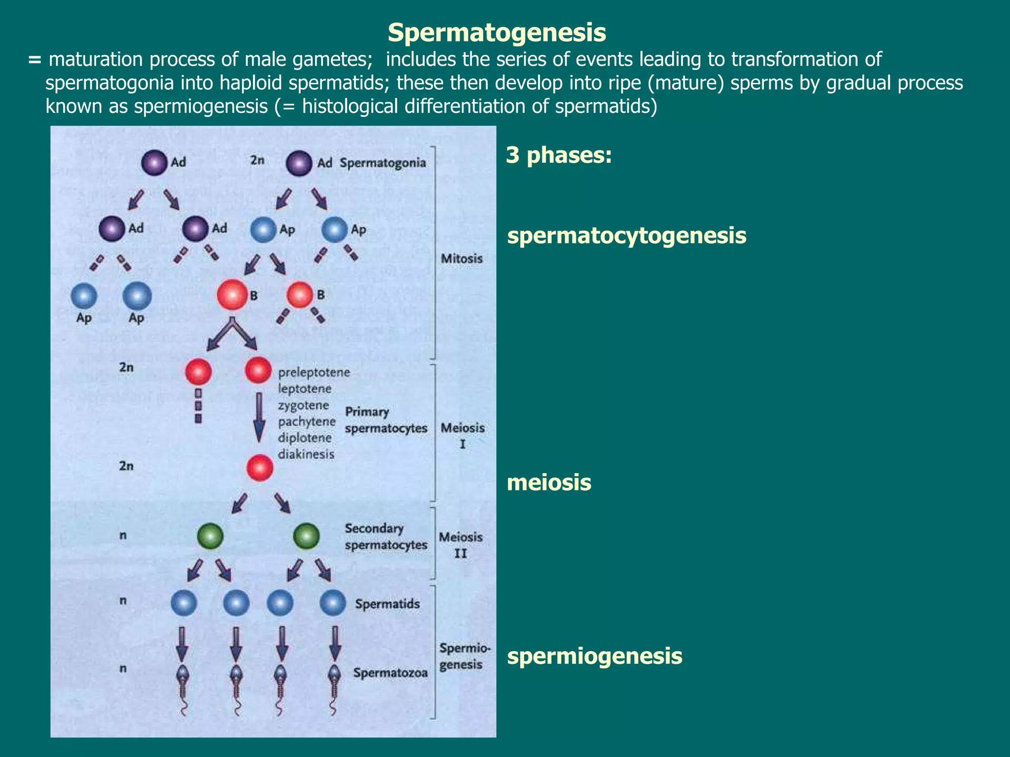
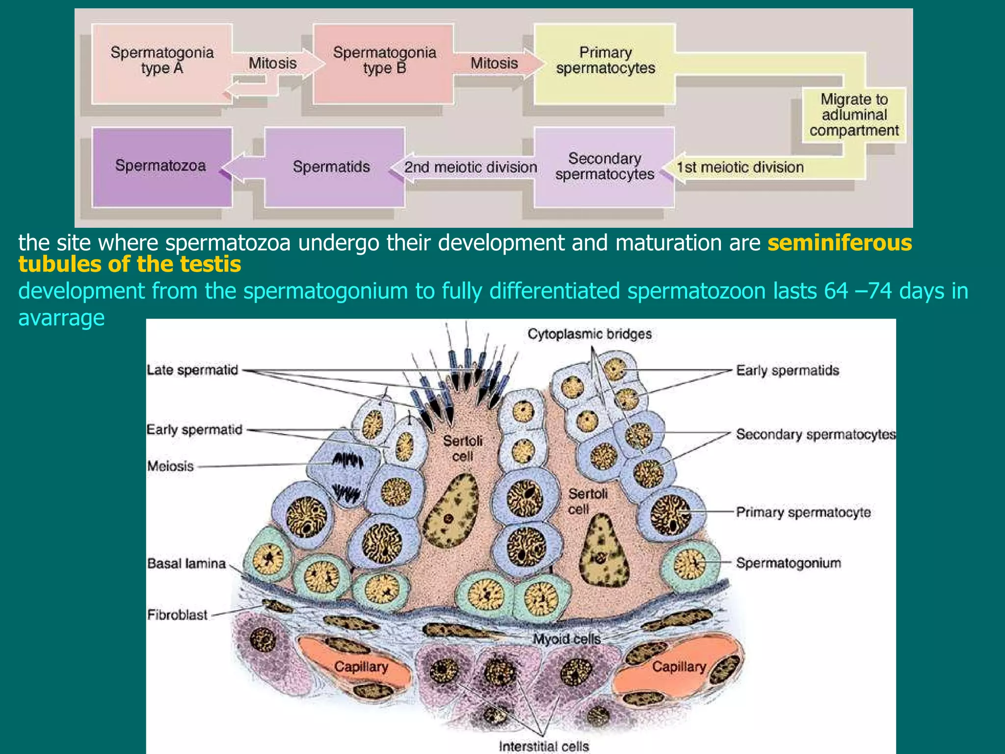
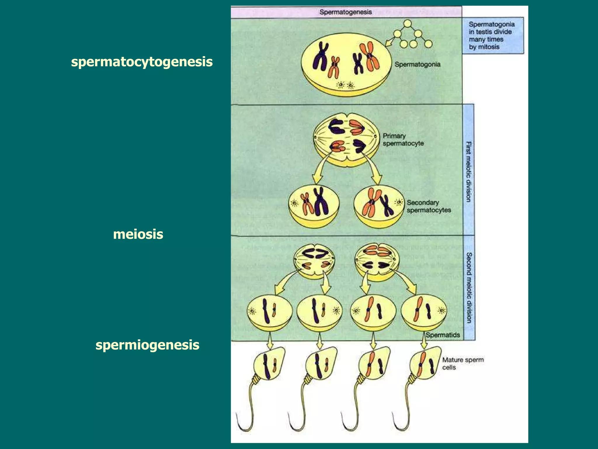
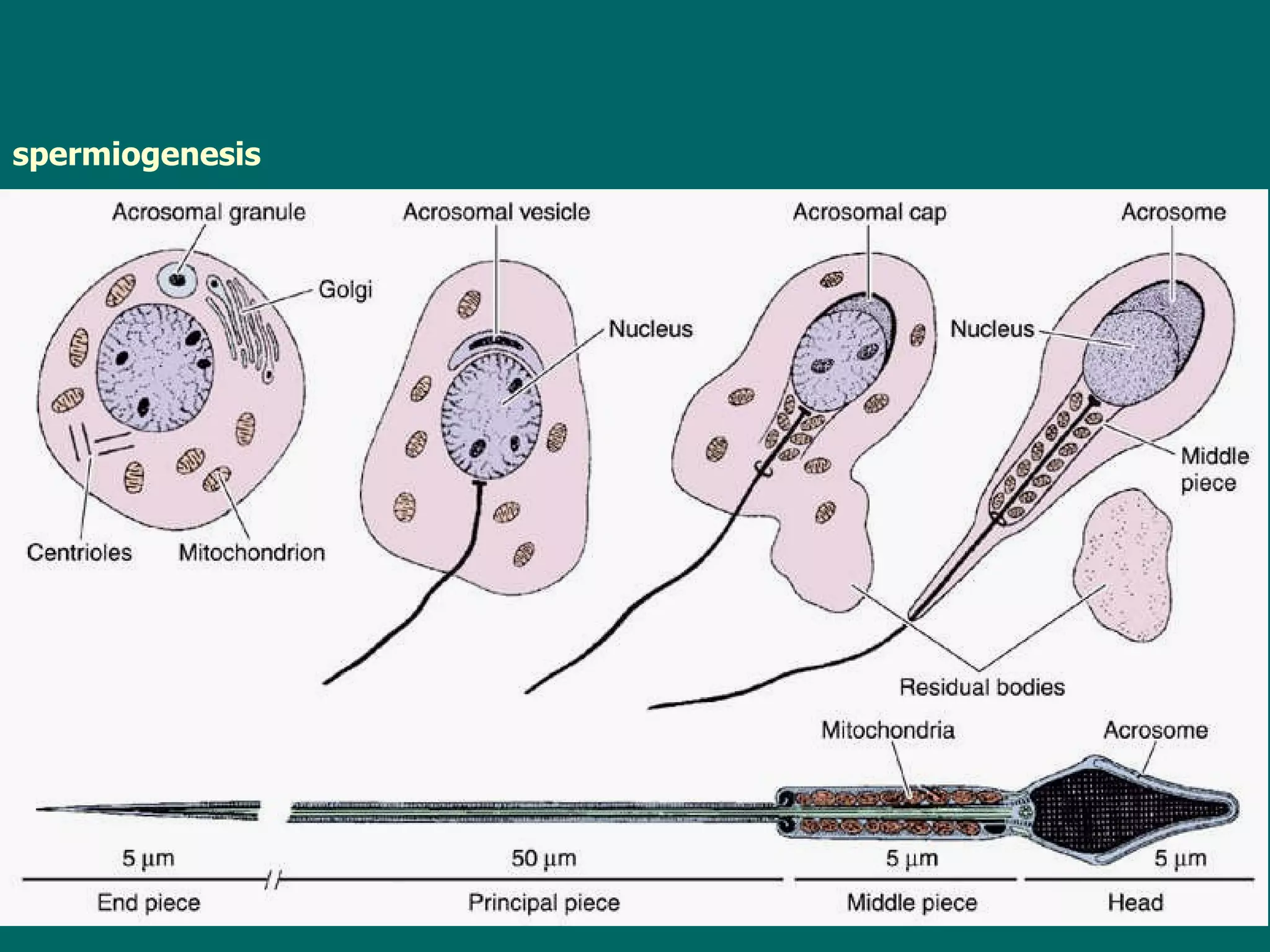
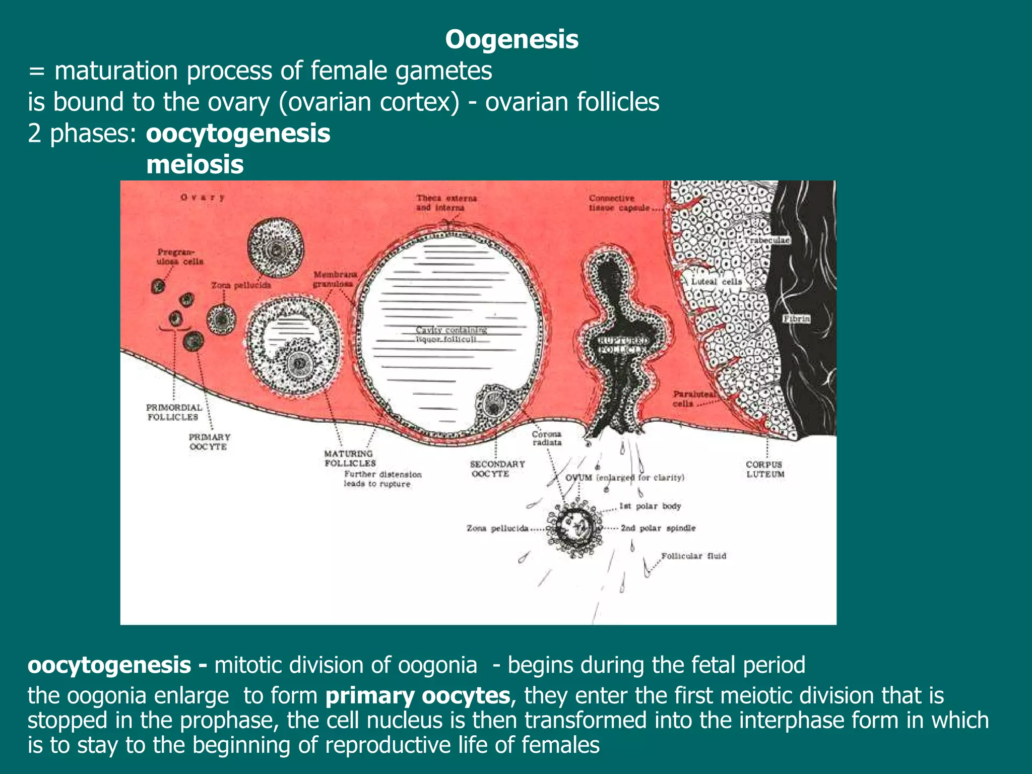
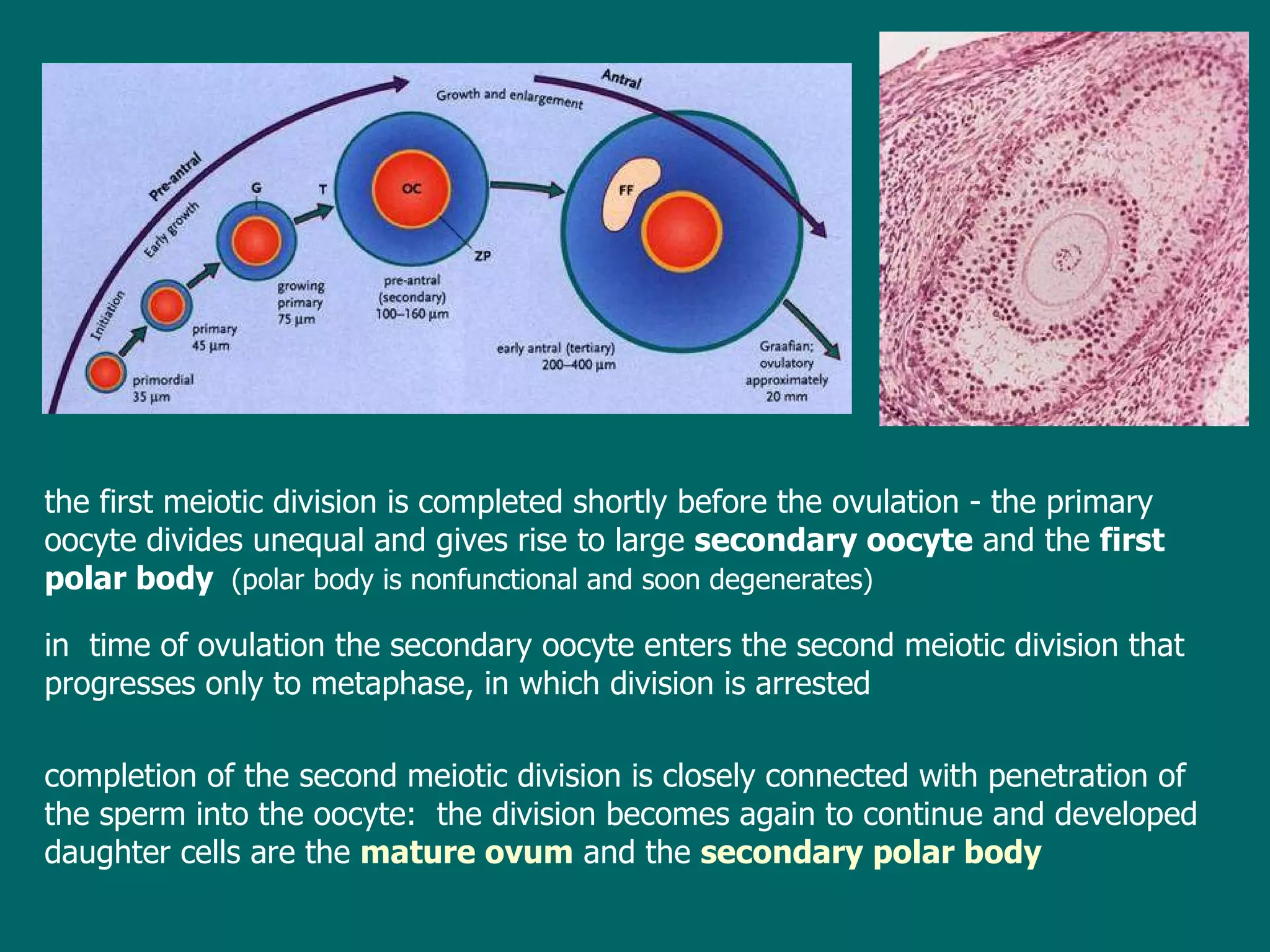
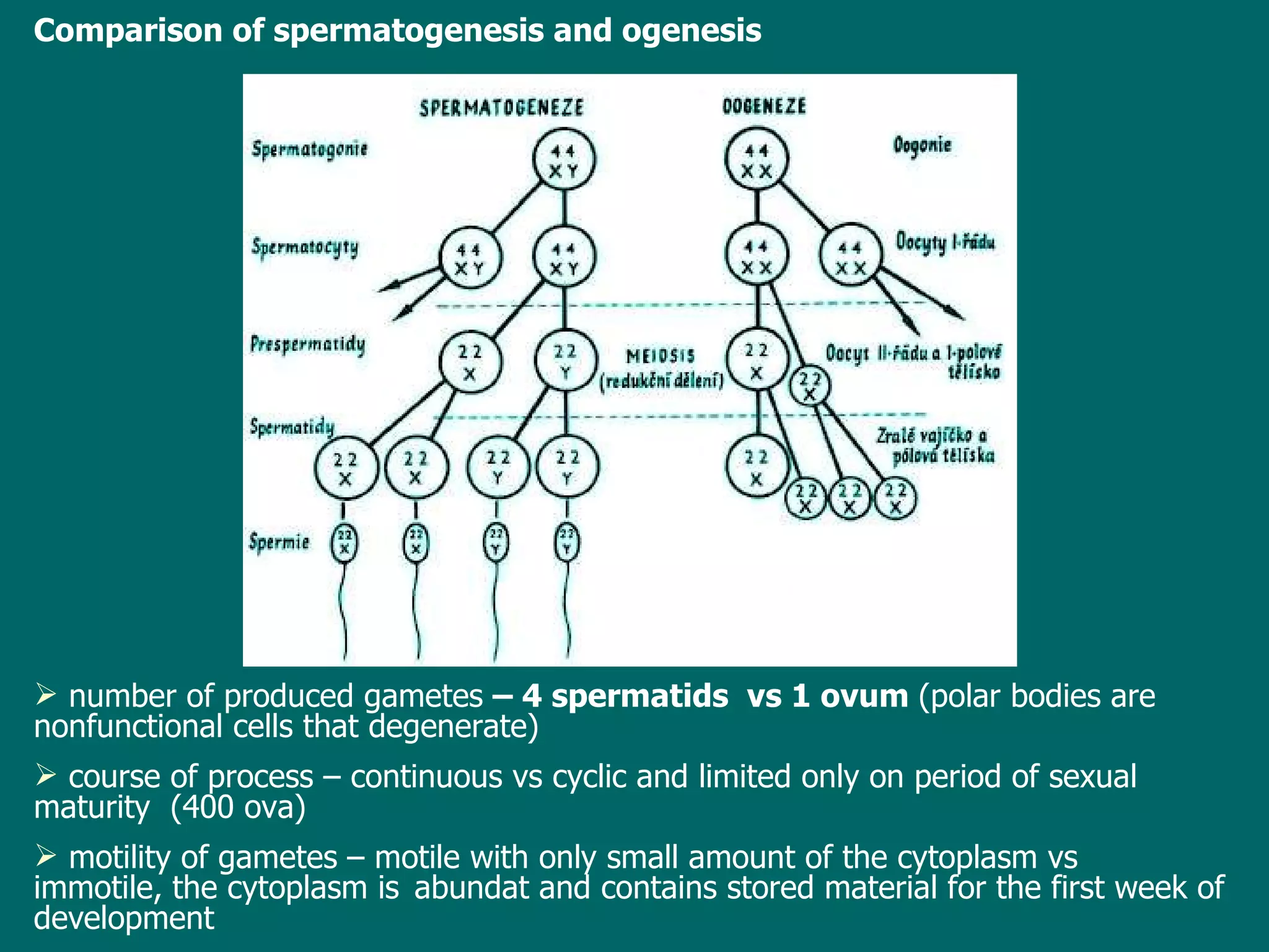
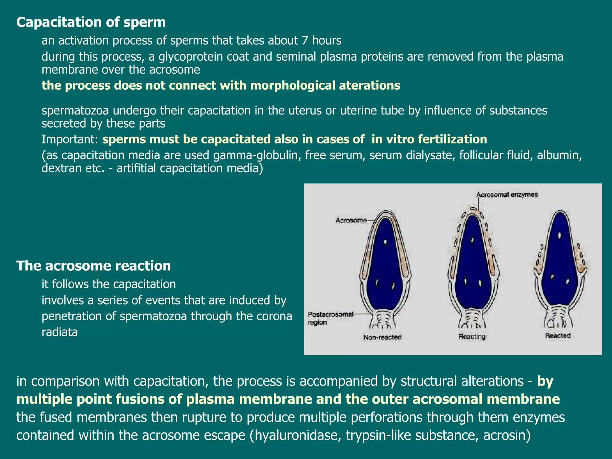
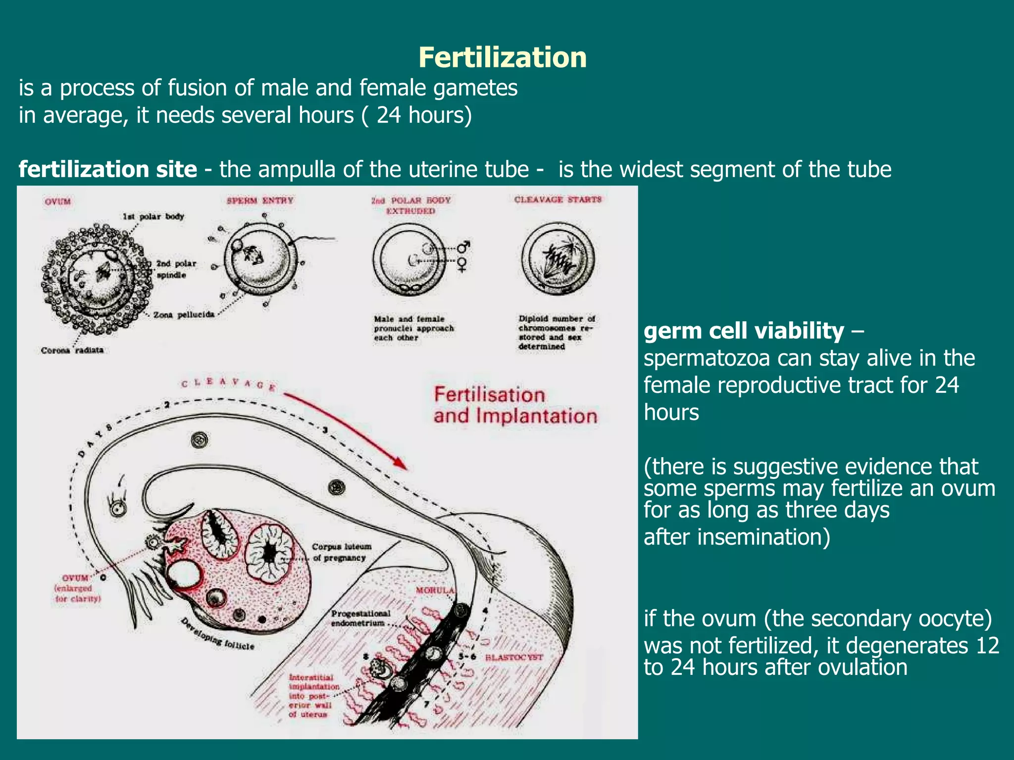
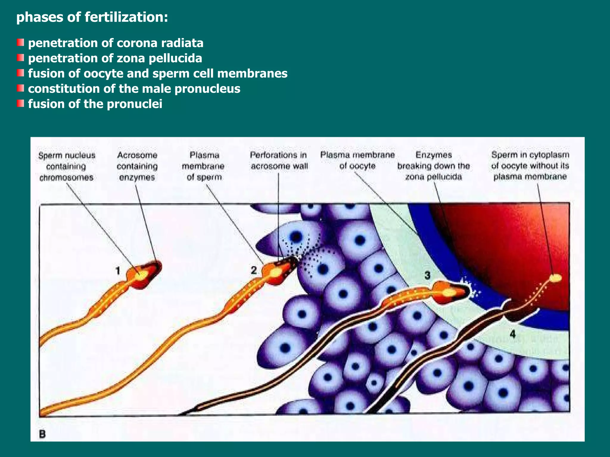
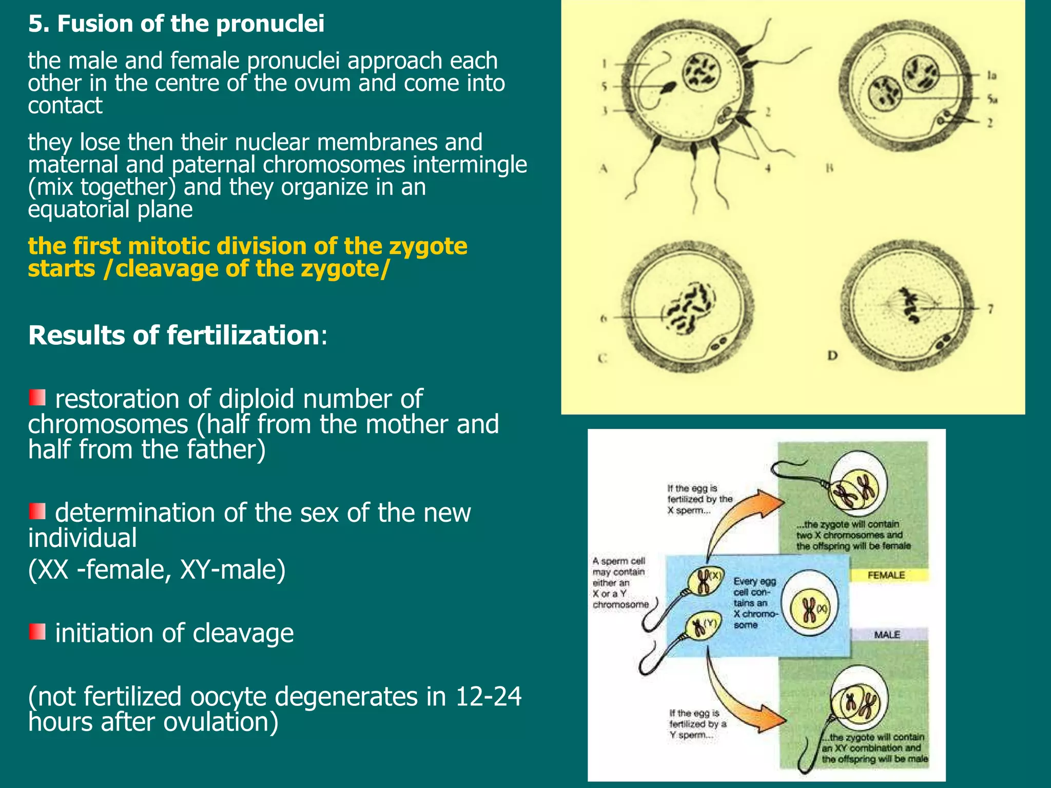
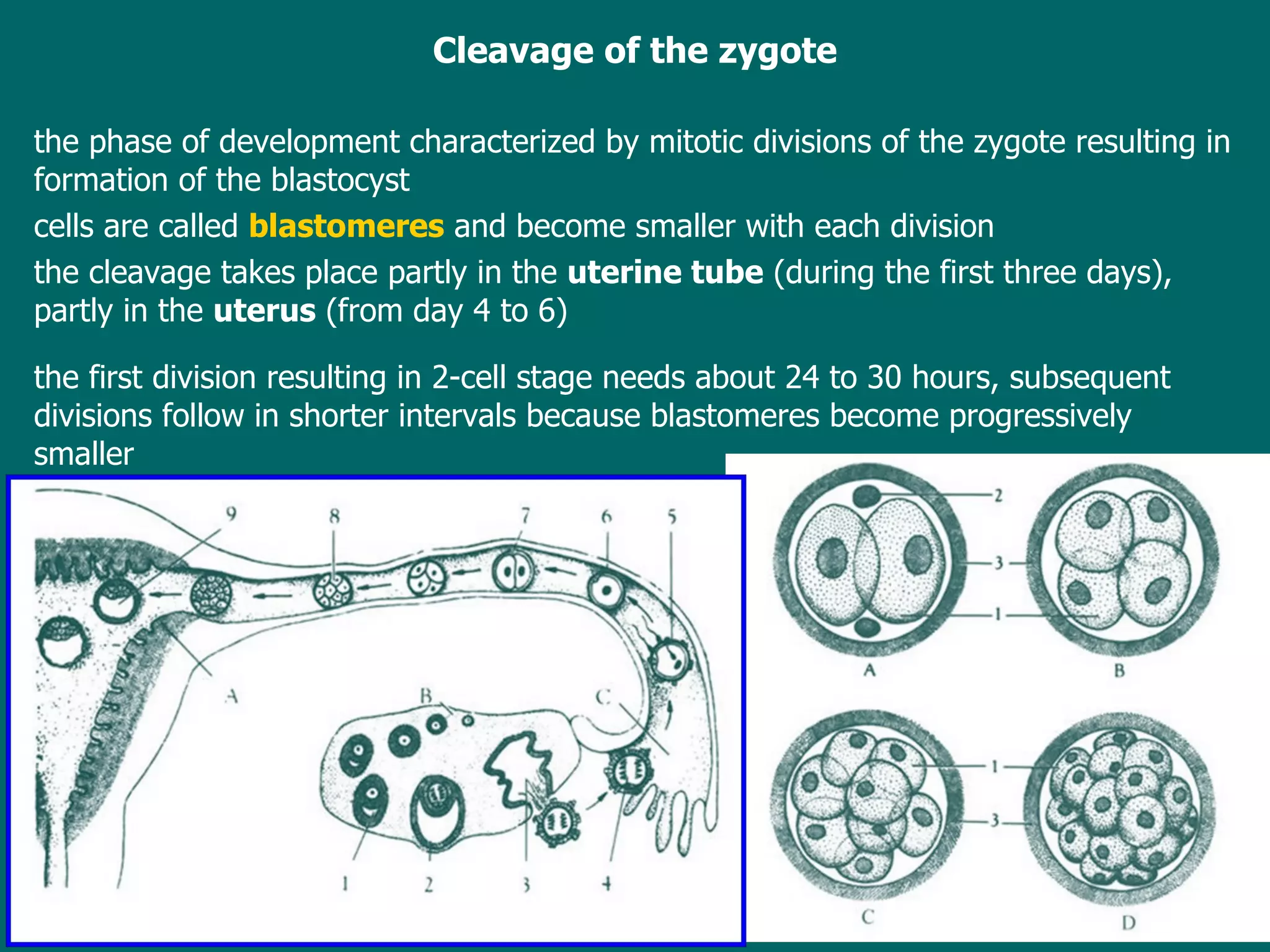
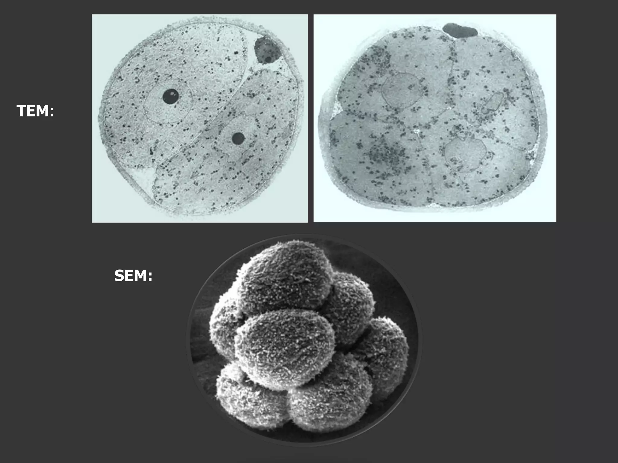
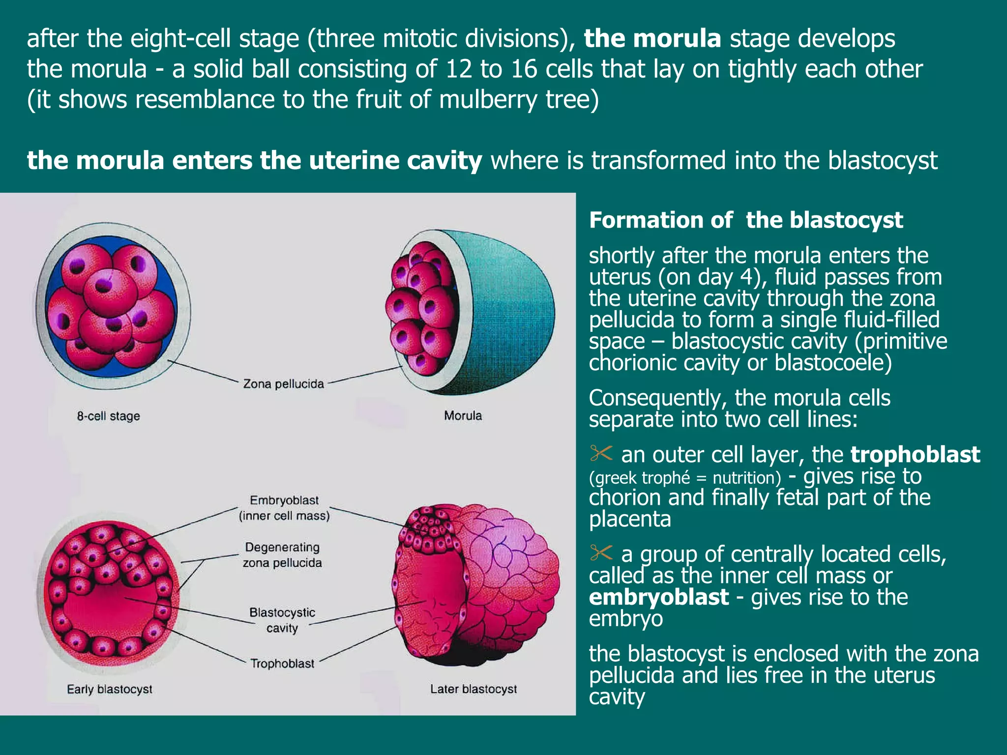
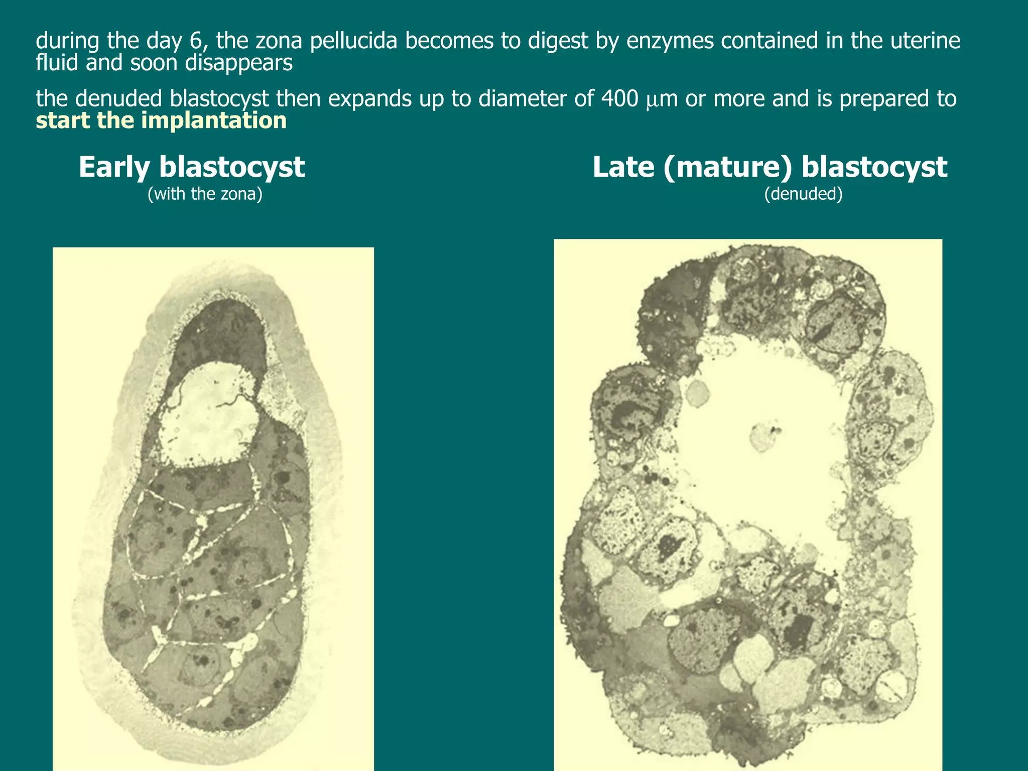
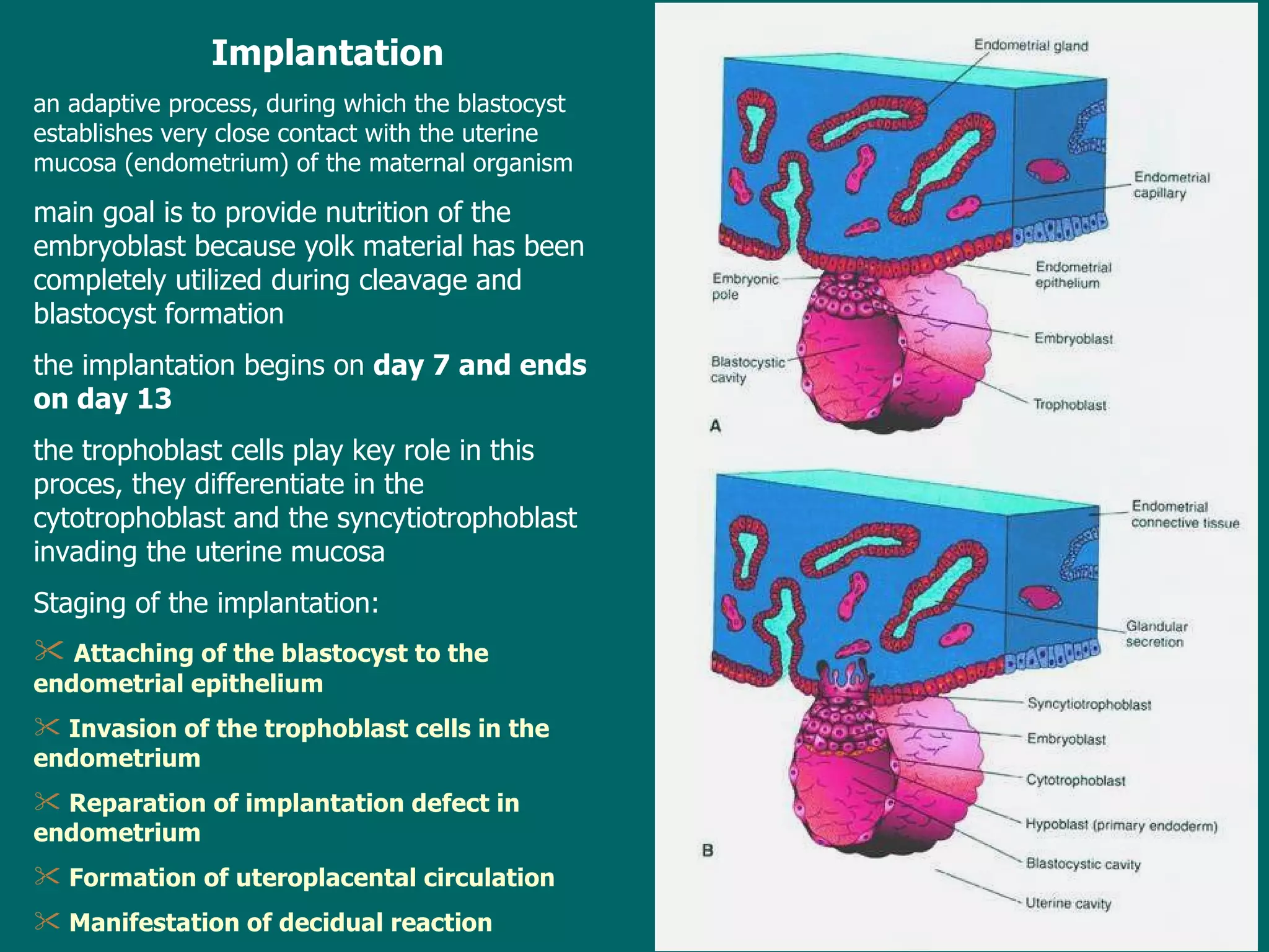
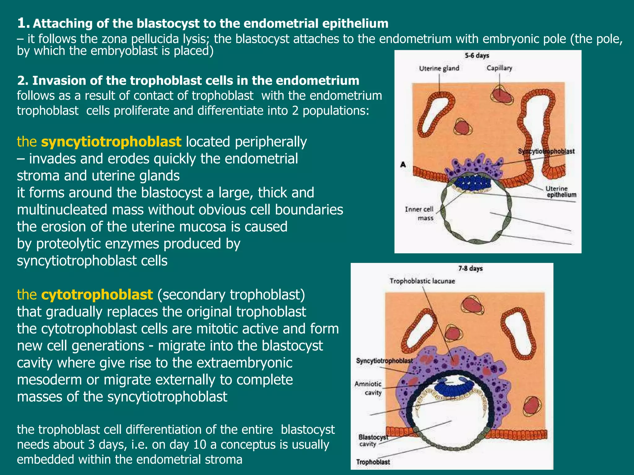
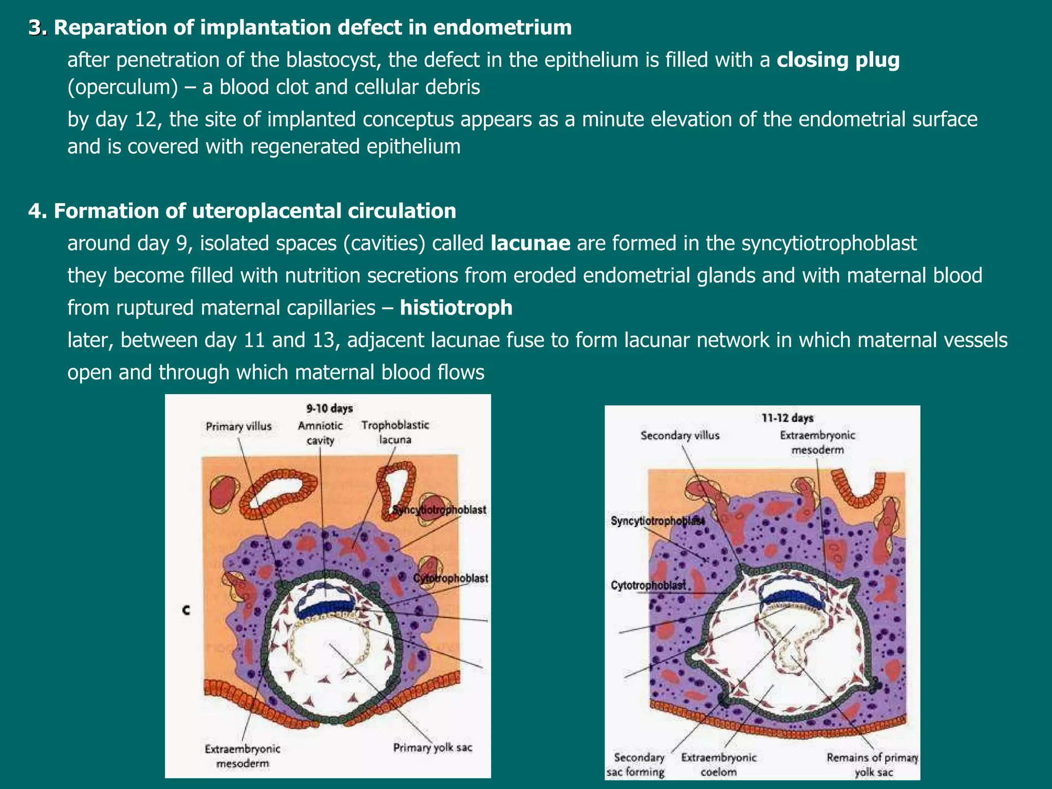
Lecture 3   ESS_2nd semester Embryology  -   d efinition ,   t he object and   significance of embryology P hases of the human ontogeny G ametes - their structure, physiology and origin (gametogenesis). Differences between spermatogenesis and oogenesis Transport ation  of gametes. Sperm capacitation  and acrosome  reaction Fertilization and cleavage,  the  morula and blastocyst Implantation of the blastocyst
d evelopment of plant  as well as  animal organism s  begins  when an ovum  is  fertilized  and ends at  the moment of  death  of the individual the time  period  from fertilization to death is called as  the   ontogeny (sometimes also as  life cycle ) in  placental mammals  inclusive the human, ontogeny involves  2 periods of different length: prenatal  period (prenatal development)  - from fertilization  of an ovum  to  delivery of fetus postnatal  period (postnatal development)  - from  the   delivery  to death of  the individuum main task of embryology in mammals is to study and describe events occuring during the  prenatal  period (phase) of development  goals and orientation of medical embryology are the same:  to describe and explain events and processes from the fertilization to  the  birth
Significance of embryology the basic theoretical medical branch good knowledge of   embryology  is  extremely  important in  many    medical disciplines: anatomy  - knowledge of the human   develop ment helps to  understand  sometimes very complicated  relationships  between many organs of the human body it is generally known  embryology  shows more  closely relationships  and bounds to  anatomy than  to  histology  ( reason s for teaching of embryology together with histology are rather practical then logical as study of the  pre-embryonic and  early  embryonic stages  needs to use and  studied only with the use of histological slides) pathology  - knowledge of embryology is  explored  in study of restorative (healing) processes and in description and classification of  neoplasms  or tumo u rs obstetrics, pediatrics and surgery  - knowledge of normal development is essential to understanding the mechanisms, symptomatology and correction of congenital malformations a  special medical  line - prenatal medicine -   was  constituted in last years , it  deal s  with diagnosis, treatment and prevention of congenital malformations
Phases of  prenatal development  in the human pre-embryonic phase   involves  the  first 3 weeks  after fertilization  of the ovum several well-defined stages are distinguished in this phase: -  zygote (fertilized ovum)   =  the cell standing at the beginning of  development of  a human being; there is a result of a fusion of the male and female sex cells or gametes - morula   ( from latin morus – mulberry ) =   the solid ball consisting of 12 to 16 blastomeres  blastomeres = cells that result in process of cleavage  of the zygote , b lastomeres  are  genetic equal  and   they do not substantially differ each other ,  prospective properties  or  developmental potency   of blastomeres are the same, each of them can  give rise to a new individuum - blastocyst   =  the stage with a cavity inside that is filled with fluid ,  2 different cell populations  differentiate  at the first time  in development : trophoblast cells + inner cell mass or embryoblast cells , during the day 5
-  gastrula   =   the  bilaminar pre-embryonic stage ,  in which the main body axis  and polarity  begin to become apparent, the  bilaminar  stage develop s   on day 7 and lasts to  the end of the  second  week after fertilization   embryonic phase lasts from the beginning of the 4th week to the end of the 8th week in  th e   phase  all the main body organs are laid down by the end of  it  the embryo is recognizabl e  human in appearance  and   measures  cca  2-3 cm from head to tail fetal phase   it lasts from the 9 week to  delivery  of fetus;  during th e   fetal  phase, anlagen of organs develop further and the fetus grows quic kly  in length and increases its weight gain, in particular during the terminal months
Phases of the   postnatal  development in the human infancy  –  from the birth to the end of the  first year of the  age  (the first two weeks of infancy are  called  as the newborn or neonatal period) childhood  - from 15 month until 12 to 13 years; the primary teeth appear and are replaced by the secondary or permanent teeth puberty  - is the period between 12-15 years in girls and 13-16 years in boys, during which secondary sexual characteristics develop adolescence  - follows the puberty and ends 3-4 years after it; the organism reaches sexual but also physical, mental and emotional maturity adulthood  -   a period in which developmental changes occur very slowly and are mostly characterized by selective loss of highly specialized cells and tissues postnatal development of the human is studied and treated with various medical branches
S tructure and origin of gametes g ametes  =  generative (germ or sex) cells i n comparison with somatic cells, gametes are highly specialized and contain only half the usual number of chromosomes the sperm or spermatozoon  (the male germ cell) the ovum or egg cell  (the female germ cell) h uman gametes similar to gametes of another species substantially differ each other in size, structure, motility, and viability The sperm, spermatozoon is a motile cell that  is   cca  60   m  long a head, a neck  (connecting piece)  and a tail t he head  has a flattened pyriform shape and measures approximately 4   m in length ,  consists of the sperm plasma membrane, condensed nucleus and acrosomal cap covering the anterior two-thirds of the nucleus the  acrosomal cap derives from the Golgi apparatus  and  contains hydrolytic enzymes (hyaluronidase, acrosin, neuraminidase)  t he neck   (= connecting piece)  is  very short sperm section  composed of segmented columns, proximal centriole, and a part of distal centriole that serves as basal body of the axoneme of tail
t he  tail   - cca  55   m in length  middle piece  –  about 5 to 9   m in length,  1-2   m in diameter ,  consists of the  axoneme  that is inserted in the distal centriole located  in the neck ,  is surrounded by 9 outer  dense fibers  (dense columns) and externally  to them by  a mitochondrial sheath principal piece  -  40-45   m in length is narrower than the middle piece contains  the axoneme, 9 outer dense  fibers and fibrous sheath end piece cca  5-10   m in length , consists of  the axoneme  and associated cytoplasm only t he neck and all pieces of the tail are covered with a membrane that is continuous with the plasma membrane of the head
the  semen  ( ejaculate ) viscid fluid expelled from the urethra during the orgasm  volume of one ejaculate  is   3  - 6 ml  it consists of  the  seminal plasma  =  secretions of accessory sex glands (seminal vesicles and prostate) formed elements: spermatozoa,  exfoliated epithelial cells form genital and urinary passages, lymfocytes, prostatic concretions average sperm density  60 -  100 million/ml  - normospermy   (density lesser than 20 million/ml = infertility) average sperm velocity is 2 - 3 mm/min under alkaline pH spermatozoa retain ability to fertilize ovum not longer than 16 to 24 hours
The ovum a  large spherical nonmotile cell  with  diameter of over 100   m (130-150   m) eccentrically located  large and pale nucleus with a well-developed nucleolus numerous f ree ribosomes and smaller mitochondria with arch-like oriented cristae  voluminous   Golgi apparatus located near the nucleus, lately near the cell surface few endoplasmic  reticulum  absence of  centrioles cortical granules under cell membrane ,  contain material of mucopolysaccharide character ( presence of  granules is the  sign of  maturation of an  ovum)  during the sperm penetration into ovum, the content of cortical granules is released  and prevents the polyspermia =  basis of the  cortical reaction, zona reaction? yolk = cell inclusions chemically, yolk involves saccharides,  especially of glycogen, lipids and proteins  that mostly occur in the form of proteinaceous  lattices amount of the yolk in human ova is low   (oligolecithal ovum)   is diffusely scattered throuhout the ooplasm   (isolecithal ovum)    greek le ki thos   =yolk
surface of the ovum is  limited by  the cell membrane, from which short microvilli project into the zona pellucida  the  zona pellucida  is a clear envelope 10 to 12   m thick it is composed of highly sulfated mucoproteins  perivitelline space =   space between the cell membrane and zona pellucida
zona pellucida
Gametogenesis process of formation and development of sex cells  t he aim is to prepare  sex  cells to their assimilation or  fusion that is a basis of  fertilization d uring gametogenesis, the  chromosome number is reduced by half and shape of cells is altered Reduction of the chromosome number is  achieved  by special type of cell division called  meiosis ; th ere are two successive meiotic divisions : the   first meiotic division :  homologous chromosomes pair during prophase and separate during anaphase, with one representative of each pair going to the respective pole ; by  the end of the first meiotic division, each new cell formed contains the haploid chromosome number, i.e. half the original number of chromosomes of proceeding cell (homologous chromosomes go well together and two always form a "matched pair" whose members are derived from each parents) - the  reduction division ;  disjunction of paired homologous chromosomes is the physical basis of segregation (the separation) of allelic genes. the  second meiotic division :  follows immediately the first division  without DNA replication  and without a normal interphase , each  chromosome divides in two chromatids that are then drawn to opposite poles ,  the  haploid number  of chromosomes is retained, and daughter cells formed during the second division really contain reduced chromosome number, with one representative of every chromosome pair.
Spermatogenesis  =  m aturation process of male gametes ;  includes the series of events leading to transformation of  spermatogonia into haploid spermatids ; t hese then develop into ripe (mature) sperms by gradual process known as spermiogenesis ( =  histological differentiation of spermatids )     3 phases: s permiogenesis   meiosis s perm atocyt ogenesis
the site where spermatozoa undergo their development and maturation are  s eminiferous tubules of the testis development from the spermatogonium to fully differentiated spermatozoon lasts 64 –74 days in avarrage
s perm atocyt ogenesis   meiosis spermiogenesis
s permiogene sis
O ogenesis   = m aturation process of female gametes  is bound to the ovary  (ovarian cortex) - ovarian follicles 2 phases:  oocytogenesis   meiosis oocytogenesis -  mitotic division of oogonia  -  begins  during the fetal period  the oogonia enlarge  to form  primary oocytes , they enter the first meiotic division that is stopped in the prophase, the cell nucleus is then transformed into the interphase form in which is to stay to the beginning of reproductive life of females
the first  meiotic  division is completed  s hortly before  the  ovulation  -  the primary oocyte divides unequal  and gives rise to  large  secondary oocyte  and  the  first polar body   ( polar body is nonfunctional and soon degenerates ) in  time of  ovulation the secondary oocyte enters the second meiotic division that progresses only to metaphase,  in which  division is arrested completion  of the second meiotic division  is closely connected with  penetration of the sperm into the oocyte:  the division becomes again to continue and developed  daughter  cells are the  mature ovum  and the  secondary polar body
Comparison of spermatogenesis and  ogenesis   number of produced gametes  – 4 spermatids  vs 1 ovum  (polar bodies are nonfunctional cells that degenerate) course of process – continuous vs cyclic and limited only on period of sexual maturity  (400 ova) motility of gametes – motile with only small amount of the cytoplasm vs  immotile, the cytoplasm is  abundat and contains stored material for the first week of development
Transportation of gametes the site of fertilisation is  the uterine tube – its dilated part  termed  the ampula nearly to the ovary Oocyte transport :  the secondary oocyte  surrounded by some granulosa cells  is expelled from the graaphian follicle and is directed  via stream of follicular fluid  under contraction of fimbriae (finger-like processes of the tube) into the lumen of the infudibulum the oocyte then passes into the ampulla by waves of peristalsis of muscle cells in the wall of oviduct  i t is supposed that oocyte reaches the fertilization site in cca  25 min Sperm transport:   a fter cohabitation, the spermatozoa deposited against the cervix  and the posterior fornix of the vagina, pass by movements of their tails through the cervical canal into the uterus and subsequently in the respective uterine tube t he ascent is caused by contractions of  smooth muscle cells   in the wall  of the uterus and   uterine tube f resh spermatozoa, upon arrival in the female genital tract, are not capable to fertilize the oocyte  because they  must undergo  capacitation  and the  acrosome reaction
Capacitation of sperm an activation process of sperms that takes about 7 hours during  th is  process ,   a  glycoprotein coat and seminal plasma proteins  are  remov ed  from the plasma membrane over the acrosome t he process does not connect with   morphological  aterations s perm atozoa  undergo their capacitation in the uterus or uterine tube by influence of substances  secreted by these parts  Important:  s perms must be capacitated also in cases  of  in vitro fertilization   (as capacitation media are used gamma-globulin, free serum, serum dialysate, follicular fluid, albumin, dextran etc. - artifitial capacitation media) The acrosome reaction it  follows the capacitation  involves   a  se ries  of events that  are induced by   penetration  of sperm atozoa  through the corona radiata in  comparison with capacitation, the process is accompanied by structural  alterations -   by  multiple point fusions  of  plasma membrane and the outer acrosomal membrane t he fused membranes then rupture  to  produc e  multiple perforations through  them  enzymes   contained   within  the acrosome escape (hyaluronidase, trypsin-like substance, acrosin)
Fertilization is a process of fusion of male and female gametes in average, it needs several hours (  24 hours ) f ertilization site  - t he ampulla of the uterine tube  -   is the widest segment of the tube  germ cell viability  –  s permatozoa   can stay alive in the  female reproductive tract for 24  hours (there is suggestive evidence that some sperms may fertilize an ovum for as long as three days  after insemination) if  the ovum (the secondary oocyte) was  not fertilized , it  degenerates 12 to 24 hours after ovulation
phases of fertilization: p enetration of corona   radiata p enetration of zon a  pellucida f usion of oocyte  and  sperm   cell   membranes c onstitution of th e  male pronucleus f usion of the pronuclei
1 .   Penetration of corona radiata the  corona radiata  is envelope from  follicular cells  that  encloses the ovulated oocyte the main goal of this phase  is d ispersal of corona radiata cells  by  enzyme hyaluronidase released from the acrosomal cap s of spermatozoa   2. Penetration of zona pellucida is facilitated by the action of enzymes released from the acrosome primary role plays  acrosin ,   enzyme causes  lysis of  the zone  (inhibition of acrosin prevents passage of sperms through the zona pellucida) o nce the first sperm passes through the zona pellucida,  so-called  a  zona reaction   starts, during which the zona pellucida becomes  impermeable to  further  spermato z oa is  supposed  that the zona reaction  is  mediate d by  cortical granules of the secondary oocyte 3. Fusion of oocyte-sperm cell membranes this phase  is characterised by  actual fusion  of  the oocyte membrane and membrane  covering  the posterior region of the sperm head , t he head and tail of the sperm enter the cytoplasm of the oocyte after fusion of membranes, t he secondary oocyte  can  complete   the second meiotic division  to give rise to the mature ovum with  haploid number  of chromosomes and  the second polar body  chromosomes in the egg nucleus decondense to form  the  female pronucleus 4. Constitution of the male pronucleus t he penetrated sperm moves in close vicinity to the female pronucleus  its nucleus becomes rapidly swollen and forms  the  male pronucleus   ( the tail of the sperm degenerates )
5. Fusion of the pronuclei t he male and female pronuclei approach each other in the centre of the ovum and come into contact they lose then their nuclear membranes  and  maternal and paternal chromosomes intermingle (mix together)  and they organize in an equatorial plane the first mitotic division of the zygote start s /cleavage of the zygote/ Results of fertilization : restoration of diploid number of chromosomes (half from the mother and half from the father) determination of the sex of the new individual  (XX -female, XY-male) initiation of cleavage  (not  fertiliz ed  oocyte degenerates  in 12- 24 hours after ovulation )
Cleavage of the zygote the phase of development  characterized by  mitotic division s  of the zygote  resulting in  formation  of  the blastocyst cells are  called  blastomeres  and become smaller with each division the  cleavage  takes place  partly in the  uterine tube  (during the first three days), partly in the  uterus  (from day 4 to 6) the  first division  resulting in  2-cell stage  needs  about 24 to 30 hours , s ubsequent divisions follow in shorter intervals  because  blastomeres become progressively smaller
TEM : SEM:
af ter  the eight-cell stage   ( three  mitotic  divisions ),   the  morula  stage develops the morula -  a solid ball  consisting of  12 to 16 cells  that lay on tightly each other (it shows resemblance to the fruit of mulberry tree) the morula enters the uterine cavity  where  is  transform ed  into the blastocyst Formation of  the b lastocyst shortly after the morula   enters the uterus (on day 4), fluid passes from the uterine cavity through the zona pellucida to form  a single  fluid-filled  space  – blastocystic cavity  (primitive chorionic cavity or blastocoele) Consequently, the morula cells separate into two cell lines: an outer cell layer, the  trophoblast   ( g reek trophé  =  nutrition)  - gives rise to chorion and finally fetal part of the placenta a group of centrally located cells, called as the inner cell mass or  embryoblast  - gives rise to the embryo t he blastocyst is en closed  with the zona pellucida and lies free in the uterus cavity
Early blastocyst      Late (mature) blastocyst (with the zona) (denuded) d uring the day 6 ,  the zona pellucida becomes to digest by enzymes  contained in the uterine fluid  and soon disappears t he denuded blastocyst then expands up to diameter of 400   m  or more  and is prepared to  start the implantation
Implantation an adaptive process, during which the blastocyst establishes very close contact with  the  uterine mucosa  ( endometrium )   of the  maternal organism main goal  is to  provid e  nutrition of the embryoblast because yolk material has been completely utilized during cleavage and blastocyst formation t he implantation  begins  on  day 7 and ends on day 13 the  trophoblast cells play  key role in  this proces, they differentiate in the  cytotrophoblast  and the  syncytiotrophoblast invading the uterine mucosa Staging of the implantation: Attaching of the blastocyst to the endometrial epithelium  Invasion of the trophoblast cells in the endometrium Reparation of implantation defect in endometrium Formation of uteroplacental circulation Manifestation of decidual reaction
1.   Attaching of the blastocyst to the endometrial epithelium   –  it follows the zona pellucida lysis; the blastocyst attaches to the endometrium with embryonic pole (the pole ,   by which  the embryoblast is placed) 2. Invasion of the trophoblast cells in the endometrium follows as a result of  contact  of trophoblast  with the endometrium trophoblast  cells proliferate and differentiate into 2  populations : the  syncytiotrophoblast  located peripherally   –  invades  and erodes  quick ly  the endometrial  stroma and uterine glands  it  forms  around the blastocyst  a large, thick and  multinucleated mass without obvious cell boundaries t he erosion of the uterine mucosa  is caused  by  proteolytic enzymes produced by  syncytiotrophoblast  cells the  cytotrophoblast  (secondary trophoblast)  that gradually replaces the original trophoblast t he  cyto trophoblast cells are mitotic active and form  new cell generations  -  migrate into the blastocyst cavity where give rise to the extraembryonic  mesoderm  or migrate externally to  complete  masses of the syncytiotrophoblast t he trophoblast cell differentiation of the  entire  blastocyst  needs about 3 days, i.e.  on  day  10  a conceptus is  usually  embedded within the endometrial stroma
3.   Reparation of implantation defect in endometrium after penetration of  the blastocyst, the defect in the epithelium is filled with a  closing plug  (operculum)  –  a blood clot and cellular debris b y day 12, the site of implanted conceptus appears as a minute elevation of the endometrial surface  and is covered with  regenerated epithelium 4. Formation of uteroplacental circulation around  day 9, isolated spaces (cavities) called  lacunae   are formed  in the syncytiotrophoblast they  become filled with nutrition secretions from eroded endometrial glands and with maternal blood  from ruptured maternal capillaries  –   histiotroph later,   b etween day 11 and 13, adjacent lacunae fuse  to  form lacunar network in which maternal vessels open and through which maternal blood flows
5. Manifestation of decidual reaction the  conceptus evokes reaction of the  entire  endometrium consists in endometrial  stromal cell changes  that  enlarge and accumulate glycogen and lipid droplets,  in  vascular alterations (capillary network around the conceptus becomes denser), and glandular alterations  – uterine  glands increase their activity changes are referred to as the  decidual reaction   and  the  decidual transformed endometrium to as  the   decidua The decidua involves 4 distinct regions as follows: basal decidua - under the conceptus marginal decidua - at the side of the conceptus capsular decidua - above the conceptus parietal decidua - has no contact with the conceptus
Implantation sites  of the blastocyst Intrauterine sites :  normally ,  the blastocyst implants on the posterior wall of the midportion of the uterus body important: nidation  near the internal ostium  – the  develop ing  placenta covers the internal ostium as  placenta previa it  cause s  severe bleeding during pregnancy ( it usually occurs  in the 28th week) or  during the labor Extrauterine sites :  i f blastocyst  achieves  the mature blastocyst  stage ( without zona pellucida) , it  starts to implant in the site where is just  -  implantation in the uterine tube ( ectopic tubal pregnancy ) - is represents about 90 % ectopic pregnancies; tubal pregnancy usually results in rupture of the uterine tube and hemorrhage during the first 8 weeks, followed by death of the embryo   - implantation in the ovary ( ectopic ovarian pregnancy ) ectopic pregnancies are connected with profuse hemorrhage

Lecture3

  • 1.
    Lecture 3 ESS_2nd semester Embryology - d efinition , t he object and significance of embryology P hases of the human ontogeny G ametes - their structure, physiology and origin (gametogenesis). Differences between spermatogenesis and oogenesis Transport ation of gametes. Sperm capacitation and acrosome reaction Fertilization and cleavage, the morula and blastocyst Implantation of the blastocyst
  • 2.
    d evelopment ofplant as well as animal organism s begins when an ovum is fertilized and ends at the moment of death of the individual the time period from fertilization to death is called as the ontogeny (sometimes also as life cycle ) in placental mammals inclusive the human, ontogeny involves 2 periods of different length: prenatal period (prenatal development) - from fertilization of an ovum to delivery of fetus postnatal period (postnatal development) - from the delivery to death of the individuum main task of embryology in mammals is to study and describe events occuring during the prenatal period (phase) of development goals and orientation of medical embryology are the same: to describe and explain events and processes from the fertilization to the birth
  • 3.
    Significance of embryologythe basic theoretical medical branch good knowledge of embryology is extremely important in many medical disciplines: anatomy - knowledge of the human develop ment helps to understand sometimes very complicated relationships between many organs of the human body it is generally known embryology shows more closely relationships and bounds to anatomy than to histology ( reason s for teaching of embryology together with histology are rather practical then logical as study of the pre-embryonic and early embryonic stages needs to use and studied only with the use of histological slides) pathology - knowledge of embryology is explored in study of restorative (healing) processes and in description and classification of neoplasms or tumo u rs obstetrics, pediatrics and surgery - knowledge of normal development is essential to understanding the mechanisms, symptomatology and correction of congenital malformations a special medical line - prenatal medicine - was constituted in last years , it deal s with diagnosis, treatment and prevention of congenital malformations
  • 4.
    Phases of prenatal development in the human pre-embryonic phase involves the first 3 weeks after fertilization of the ovum several well-defined stages are distinguished in this phase: - zygote (fertilized ovum) = the cell standing at the beginning of development of a human being; there is a result of a fusion of the male and female sex cells or gametes - morula ( from latin morus – mulberry ) = the solid ball consisting of 12 to 16 blastomeres blastomeres = cells that result in process of cleavage of the zygote , b lastomeres are genetic equal and they do not substantially differ each other , prospective properties or developmental potency of blastomeres are the same, each of them can give rise to a new individuum - blastocyst = the stage with a cavity inside that is filled with fluid , 2 different cell populations differentiate at the first time in development : trophoblast cells + inner cell mass or embryoblast cells , during the day 5
  • 5.
    - gastrula = the bilaminar pre-embryonic stage , in which the main body axis and polarity begin to become apparent, the bilaminar stage develop s on day 7 and lasts to the end of the second week after fertilization embryonic phase lasts from the beginning of the 4th week to the end of the 8th week in th e phase all the main body organs are laid down by the end of it the embryo is recognizabl e human in appearance and measures cca 2-3 cm from head to tail fetal phase it lasts from the 9 week to delivery of fetus; during th e fetal phase, anlagen of organs develop further and the fetus grows quic kly in length and increases its weight gain, in particular during the terminal months
  • 6.
    Phases of the postnatal development in the human infancy – from the birth to the end of the first year of the age (the first two weeks of infancy are called as the newborn or neonatal period) childhood - from 15 month until 12 to 13 years; the primary teeth appear and are replaced by the secondary or permanent teeth puberty - is the period between 12-15 years in girls and 13-16 years in boys, during which secondary sexual characteristics develop adolescence - follows the puberty and ends 3-4 years after it; the organism reaches sexual but also physical, mental and emotional maturity adulthood - a period in which developmental changes occur very slowly and are mostly characterized by selective loss of highly specialized cells and tissues postnatal development of the human is studied and treated with various medical branches
  • 7.
    S tructure andorigin of gametes g ametes = generative (germ or sex) cells i n comparison with somatic cells, gametes are highly specialized and contain only half the usual number of chromosomes the sperm or spermatozoon (the male germ cell) the ovum or egg cell (the female germ cell) h uman gametes similar to gametes of another species substantially differ each other in size, structure, motility, and viability The sperm, spermatozoon is a motile cell that is cca 60  m long a head, a neck (connecting piece) and a tail t he head has a flattened pyriform shape and measures approximately 4  m in length , consists of the sperm plasma membrane, condensed nucleus and acrosomal cap covering the anterior two-thirds of the nucleus the acrosomal cap derives from the Golgi apparatus and contains hydrolytic enzymes (hyaluronidase, acrosin, neuraminidase) t he neck (= connecting piece) is very short sperm section composed of segmented columns, proximal centriole, and a part of distal centriole that serves as basal body of the axoneme of tail
  • 8.
    t he tail - cca 55  m in length middle piece – about 5 to 9  m in length, 1-2  m in diameter , consists of the axoneme that is inserted in the distal centriole located in the neck , is surrounded by 9 outer dense fibers (dense columns) and externally to them by a mitochondrial sheath principal piece - 40-45  m in length is narrower than the middle piece contains the axoneme, 9 outer dense fibers and fibrous sheath end piece cca 5-10  m in length , consists of the axoneme and associated cytoplasm only t he neck and all pieces of the tail are covered with a membrane that is continuous with the plasma membrane of the head
  • 9.
    the semen ( ejaculate ) viscid fluid expelled from the urethra during the orgasm volume of one ejaculate is 3 - 6 ml it consists of the seminal plasma = secretions of accessory sex glands (seminal vesicles and prostate) formed elements: spermatozoa, exfoliated epithelial cells form genital and urinary passages, lymfocytes, prostatic concretions average sperm density 60 - 100 million/ml - normospermy (density lesser than 20 million/ml = infertility) average sperm velocity is 2 - 3 mm/min under alkaline pH spermatozoa retain ability to fertilize ovum not longer than 16 to 24 hours
  • 10.
    The ovum a large spherical nonmotile cell with diameter of over 100  m (130-150  m) eccentrically located large and pale nucleus with a well-developed nucleolus numerous f ree ribosomes and smaller mitochondria with arch-like oriented cristae voluminous Golgi apparatus located near the nucleus, lately near the cell surface few endoplasmic reticulum absence of centrioles cortical granules under cell membrane , contain material of mucopolysaccharide character ( presence of granules is the sign of maturation of an ovum) during the sperm penetration into ovum, the content of cortical granules is released and prevents the polyspermia = basis of the cortical reaction, zona reaction? yolk = cell inclusions chemically, yolk involves saccharides, especially of glycogen, lipids and proteins that mostly occur in the form of proteinaceous lattices amount of the yolk in human ova is low (oligolecithal ovum) is diffusely scattered throuhout the ooplasm (isolecithal ovum) greek le ki thos =yolk
  • 11.
    surface of theovum is limited by the cell membrane, from which short microvilli project into the zona pellucida the zona pellucida is a clear envelope 10 to 12  m thick it is composed of highly sulfated mucoproteins perivitelline space = space between the cell membrane and zona pellucida
  • 12.
  • 13.
    Gametogenesis process offormation and development of sex cells t he aim is to prepare sex cells to their assimilation or fusion that is a basis of fertilization d uring gametogenesis, the chromosome number is reduced by half and shape of cells is altered Reduction of the chromosome number is achieved by special type of cell division called meiosis ; th ere are two successive meiotic divisions : the first meiotic division : homologous chromosomes pair during prophase and separate during anaphase, with one representative of each pair going to the respective pole ; by the end of the first meiotic division, each new cell formed contains the haploid chromosome number, i.e. half the original number of chromosomes of proceeding cell (homologous chromosomes go well together and two always form a "matched pair" whose members are derived from each parents) - the reduction division ; disjunction of paired homologous chromosomes is the physical basis of segregation (the separation) of allelic genes. the second meiotic division : follows immediately the first division without DNA replication and without a normal interphase , each chromosome divides in two chromatids that are then drawn to opposite poles , the haploid number of chromosomes is retained, and daughter cells formed during the second division really contain reduced chromosome number, with one representative of every chromosome pair.
  • 14.
    Spermatogenesis = m aturation process of male gametes ; includes the series of events leading to transformation of spermatogonia into haploid spermatids ; t hese then develop into ripe (mature) sperms by gradual process known as spermiogenesis ( = histological differentiation of spermatids ) 3 phases: s permiogenesis meiosis s perm atocyt ogenesis
  • 15.
    the site wherespermatozoa undergo their development and maturation are s eminiferous tubules of the testis development from the spermatogonium to fully differentiated spermatozoon lasts 64 –74 days in avarrage
  • 16.
    s perm atocytogenesis meiosis spermiogenesis
  • 17.
  • 18.
    O ogenesis = m aturation process of female gametes is bound to the ovary (ovarian cortex) - ovarian follicles 2 phases: oocytogenesis meiosis oocytogenesis - mitotic division of oogonia - begins during the fetal period the oogonia enlarge to form primary oocytes , they enter the first meiotic division that is stopped in the prophase, the cell nucleus is then transformed into the interphase form in which is to stay to the beginning of reproductive life of females
  • 19.
    the first meiotic division is completed s hortly before the ovulation - the primary oocyte divides unequal and gives rise to large secondary oocyte and the first polar body ( polar body is nonfunctional and soon degenerates ) in time of ovulation the secondary oocyte enters the second meiotic division that progresses only to metaphase, in which division is arrested completion of the second meiotic division is closely connected with penetration of the sperm into the oocyte: the division becomes again to continue and developed daughter cells are the mature ovum and the secondary polar body
  • 20.
    Comparison of spermatogenesisand ogenesis number of produced gametes – 4 spermatids vs 1 ovum (polar bodies are nonfunctional cells that degenerate) course of process – continuous vs cyclic and limited only on period of sexual maturity (400 ova) motility of gametes – motile with only small amount of the cytoplasm vs immotile, the cytoplasm is abundat and contains stored material for the first week of development
  • 21.
    Transportation of gametesthe site of fertilisation is the uterine tube – its dilated part termed the ampula nearly to the ovary Oocyte transport : the secondary oocyte surrounded by some granulosa cells is expelled from the graaphian follicle and is directed via stream of follicular fluid under contraction of fimbriae (finger-like processes of the tube) into the lumen of the infudibulum the oocyte then passes into the ampulla by waves of peristalsis of muscle cells in the wall of oviduct i t is supposed that oocyte reaches the fertilization site in cca 25 min Sperm transport: a fter cohabitation, the spermatozoa deposited against the cervix and the posterior fornix of the vagina, pass by movements of their tails through the cervical canal into the uterus and subsequently in the respective uterine tube t he ascent is caused by contractions of smooth muscle cells in the wall of the uterus and uterine tube f resh spermatozoa, upon arrival in the female genital tract, are not capable to fertilize the oocyte because they must undergo capacitation and the acrosome reaction
  • 22.
    Capacitation of sperman activation process of sperms that takes about 7 hours during th is process , a glycoprotein coat and seminal plasma proteins are remov ed from the plasma membrane over the acrosome t he process does not connect with morphological aterations s perm atozoa undergo their capacitation in the uterus or uterine tube by influence of substances secreted by these parts Important: s perms must be capacitated also in cases of in vitro fertilization (as capacitation media are used gamma-globulin, free serum, serum dialysate, follicular fluid, albumin, dextran etc. - artifitial capacitation media) The acrosome reaction it follows the capacitation involves a se ries of events that are induced by penetration of sperm atozoa through the corona radiata in comparison with capacitation, the process is accompanied by structural alterations - by multiple point fusions of plasma membrane and the outer acrosomal membrane t he fused membranes then rupture to produc e multiple perforations through them enzymes contained within the acrosome escape (hyaluronidase, trypsin-like substance, acrosin)
  • 23.
    Fertilization is aprocess of fusion of male and female gametes in average, it needs several hours ( 24 hours ) f ertilization site - t he ampulla of the uterine tube - is the widest segment of the tube germ cell viability – s permatozoa can stay alive in the female reproductive tract for 24 hours (there is suggestive evidence that some sperms may fertilize an ovum for as long as three days after insemination) if the ovum (the secondary oocyte) was not fertilized , it degenerates 12 to 24 hours after ovulation
  • 24.
    phases of fertilization:p enetration of corona radiata p enetration of zon a pellucida f usion of oocyte and sperm cell membranes c onstitution of th e male pronucleus f usion of the pronuclei
  • 25.
    1 . Penetration of corona radiata the corona radiata is envelope from follicular cells that encloses the ovulated oocyte the main goal of this phase is d ispersal of corona radiata cells by enzyme hyaluronidase released from the acrosomal cap s of spermatozoa 2. Penetration of zona pellucida is facilitated by the action of enzymes released from the acrosome primary role plays acrosin , enzyme causes lysis of the zone (inhibition of acrosin prevents passage of sperms through the zona pellucida) o nce the first sperm passes through the zona pellucida, so-called a zona reaction starts, during which the zona pellucida becomes impermeable to further spermato z oa is supposed that the zona reaction is mediate d by cortical granules of the secondary oocyte 3. Fusion of oocyte-sperm cell membranes this phase is characterised by actual fusion of the oocyte membrane and membrane covering the posterior region of the sperm head , t he head and tail of the sperm enter the cytoplasm of the oocyte after fusion of membranes, t he secondary oocyte can complete the second meiotic division to give rise to the mature ovum with haploid number of chromosomes and the second polar body chromosomes in the egg nucleus decondense to form the female pronucleus 4. Constitution of the male pronucleus t he penetrated sperm moves in close vicinity to the female pronucleus its nucleus becomes rapidly swollen and forms the male pronucleus ( the tail of the sperm degenerates )
  • 26.
    5. Fusion ofthe pronuclei t he male and female pronuclei approach each other in the centre of the ovum and come into contact they lose then their nuclear membranes and maternal and paternal chromosomes intermingle (mix together) and they organize in an equatorial plane the first mitotic division of the zygote start s /cleavage of the zygote/ Results of fertilization : restoration of diploid number of chromosomes (half from the mother and half from the father) determination of the sex of the new individual (XX -female, XY-male) initiation of cleavage (not fertiliz ed oocyte degenerates in 12- 24 hours after ovulation )
  • 27.
    Cleavage of thezygote the phase of development characterized by mitotic division s of the zygote resulting in formation of the blastocyst cells are called blastomeres and become smaller with each division the cleavage takes place partly in the uterine tube (during the first three days), partly in the uterus (from day 4 to 6) the first division resulting in 2-cell stage needs about 24 to 30 hours , s ubsequent divisions follow in shorter intervals because blastomeres become progressively smaller
  • 28.
  • 29.
    af ter the eight-cell stage ( three mitotic divisions ), the morula stage develops the morula - a solid ball consisting of 12 to 16 cells that lay on tightly each other (it shows resemblance to the fruit of mulberry tree) the morula enters the uterine cavity where is transform ed into the blastocyst Formation of the b lastocyst shortly after the morula enters the uterus (on day 4), fluid passes from the uterine cavity through the zona pellucida to form a single fluid-filled space – blastocystic cavity (primitive chorionic cavity or blastocoele) Consequently, the morula cells separate into two cell lines: an outer cell layer, the trophoblast ( g reek trophé = nutrition) - gives rise to chorion and finally fetal part of the placenta a group of centrally located cells, called as the inner cell mass or embryoblast - gives rise to the embryo t he blastocyst is en closed with the zona pellucida and lies free in the uterus cavity
  • 30.
    Early blastocyst Late (mature) blastocyst (with the zona) (denuded) d uring the day 6 , the zona pellucida becomes to digest by enzymes contained in the uterine fluid and soon disappears t he denuded blastocyst then expands up to diameter of 400  m or more and is prepared to start the implantation
  • 31.
    Implantation an adaptiveprocess, during which the blastocyst establishes very close contact with the uterine mucosa ( endometrium ) of the maternal organism main goal is to provid e nutrition of the embryoblast because yolk material has been completely utilized during cleavage and blastocyst formation t he implantation begins on day 7 and ends on day 13 the trophoblast cells play key role in this proces, they differentiate in the cytotrophoblast and the syncytiotrophoblast invading the uterine mucosa Staging of the implantation: Attaching of the blastocyst to the endometrial epithelium Invasion of the trophoblast cells in the endometrium Reparation of implantation defect in endometrium Formation of uteroplacental circulation Manifestation of decidual reaction
  • 32.
    1. Attaching of the blastocyst to the endometrial epithelium – it follows the zona pellucida lysis; the blastocyst attaches to the endometrium with embryonic pole (the pole , by which the embryoblast is placed) 2. Invasion of the trophoblast cells in the endometrium follows as a result of contact of trophoblast with the endometrium trophoblast cells proliferate and differentiate into 2 populations : the syncytiotrophoblast located peripherally – invades and erodes quick ly the endometrial stroma and uterine glands it forms around the blastocyst a large, thick and multinucleated mass without obvious cell boundaries t he erosion of the uterine mucosa is caused by proteolytic enzymes produced by syncytiotrophoblast cells the cytotrophoblast (secondary trophoblast) that gradually replaces the original trophoblast t he cyto trophoblast cells are mitotic active and form new cell generations - migrate into the blastocyst cavity where give rise to the extraembryonic mesoderm or migrate externally to complete masses of the syncytiotrophoblast t he trophoblast cell differentiation of the entire blastocyst needs about 3 days, i.e. on day 10 a conceptus is usually embedded within the endometrial stroma
  • 33.
    3. Reparation of implantation defect in endometrium after penetration of the blastocyst, the defect in the epithelium is filled with a closing plug (operculum) – a blood clot and cellular debris b y day 12, the site of implanted conceptus appears as a minute elevation of the endometrial surface and is covered with regenerated epithelium 4. Formation of uteroplacental circulation around day 9, isolated spaces (cavities) called lacunae are formed in the syncytiotrophoblast they become filled with nutrition secretions from eroded endometrial glands and with maternal blood from ruptured maternal capillaries – histiotroph later, b etween day 11 and 13, adjacent lacunae fuse to form lacunar network in which maternal vessels open and through which maternal blood flows
  • 34.
    5. Manifestation ofdecidual reaction the conceptus evokes reaction of the entire endometrium consists in endometrial stromal cell changes that enlarge and accumulate glycogen and lipid droplets, in vascular alterations (capillary network around the conceptus becomes denser), and glandular alterations – uterine glands increase their activity changes are referred to as the decidual reaction and the decidual transformed endometrium to as the decidua The decidua involves 4 distinct regions as follows: basal decidua - under the conceptus marginal decidua - at the side of the conceptus capsular decidua - above the conceptus parietal decidua - has no contact with the conceptus
  • 35.
    Implantation sites of the blastocyst Intrauterine sites : normally , the blastocyst implants on the posterior wall of the midportion of the uterus body important: nidation near the internal ostium – the develop ing placenta covers the internal ostium as placenta previa it cause s severe bleeding during pregnancy ( it usually occurs in the 28th week) or during the labor Extrauterine sites : i f blastocyst achieves the mature blastocyst stage ( without zona pellucida) , it starts to implant in the site where is just - implantation in the uterine tube ( ectopic tubal pregnancy ) - is represents about 90 % ectopic pregnancies; tubal pregnancy usually results in rupture of the uterine tube and hemorrhage during the first 8 weeks, followed by death of the embryo - implantation in the ovary ( ectopic ovarian pregnancy ) ectopic pregnancies are connected with profuse hemorrhage

Editor's Notes

  • #19 5th month: 7 million oogonia ////// Beginning of 3rd month the cells start to enter the prophase of the first meiotic division. Primary oocyte are lost during development and around puberty the women have 300000 oocytes. Reproductive life 30-40 years. Menstruation cycle 28 days……450 eggs are released