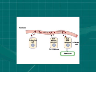
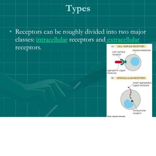
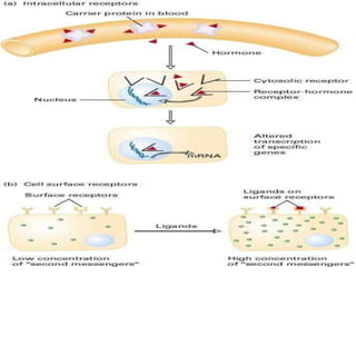
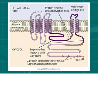
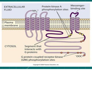
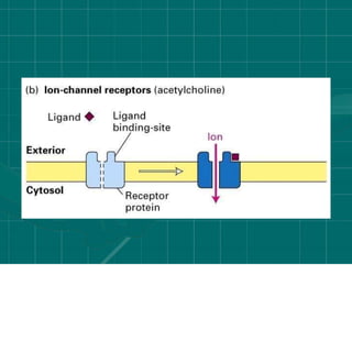
Cell signalling
Prepared By:
Anjali A Sinha
• Cellular Signaling
• Many living organisms contain billions of cells
that carry out diverse functions. In order for the
cells to cooperate, cells need to be able to
communicate with each other. Many of the
genes that cells are capable of synthesizing are
thought to be involved in cellular signaling.
Environmental stimuli
• With single-celled organisms, the variety of signal
transduction processes influence its reaction to its
environment.
• With multicellular organisms, numerous processes are
required for coordinating individual cells to support
the organism as a whole; the complexity of these
processes tend to increase with the complexity of the
organism. Sensing of environments at the cellular level
relies on signal transduction; many disease processes,
such as diabetes and heart disease arise from defects in
these pathways, highlighting the importance of this
process in biology and medicine.
• Various environmental stimuli exist that initiate
signal transmission processes in multicellular
organisms; examples include photons hitting
cells in the retina of the eye, and odorants
binding to odorant receptors in the
nasal epithelium. Certain microbial molecules,
such as viral nucleotides and protein antigens,
can elicit an immune system response against
invading pathogens mediated by signal
transduction processes.
Types of cellular signaling
• Extra cellular signaling or chemical signaling
• Cell”s direct signaling or intracellular signalling
Ectracellular signaling
• signaling by extracellular, secreted molecules can
be classified into three types — endocrine,
paracrine, or autocrine — based on the distance
over which the signal acts.
• In endocrine signaling, signaling molecules,
called hormones, act on target cells distant from
their site of synthesis by cells of endocrine
organs. In animals, an endocrine hormone
usually is carried by the blood from its site of
release to its target.
• In paracrine signaling, the signaling molecules
released by a cell only affect target cells in close
proximity to it. The conduction of an electric
impulse from one nerve cell to another or from
a nerve cell to a muscle cell (inducing or
inhibiting muscle contraction) occurs via
paracrine signaling.
autocrine signaling,
• cells respond to substances that they themselves
release. Many growth factors act in this fashion,
and cultured cells often secrete growth factors
that stimulate their own growth and
proliferation. This type of signaling is
particularly common in tumor cells, many of
which overproduce and release growth factors
that stimulate inappropriate, unregulated
proliferation of themselves as well as adjacent
nontumor cells; this process may lead to
formation of tumor mass.
Cell direct contact signalling
• Three types
• Gap junctions
• Surface protein interactions
• Receptors
Receptors
• In biochemistry, a receptor is a molecule found on the
surface of a cell, which receives specific chemical
signals from neighbouring cells or the wider
environment within an organism. These signals tell a
cell to do something—for example to divide or die, or
to allow certain molecules to enter or exit the cell.
• Receptors are protein molecules, embedded in either
the plasma membrane (cell surface receptors) or the
cytoplasm (nuclear receptors) of a cell, to which one
or more specific kinds ofsignaling molecules may
attach.
• A molecule which binds (attaches) to a receptor
is called a ligand, and may be a peptide (short
protein) or other small molecule, such as a
neurotransmitter, a hormone, a pharmaceutical
drug, or a toxin. Each kind of receptor can bind
only certain ligand shapes. Each cell typically has
many receptors, of many different kinds. Simply
put, a receptor functions as a keyhole that opens
a biochemical pathway when the proper ligand is
inserted.
Structure
• The shapes and actions of receptors are
studied by X-ray crystallography,
dual polarisation interferometry,
computer modelling, and structure-function
studies, which have advanced the understanding
of drug action at the binding sites of receptors.
Structure activity relationships correlate induced
conformational changes with biomolecular
activity, and are studied using dynamic
techniques such as circular dichroism and
dual polarisation interferometry.
Binding and activation
• Ligand binding is an equilibrium process.
Ligands bind to receptors and dissociate from
them according to the law of mass action.
One measure of how well a molecule fits a
receptor is the binding affinity, which is
inversely related to the dissociation constant Kd.
A good fit corresponds with high affinity and
low Kd. The final biological response (e.g.
second messenger cascade, muscle contraction),
is only achieved after a significant number of
receptors are activated.
• The receptor-ligand affinity is greater than
enzyme-substrate affinity. Whilst both
interactions are specific and reversible, there is
no chemical modification of the ligand as
seen with the substrate upon binding to its
enzyme.
Constitutive activity
• A receptor which is capable of producing its biological
response in the absence of a bound ligand is said to
display "constitutive activity". The constitutive activity
of receptors may be blocked by inverse agonist binding.
Mutations in receptors that result in increased
constitutive activity underlie some inherited diseases,
such as precocious puberty (due to mutations in
luteinizing hormone receptors) and hyperthyroidism
(due to mutations in thyroid-stimulating hormone
receptors).
Ligands
• (Full) agonists are able to activate the receptor and result
in a maximal biological response. Most natural ligands
are full agonists.
• Partial agonists do not activate receptors thoroughly,
causing responses which are partial compared to those
of full agonists.
• Antagonists bind to receptors but do not activate them.
This results in receptor blockage, inhibiting the binding
of other agonists.
• Inverse agonists reduce the activity of receptors
by inhibiting their constitutive activity.
Cell surface receptor
• Cell surface receptors (membrane
receptors, transmembrane receptors) are
specialized integral membrane proteins that take
part in communication between the cell and the
outside world. Extracellular signaling molecules
(usually hormones,neurotransmitters, cytokines
, growth factors or cell recognition molecules)
attach to the receptor, triggering changes in the
function of the cell. This process is called
signal transduction:
• The binding initiates a chemical change on the
intracellular side of the membrane. In this way
the receptors play a unique and important role in
cellular communications and signal transduction.
Types
• Receptors can be roughly divided into two major
classes: intracellular receptors and extracellular
receptors.
Extracellular receptors
• Extracellular receptors are integral transmembrane proteins and
make up most receptors. They span the plasma membrane of the
cell, with one part of the receptor on the outside of the cell and
the other on the inside. Signal transduction occurs as a result of a
ligand binding to the outside; the molecule does not pass
through the membrane. This binding stimulates a series of events
inside the cell; different types of receptor stimulate different
responses and receptors typically respond to only the binding of
a specific ligand. Upon binding, the ligand induces a change in
the conformation of the inside part of the receptor. These result
in either the activation of an enzyme in the receptor or the
exposure of a binding site for other intracellular signaling
proteins within the cell, eventually propagating the signal
through the cytoplasm.
• These are transmembrane recptors of
various types
• Having 3 domains
The extracellular domain
• The extracellular domain is the part of the
receptor that sticks out of the membrane on
the outside of the cell or organelle. If the
polypeptide chain of the receptor crosses the
bilayer several times, the external domain can
comprise several "loops" sticking out of the
membrane.
the transmembrane domains
• In the majority of receptors for which
structural evidence exists, transmembrane
alpha helices make up most of the
transmembrane domain.
In certain receptors, such as the
nicotinic acetylcholine receptor, the
transmembrane domain forms a protein-lined
pore through the membrane, or ion channel.
Upon activation of an extracellular domain
by binding of the appropriate ligand, the
pore becomes accessible to ions, which then
pass through.
• In other receptors, the transmembrane domains
are presumed to undergo a conformational
change upon binding, which exerts an effect
intracellularly. In some receptors, such as
members of the 7TM superfamily, the
transmembrane domain may contain the ligand
binding pocket
intracellular (or cytoplasmic)
domain
• The intracellular (or cytoplasmic) domain of the
receptor interacts with the interior of the cell or
organelle, relaying the signal. There are two
fundamentally different ways for this interaction:
• The intracellular domain communicates via specific
protein-protein-interactions with effector proteins, which
in turn send the signal along a signal chain to its
destination.
• With enzyme-linked receptors, the intracellular domain
has enzymatic activity. Often, this is a tyrosine kinase
activity. The enzymatic activity can also be located on
an enzyme associated with the intracellular domain.
• processes through membrane receptors involve
the External Reactions, in which the ligand
binds to a membrane receptor, and the Internal
Reactions, in which intracellular response is
triggered.
• Based on structural and functional similarities,
membrane receptors are mainly divided into 3
classes: The ion channel-linked receptor; The
enzyme-linked receptor and
G protein-coupled receptor.
Ion channel linked receptors
• Ion channel linked receptors are ion-channels
(including cation-channels and anion-channels)
themselves and constitute a large family of
multipass transmembrane proteins. They are
involved in rapid signaling events most generally
found in electrically excitable cells such as
neurons and are also called
ligand-gated ion channels. Opening and closing
of Ion channels are controlled by
neurotransmitters.
Enzyme-linked receptors
• Enzyme-linked receptors are either enzymes
themselves, or are directly associated with the
enzymes that they activate. These are usually
single-pass transmembrane receptors, with the
enzymatic portion of the receptor being
intracellular. The majority of enzyme-lined
receptors are protein kinases, or associate with
protein kinases.
G protein-coupled receptors
• G protein-coupled receptors are integral membrane
proteins that possess seven membrane-spanning
domains or transmembrane helices. These receptors
activate a G protein ligand binding. G-protein is a
trimeric protein. The 3 subunits are called α 、 β and γ.
The α subunit can bind with guanosine diphosphate,
GDP. This causesphosphorylation of the GDP to
guanosine triphosphate, GTP, and activates the α
subunit, which then dissociates from the β and γ
subunits. The activated α subunit can further affect
intracellular signaling proteins or target functional
proteins directly.
G Protein-Linked
Receptors
• Signal transduction through membrane
receptors usually requires four characters:
• Extracellular signal molecule: an extracellular
signal molecule is produced by one cell and is
capable of traveling to neighboring cells, or
to cells that may be far away.
• Receptor protein: the cells in an organism must
have cell surface receptor proteins that bind to
the signal molecule and communicate its
presence inward into the cell.
four Stages of Signal Transductio
• Intracellular signaling proteins: these distribute
the signal to the appropriate parts of the cell.
The binding of the signal molecule to the
receptor protein will activate intracellular
signaling proteins that initiate a signaling cascade
(a series of intracellular signaling molecules that
act sequentially).
• Target proteins: the conformations or other
properties of the target proteins are altered
when a signaling pathway is active and changes
the behavior of the cell.
Three Stages of Signal
Transduction
NEXT
• Detailed role of G-protein in signal
transduction
•Thanks

Lecture 3 cellsignalling

  • 1.
  • 2.
    • Cellular Signaling •Many living organisms contain billions of cells that carry out diverse functions. In order for the cells to cooperate, cells need to be able to communicate with each other. Many of the genes that cells are capable of synthesizing are thought to be involved in cellular signaling.
  • 3.
    Environmental stimuli • Withsingle-celled organisms, the variety of signal transduction processes influence its reaction to its environment. • With multicellular organisms, numerous processes are required for coordinating individual cells to support the organism as a whole; the complexity of these processes tend to increase with the complexity of the organism. Sensing of environments at the cellular level relies on signal transduction; many disease processes, such as diabetes and heart disease arise from defects in these pathways, highlighting the importance of this process in biology and medicine.
  • 4.
    • Various environmentalstimuli exist that initiate signal transmission processes in multicellular organisms; examples include photons hitting cells in the retina of the eye, and odorants binding to odorant receptors in the nasal epithelium. Certain microbial molecules, such as viral nucleotides and protein antigens, can elicit an immune system response against invading pathogens mediated by signal transduction processes.
  • 5.
    Types of cellularsignaling • Extra cellular signaling or chemical signaling • Cell”s direct signaling or intracellular signalling
  • 6.
    Ectracellular signaling • signalingby extracellular, secreted molecules can be classified into three types — endocrine, paracrine, or autocrine — based on the distance over which the signal acts.
  • 7.
    • In endocrinesignaling, signaling molecules, called hormones, act on target cells distant from their site of synthesis by cells of endocrine organs. In animals, an endocrine hormone usually is carried by the blood from its site of release to its target.
  • 9.
    • In paracrinesignaling, the signaling molecules released by a cell only affect target cells in close proximity to it. The conduction of an electric impulse from one nerve cell to another or from a nerve cell to a muscle cell (inducing or inhibiting muscle contraction) occurs via paracrine signaling.
  • 11.
    autocrine signaling, • cellsrespond to substances that they themselves release. Many growth factors act in this fashion, and cultured cells often secrete growth factors that stimulate their own growth and proliferation. This type of signaling is particularly common in tumor cells, many of which overproduce and release growth factors that stimulate inappropriate, unregulated proliferation of themselves as well as adjacent nontumor cells; this process may lead to formation of tumor mass.
  • 14.
    Cell direct contactsignalling • Three types • Gap junctions • Surface protein interactions • Receptors
  • 15.
    Receptors • In biochemistry,a receptor is a molecule found on the surface of a cell, which receives specific chemical signals from neighbouring cells or the wider environment within an organism. These signals tell a cell to do something—for example to divide or die, or to allow certain molecules to enter or exit the cell. • Receptors are protein molecules, embedded in either the plasma membrane (cell surface receptors) or the cytoplasm (nuclear receptors) of a cell, to which one or more specific kinds ofsignaling molecules may attach.
  • 16.
    • A moleculewhich binds (attaches) to a receptor is called a ligand, and may be a peptide (short protein) or other small molecule, such as a neurotransmitter, a hormone, a pharmaceutical drug, or a toxin. Each kind of receptor can bind only certain ligand shapes. Each cell typically has many receptors, of many different kinds. Simply put, a receptor functions as a keyhole that opens a biochemical pathway when the proper ligand is inserted.
  • 17.
    Structure • The shapesand actions of receptors are studied by X-ray crystallography, dual polarisation interferometry, computer modelling, and structure-function studies, which have advanced the understanding of drug action at the binding sites of receptors. Structure activity relationships correlate induced conformational changes with biomolecular activity, and are studied using dynamic techniques such as circular dichroism and dual polarisation interferometry.
  • 18.
    Binding and activation •Ligand binding is an equilibrium process. Ligands bind to receptors and dissociate from them according to the law of mass action. One measure of how well a molecule fits a receptor is the binding affinity, which is inversely related to the dissociation constant Kd. A good fit corresponds with high affinity and low Kd. The final biological response (e.g. second messenger cascade, muscle contraction), is only achieved after a significant number of receptors are activated.
  • 19.
    • The receptor-ligandaffinity is greater than enzyme-substrate affinity. Whilst both interactions are specific and reversible, there is no chemical modification of the ligand as seen with the substrate upon binding to its enzyme.
  • 20.
    Constitutive activity • Areceptor which is capable of producing its biological response in the absence of a bound ligand is said to display "constitutive activity". The constitutive activity of receptors may be blocked by inverse agonist binding. Mutations in receptors that result in increased constitutive activity underlie some inherited diseases, such as precocious puberty (due to mutations in luteinizing hormone receptors) and hyperthyroidism (due to mutations in thyroid-stimulating hormone receptors).
  • 21.
    Ligands • (Full) agonistsare able to activate the receptor and result in a maximal biological response. Most natural ligands are full agonists. • Partial agonists do not activate receptors thoroughly, causing responses which are partial compared to those of full agonists. • Antagonists bind to receptors but do not activate them. This results in receptor blockage, inhibiting the binding of other agonists. • Inverse agonists reduce the activity of receptors by inhibiting their constitutive activity.
  • 22.
    Cell surface receptor •Cell surface receptors (membrane receptors, transmembrane receptors) are specialized integral membrane proteins that take part in communication between the cell and the outside world. Extracellular signaling molecules (usually hormones,neurotransmitters, cytokines , growth factors or cell recognition molecules) attach to the receptor, triggering changes in the function of the cell. This process is called signal transduction:
  • 23.
    • The bindinginitiates a chemical change on the intracellular side of the membrane. In this way the receptors play a unique and important role in cellular communications and signal transduction.
  • 24.
    Types • Receptors canbe roughly divided into two major classes: intracellular receptors and extracellular receptors.
  • 25.
    Extracellular receptors • Extracellularreceptors are integral transmembrane proteins and make up most receptors. They span the plasma membrane of the cell, with one part of the receptor on the outside of the cell and the other on the inside. Signal transduction occurs as a result of a ligand binding to the outside; the molecule does not pass through the membrane. This binding stimulates a series of events inside the cell; different types of receptor stimulate different responses and receptors typically respond to only the binding of a specific ligand. Upon binding, the ligand induces a change in the conformation of the inside part of the receptor. These result in either the activation of an enzyme in the receptor or the exposure of a binding site for other intracellular signaling proteins within the cell, eventually propagating the signal through the cytoplasm.
  • 27.
    • These aretransmembrane recptors of various types • Having 3 domains
  • 28.
    The extracellular domain •The extracellular domain is the part of the receptor that sticks out of the membrane on the outside of the cell or organelle. If the polypeptide chain of the receptor crosses the bilayer several times, the external domain can comprise several "loops" sticking out of the membrane.
  • 30.
    the transmembrane domains •In the majority of receptors for which structural evidence exists, transmembrane alpha helices make up most of the transmembrane domain. In certain receptors, such as the nicotinic acetylcholine receptor, the transmembrane domain forms a protein-lined pore through the membrane, or ion channel. Upon activation of an extracellular domain by binding of the appropriate ligand, the pore becomes accessible to ions, which then pass through.
  • 32.
    • In otherreceptors, the transmembrane domains are presumed to undergo a conformational change upon binding, which exerts an effect intracellularly. In some receptors, such as members of the 7TM superfamily, the transmembrane domain may contain the ligand binding pocket
  • 33.
    intracellular (or cytoplasmic) domain •The intracellular (or cytoplasmic) domain of the receptor interacts with the interior of the cell or organelle, relaying the signal. There are two fundamentally different ways for this interaction: • The intracellular domain communicates via specific protein-protein-interactions with effector proteins, which in turn send the signal along a signal chain to its destination. • With enzyme-linked receptors, the intracellular domain has enzymatic activity. Often, this is a tyrosine kinase activity. The enzymatic activity can also be located on an enzyme associated with the intracellular domain.
  • 35.
    • processes throughmembrane receptors involve the External Reactions, in which the ligand binds to a membrane receptor, and the Internal Reactions, in which intracellular response is triggered.
  • 36.
    • Based onstructural and functional similarities, membrane receptors are mainly divided into 3 classes: The ion channel-linked receptor; The enzyme-linked receptor and G protein-coupled receptor.
  • 37.
    Ion channel linkedreceptors • Ion channel linked receptors are ion-channels (including cation-channels and anion-channels) themselves and constitute a large family of multipass transmembrane proteins. They are involved in rapid signaling events most generally found in electrically excitable cells such as neurons and are also called ligand-gated ion channels. Opening and closing of Ion channels are controlled by neurotransmitters.
  • 39.
    Enzyme-linked receptors • Enzyme-linkedreceptors are either enzymes themselves, or are directly associated with the enzymes that they activate. These are usually single-pass transmembrane receptors, with the enzymatic portion of the receptor being intracellular. The majority of enzyme-lined receptors are protein kinases, or associate with protein kinases.
  • 42.
    G protein-coupled receptors •G protein-coupled receptors are integral membrane proteins that possess seven membrane-spanning domains or transmembrane helices. These receptors activate a G protein ligand binding. G-protein is a trimeric protein. The 3 subunits are called α 、 β and γ. The α subunit can bind with guanosine diphosphate, GDP. This causesphosphorylation of the GDP to guanosine triphosphate, GTP, and activates the α subunit, which then dissociates from the β and γ subunits. The activated α subunit can further affect intracellular signaling proteins or target functional proteins directly.
  • 43.
  • 46.
    • Signal transductionthrough membrane receptors usually requires four characters: • Extracellular signal molecule: an extracellular signal molecule is produced by one cell and is capable of traveling to neighboring cells, or to cells that may be far away. • Receptor protein: the cells in an organism must have cell surface receptor proteins that bind to the signal molecule and communicate its presence inward into the cell. four Stages of Signal Transductio
  • 47.
    • Intracellular signalingproteins: these distribute the signal to the appropriate parts of the cell. The binding of the signal molecule to the receptor protein will activate intracellular signaling proteins that initiate a signaling cascade (a series of intracellular signaling molecules that act sequentially). • Target proteins: the conformations or other properties of the target proteins are altered when a signaling pathway is active and changes the behavior of the cell.
  • 48.
    Three Stages ofSignal Transduction
  • 49.
    NEXT • Detailed roleof G-protein in signal transduction
  • 50.