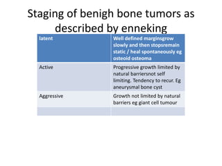
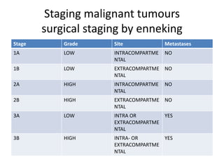
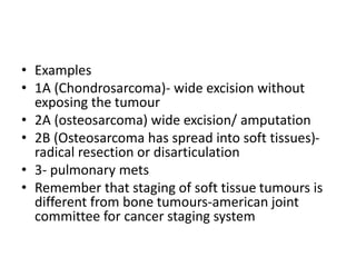
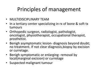
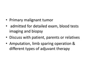
BONE TUMOURS
• Discussion on:
• Tumours
• Tumour –like lesions
• Cysts
• Benigh lesion are quite common
• Primary malignant are rare
• Often they mimick each other
classification
• Based on recognition of dominant tx in the
various lesions
• Assignment: Find Modified WHO
classification;
Clinical features
History
• Usually prolonged & there is delay in
obtaining rx
• Asymptomatic –abnormality seen on X-ray/
more likely benigh.
– Mostly benigh in children. Rare after 30yrs. So
resolve on their own/spontaneously eg. fibroma
– Malignant- slow growing or situated where there
is room incospicuous expansion (cavity eg. pelvis)
Age; The age of the pt gives a useful clue
– childhood & adolescents- benigh. Also some
malignant primary tumors eg. ewings sarcoma, &
osteosarcoma.
– 4th and 6th decade; Chondrosarcoma &
fibrosarcoma- typically occur occur in older
people.
Myeloma – the commonest of all primary
tumors is seldom seen before 6th decade.
> 70 years- metastatic bone tumors are more
common than all primary tumors together .
Pain- mmemonic = socrates
• Common
• Non-specific; cannot tell nature of lesion
• Severe pain of rapid onset
– Rapid expansion with stretching of surrounding txs
– Central hemorrhage or degeneration in the tumor.
– Pathological #.
– Tiny lesion can be encapsulated in dense bone e.g osteoid
osteoma.
Swelling- lump,
– patient seek advice when its painful or continues to grow.
Trauma
• is usually a ‘red herring’.
• ?initiate a pathological change or draw attention
Neurological symptoms –
• paraesthesia/ numbness may present upon or
stretching of a peripheral nerve.
• Progressive dysfxn is more ominous & suggest
invasion by an aggressive tumor.
Pathological #s may be the first & only clinical
symptom
• Suspicion- slight injury
• Elderly with midshaft #.
Exam;
Lump;
• Where does it arise
• Is it discrete or ill defined
• Soft or hard
• Pulsatile
• Tender
Swelling-? haematoma / infection. Diffuse overlying skin is warm &
inflamed.
Near joint-cause effusion & limitation of movement.
Spinal lesions- cause muscle spasms, back stiffness or painful scoliosis.
Limited R.O.M.
Lymphatic drainage-pelvis abdomen chest or spine.
Radiological exam
• X-ray
• Radionuclide scanning
• CT SCAN-More accurate( intra & extra-osseous
extension)& relationship to the surrounding
structures
– Inaccessible sites eg. spine
– Pulmonary mets –more reliable.
MRI
Assessment of tumor spread
a. Within the bone
b. Into nearby jt
c. Into soft txs
Blood vessels & perivascular tx relationship
Soft tx tumors and cartilage lesions.
Radiographs/ xray
Plain x-rays are still the most useful of all
imaging techniques
Abnormality (obvious) in the bone:
Cortical thickening
Discrete lump
Cyst
Ill- defined destruction
Where: metaphysis or diaphysis
• Solitary or multiple lesions/ skip lesions
• Margins: well defined- cyst, ill defined-
malignant
• Note: fibroma or chondroma may look like a
cyst
• Stipled calcification within a cystic area is xtic
of cartilage tumors
• Bone surface-
– periosteal new bone formation
– Extension of tumor into soft txs
• Muscles: muscle planes/calcification
• Bone scans, CT & MRI before undertaking a
biopsy
Radionuclide scanning/ scintigraphy
• Small tumors e.g osteoid osteoma
• Skip lesions or silent secondary deposits.
LABORATORY EXAM/ BLOOD TESTS
Used to exclude other conditions e.g infection or
metabolic bone disorders, or brown tumor in
hyperthyroidism.
Anemia
Raised esr
Raised serum alkaline phosphatase
• Not specific but Levels can differentiate btn malignant
& benigh
Serum protein electrophoresis , Bence jones proteins-
myeloma
Raised Serum acid phosphatase in ca prostate
Biopsy
Needle biopsy- may be u/s or ct guided
Sample for histologic diagnosis or microbiology
OPEN BIOPSY; incisional biopsy.
More reliable to obtain representative sample
As little as possible of the tumor is exposed
Block of tx is removed from the boundary zone;
normal tx, pseudocapsule, abnormal tissue.
Benigh tumor- excision biopsy.
Differential diagnosis
Diseases that mimick tumors
• Clinically
• Radiologically
• Histopathology difficult to interpret
Soft tx hematoma
Myositis ossificans
Stress #.
Tendon avulsion injury- osgood-Schlatter dx
Bone infection
Gout
Other bone -Fibrous cortical / medullary infarct & bone
islands
Staging of benigh tumours;
enneking staging
BY enneking in 1986
Treating tumours
• The lesion must be removed widely enough to prevent recurrence
• Damage must be kept minimum
Treatment depend on how tumor usually behaves;(aggressiveness)
• Cytological
• Clinical behaviour
How far it has spread
Benigh; latent, active, aggressive.
Least aggressive disappear spontaneously
Most aggressive undergo malignant change
MALIGNANT;
• Low grade- moderate aggressive, take long to
metastasize eg. chondrosarcoma, parosteal
osteosarcoma
• High grade- very aggressive- metastasize early
eg osteosarcoma, fibrosarcoma.
spread
• If there are no mets, local spread determine
how much to be removed
• Intracompartmental- confined to an enclosed
space eg a bone, jt cavity, or a muscle group
within a fascial envelope
• Extracompartmental;- those that extend into
interfascial or extrafascial planes with no
natural barrier to proximal or distal spread
• Eg perivascular sheath, pelvis, axilla
• Ct and mri are important to determine spread
• Skip lesions are revealed by scintigraphy
Staging of benigh bone tumors as
described by enneking
latent Well defined marginsgrow
slowly and then stopsremain
static / heal spontaneously eg
osteoid osteoma
Active Progressive growth limited by
natural barriersnot self
limiting. Tendency to recur. Eg
aneurysmal bone cyst
Aggressive Growth not limited by natural
barriers eg giant cell tumour
Surgical stage
• Staging the tumour is an important step towards
selecting the operation best suited to the
particular pt and carrying a low risk of recurrence
• Bone sarcomas are broadly divided as follows
• Stage1 all low grade sarcomas
• Stage2; histologically high grade lesions
• Stage 3 sarcomas which have metastasized
• Each category is further subdivided into type A
INTRACOMPARTMENTAL
• & type B -EXTRACOMPARTMENTAL
Staging malignant tumours
surgical staging by enneking
Stage Grade Site Metastases
1A LOW INTRACOMPARTME
NTAL
NO
1B LOW EXTRACOMPARTME
NTAL
NO
2A HIGH INTRACOMPARTME
NTAL
NO
2B HIGH EXTRACOMPARTME
NTAL
NO
3A LOW INTRA OR
EXTRACOMPARTME
NTAL
YES
3B HIGH INTRA- OR
EXTRACOMPARTME
NTAL
YES
• Examples
• 1A (Chondrosarcoma)- wide excision without
exposing the tumour
• 2A (osteosarcoma) wide excision/ amputation
• 2B (Osteosarcoma has spread into soft tissues)-
radical resection or disarticulation
• 3- pulmonary mets
• Remember that staging of soft tissue tumours is
different from bone tumours-american joint
committee for cancer staging system
Principles of management
• MULTIDISCIPLINARY TEAM
• In a tertiary center specializing in rx of bone & soft tx
tumours
• Orthopedic surgeon, radiologist, pathologist,
oncologist, physiotherapist, occupational therapist,
prosthetist.
• Benigh asymptomatic lesion- diagnosis beyond doubt;
no treatment. If not clear diagnosis,biopsy by excision
or curretage.
• Benigh symptomatic or enlarging- removal by
local(marginal excision) or curretage
• Suspected malignant tumour
• Primary malignant tumor
• admitted for detailed exam, blood tests
imaging and biopsy
• Discuss with patient, parents or relatives
• Amputation, limb sparing operation &
different types of adjuvant therapy
Methods of treatment
• Tumor excision; the more aggressive the lesion, the
more widely does it to be excised.
• Intracapsular (intralesional) excision and curretage-
incomplete form of tumor ablation. Applicable to
benigh lesions- low risk of recurrence or incurable
tomor- debulking to relieve local symptoms. Acrylic
cement prevent recurrence.
• MARGINAL EXCISION- goes beyond the tumor. Through
reactive zone, there is risk of recurrence(50%).
Therefore benigh lesions. Resulting cavity is filled with
bone graft.
• WIDE EXCISION: dissection is carried out ell clear
of the tumor, through normal tissues
• Good for grade 1A, providing risk of recurrence
below 10%
• Wide excision +chemotherapy for grade 2A
• RADICAL EXCISION: the entire compartment in
which the tumor lies is removed without
exposing the lesion
• Can also be achieved through amputation. Grade
2A&2B.
• LIMB SALVAGE: amputation is not automatic
choice for grade 2 sarcomas. Advances in imaging
& chemotherapy have made limb salvage the
treatment of choice for many patients. Bone
grafting &end prosthetic replacement,
arthrodesis, distraction osteosynthesis.
• AMPUTATION: amputation and early
rehabilitation may be the wisest option. May be
curative or for local control of aggressive tumor.
• MULTI-AGENT CHEMOTHERAPY: neo-adjuvant &
adjuvant treatment for malignant bone and soft
tissue tumors. Reduce the size of primary lesions.
Prevent metastatic seeding & improve the chance
of survival. Methotrexatre,
doxorubicin(adriamycin) cyclophosphamide,
vincristine & cis-platinum. Rx is started 8-12
weeks pre-op. continued 6-12 months post-op
• Radiotherapy restricted to highly sensitive tumors
e.g Ewing’s sarcoma
complications
• Post-irradiation spindle cell sarcoma
• Pathological #s

LEC 1; INTRODUCTION TO BONE TUMOURS.pptx

  • 1.
  • 2.
    • Discussion on: •Tumours • Tumour –like lesions • Cysts • Benigh lesion are quite common • Primary malignant are rare • Often they mimick each other
  • 3.
    classification • Based onrecognition of dominant tx in the various lesions • Assignment: Find Modified WHO classification;
  • 4.
    Clinical features History • Usuallyprolonged & there is delay in obtaining rx • Asymptomatic –abnormality seen on X-ray/ more likely benigh. – Mostly benigh in children. Rare after 30yrs. So resolve on their own/spontaneously eg. fibroma – Malignant- slow growing or situated where there is room incospicuous expansion (cavity eg. pelvis)
  • 5.
    Age; The ageof the pt gives a useful clue – childhood & adolescents- benigh. Also some malignant primary tumors eg. ewings sarcoma, & osteosarcoma. – 4th and 6th decade; Chondrosarcoma & fibrosarcoma- typically occur occur in older people. Myeloma – the commonest of all primary tumors is seldom seen before 6th decade. > 70 years- metastatic bone tumors are more common than all primary tumors together .
  • 6.
    Pain- mmemonic =socrates • Common • Non-specific; cannot tell nature of lesion • Severe pain of rapid onset – Rapid expansion with stretching of surrounding txs – Central hemorrhage or degeneration in the tumor. – Pathological #. – Tiny lesion can be encapsulated in dense bone e.g osteoid osteoma. Swelling- lump, – patient seek advice when its painful or continues to grow.
  • 7.
    Trauma • is usuallya ‘red herring’. • ?initiate a pathological change or draw attention Neurological symptoms – • paraesthesia/ numbness may present upon or stretching of a peripheral nerve. • Progressive dysfxn is more ominous & suggest invasion by an aggressive tumor. Pathological #s may be the first & only clinical symptom • Suspicion- slight injury • Elderly with midshaft #.
  • 8.
    Exam; Lump; • Where doesit arise • Is it discrete or ill defined • Soft or hard • Pulsatile • Tender Swelling-? haematoma / infection. Diffuse overlying skin is warm & inflamed. Near joint-cause effusion & limitation of movement. Spinal lesions- cause muscle spasms, back stiffness or painful scoliosis. Limited R.O.M. Lymphatic drainage-pelvis abdomen chest or spine.
  • 9.
    Radiological exam • X-ray •Radionuclide scanning • CT SCAN-More accurate( intra & extra-osseous extension)& relationship to the surrounding structures – Inaccessible sites eg. spine – Pulmonary mets –more reliable.
  • 10.
    MRI Assessment of tumorspread a. Within the bone b. Into nearby jt c. Into soft txs Blood vessels & perivascular tx relationship Soft tx tumors and cartilage lesions.
  • 11.
    Radiographs/ xray Plain x-raysare still the most useful of all imaging techniques Abnormality (obvious) in the bone: Cortical thickening Discrete lump Cyst Ill- defined destruction Where: metaphysis or diaphysis
  • 12.
    • Solitary ormultiple lesions/ skip lesions • Margins: well defined- cyst, ill defined- malignant • Note: fibroma or chondroma may look like a cyst • Stipled calcification within a cystic area is xtic of cartilage tumors
  • 13.
    • Bone surface- –periosteal new bone formation – Extension of tumor into soft txs • Muscles: muscle planes/calcification • Bone scans, CT & MRI before undertaking a biopsy
  • 14.
    Radionuclide scanning/ scintigraphy •Small tumors e.g osteoid osteoma • Skip lesions or silent secondary deposits.
  • 15.
    LABORATORY EXAM/ BLOODTESTS Used to exclude other conditions e.g infection or metabolic bone disorders, or brown tumor in hyperthyroidism. Anemia Raised esr Raised serum alkaline phosphatase • Not specific but Levels can differentiate btn malignant & benigh Serum protein electrophoresis , Bence jones proteins- myeloma Raised Serum acid phosphatase in ca prostate
  • 16.
    Biopsy Needle biopsy- maybe u/s or ct guided Sample for histologic diagnosis or microbiology OPEN BIOPSY; incisional biopsy. More reliable to obtain representative sample As little as possible of the tumor is exposed Block of tx is removed from the boundary zone; normal tx, pseudocapsule, abnormal tissue. Benigh tumor- excision biopsy.
  • 17.
    Differential diagnosis Diseases thatmimick tumors • Clinically • Radiologically • Histopathology difficult to interpret Soft tx hematoma Myositis ossificans Stress #. Tendon avulsion injury- osgood-Schlatter dx Bone infection Gout Other bone -Fibrous cortical / medullary infarct & bone islands
  • 18.
    Staging of benightumours; enneking staging BY enneking in 1986 Treating tumours • The lesion must be removed widely enough to prevent recurrence • Damage must be kept minimum Treatment depend on how tumor usually behaves;(aggressiveness) • Cytological • Clinical behaviour How far it has spread Benigh; latent, active, aggressive. Least aggressive disappear spontaneously Most aggressive undergo malignant change
  • 19.
    MALIGNANT; • Low grade-moderate aggressive, take long to metastasize eg. chondrosarcoma, parosteal osteosarcoma • High grade- very aggressive- metastasize early eg osteosarcoma, fibrosarcoma.
  • 20.
    spread • If thereare no mets, local spread determine how much to be removed • Intracompartmental- confined to an enclosed space eg a bone, jt cavity, or a muscle group within a fascial envelope • Extracompartmental;- those that extend into interfascial or extrafascial planes with no natural barrier to proximal or distal spread • Eg perivascular sheath, pelvis, axilla
  • 21.
    • Ct andmri are important to determine spread • Skip lesions are revealed by scintigraphy
  • 22.
    Staging of benighbone tumors as described by enneking latent Well defined marginsgrow slowly and then stopsremain static / heal spontaneously eg osteoid osteoma Active Progressive growth limited by natural barriersnot self limiting. Tendency to recur. Eg aneurysmal bone cyst Aggressive Growth not limited by natural barriers eg giant cell tumour
  • 23.
    Surgical stage • Stagingthe tumour is an important step towards selecting the operation best suited to the particular pt and carrying a low risk of recurrence • Bone sarcomas are broadly divided as follows • Stage1 all low grade sarcomas • Stage2; histologically high grade lesions • Stage 3 sarcomas which have metastasized • Each category is further subdivided into type A INTRACOMPARTMENTAL • & type B -EXTRACOMPARTMENTAL
  • 24.
    Staging malignant tumours surgicalstaging by enneking Stage Grade Site Metastases 1A LOW INTRACOMPARTME NTAL NO 1B LOW EXTRACOMPARTME NTAL NO 2A HIGH INTRACOMPARTME NTAL NO 2B HIGH EXTRACOMPARTME NTAL NO 3A LOW INTRA OR EXTRACOMPARTME NTAL YES 3B HIGH INTRA- OR EXTRACOMPARTME NTAL YES
  • 25.
    • Examples • 1A(Chondrosarcoma)- wide excision without exposing the tumour • 2A (osteosarcoma) wide excision/ amputation • 2B (Osteosarcoma has spread into soft tissues)- radical resection or disarticulation • 3- pulmonary mets • Remember that staging of soft tissue tumours is different from bone tumours-american joint committee for cancer staging system
  • 26.
    Principles of management •MULTIDISCIPLINARY TEAM • In a tertiary center specializing in rx of bone & soft tx tumours • Orthopedic surgeon, radiologist, pathologist, oncologist, physiotherapist, occupational therapist, prosthetist. • Benigh asymptomatic lesion- diagnosis beyond doubt; no treatment. If not clear diagnosis,biopsy by excision or curretage. • Benigh symptomatic or enlarging- removal by local(marginal excision) or curretage • Suspected malignant tumour
  • 27.
    • Primary malignanttumor • admitted for detailed exam, blood tests imaging and biopsy • Discuss with patient, parents or relatives • Amputation, limb sparing operation & different types of adjuvant therapy
  • 28.
    Methods of treatment •Tumor excision; the more aggressive the lesion, the more widely does it to be excised. • Intracapsular (intralesional) excision and curretage- incomplete form of tumor ablation. Applicable to benigh lesions- low risk of recurrence or incurable tomor- debulking to relieve local symptoms. Acrylic cement prevent recurrence. • MARGINAL EXCISION- goes beyond the tumor. Through reactive zone, there is risk of recurrence(50%). Therefore benigh lesions. Resulting cavity is filled with bone graft.
  • 29.
    • WIDE EXCISION:dissection is carried out ell clear of the tumor, through normal tissues • Good for grade 1A, providing risk of recurrence below 10% • Wide excision +chemotherapy for grade 2A • RADICAL EXCISION: the entire compartment in which the tumor lies is removed without exposing the lesion • Can also be achieved through amputation. Grade 2A&2B.
  • 30.
    • LIMB SALVAGE:amputation is not automatic choice for grade 2 sarcomas. Advances in imaging & chemotherapy have made limb salvage the treatment of choice for many patients. Bone grafting &end prosthetic replacement, arthrodesis, distraction osteosynthesis. • AMPUTATION: amputation and early rehabilitation may be the wisest option. May be curative or for local control of aggressive tumor.
  • 31.
    • MULTI-AGENT CHEMOTHERAPY:neo-adjuvant & adjuvant treatment for malignant bone and soft tissue tumors. Reduce the size of primary lesions. Prevent metastatic seeding & improve the chance of survival. Methotrexatre, doxorubicin(adriamycin) cyclophosphamide, vincristine & cis-platinum. Rx is started 8-12 weeks pre-op. continued 6-12 months post-op • Radiotherapy restricted to highly sensitive tumors e.g Ewing’s sarcoma
  • 32.
    complications • Post-irradiation spindlecell sarcoma • Pathological #s