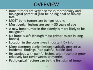
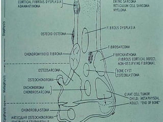
Benign bone tumors are diverse in morphology and biological potential. Most bone tumors are benign lesions seen in patients under 30 years old. Radiology is important for determining the exact location, extent of growth, and aggressiveness of bone tumors. The best test for diagnosis is a biopsy, as it confirms if a tumor is malignant or benign and determines the bone cancer type and stage. There are several classification systems for bone tumors, including the Enneking and Campanacci staging systems, which help determine prognosis and appropriate treatment.
8% of all bone tumors present in spine
25-30% of bone tumors are benign
Peak age: 2-3rd decade
Posterior element involved: osteoid osteoma, osteoblastoma, aneurysmal bone cyst
Anterior element involved: giant cell tumor, hemangioma, eosinophilic granuloma
8% of all bone tumors present in spine
25-30% of bone tumors are benign
Peak age: 2-3rd decade
Posterior element involved: osteoid osteoma, osteoblastoma, aneurysmal bone cyst
Anterior element involved: giant cell tumor, hemangioma, eosinophilic granuloma
Knee anatomy and clinical tests 2024.pdfvimalpl1234
This includes all relevant anatomy and clinical tests compiled from standard textbooks, Campbell,netter etc..It is comprehensive and best suited for orthopaedicians and orthopaedic residents.
ARTIFICIAL INTELLIGENCE IN HEALTHCARE.pdfAnujkumaranit
Artificial intelligence (AI) refers to the simulation of human intelligence processes by machines, especially computer systems. It encompasses tasks such as learning, reasoning, problem-solving, perception, and language understanding. AI technologies are revolutionizing various fields, from healthcare to finance, by enabling machines to perform tasks that typically require human intelligence.
These lecture slides, by Dr Sidra Arshad, offer a quick overview of physiological basis of a normal electrocardiogram.
Learning objectives:
1. Define an electrocardiogram (ECG) and electrocardiography
2. Describe how dipoles generated by the heart produce the waveforms of the ECG
3. Describe the components of a normal electrocardiogram of a typical bipolar leads (limb II)
4. Differentiate between intervals and segments
5. Enlist some common indications for obtaining an ECG
Study Resources:
1. Chapter 11, Guyton and Hall Textbook of Medical Physiology, 14th edition
2. Chapter 9, Human Physiology - From Cells to Systems, Lauralee Sherwood, 9th edition
3. Chapter 29, Ganong’s Review of Medical Physiology, 26th edition
4. Electrocardiogram, StatPearls - https://www.ncbi.nlm.nih.gov/books/NBK549803/
5. ECG in Medical Practice by ABM Abdullah, 4th edition
6. ECG Basics, http://www.nataliescasebook.com/tag/e-c-g-basics
These simplified slides by Dr. Sidra Arshad present an overview of the non-respiratory functions of the respiratory tract.
Learning objectives:
1. Enlist the non-respiratory functions of the respiratory tract
2. Briefly explain how these functions are carried out
3. Discuss the significance of dead space
4. Differentiate between minute ventilation and alveolar ventilation
5. Describe the cough and sneeze reflexes
Study Resources:
1. Chapter 39, Guyton and Hall Textbook of Medical Physiology, 14th edition
2. Chapter 34, Ganong’s Review of Medical Physiology, 26th edition
3. Chapter 17, Human Physiology by Lauralee Sherwood, 9th edition
4. Non-respiratory functions of the lungs https://academic.oup.com/bjaed/article/13/3/98/278874
Explore natural remedies for syphilis treatment in Singapore. Discover alternative therapies, herbal remedies, and lifestyle changes that may complement conventional treatments. Learn about holistic approaches to managing syphilis symptoms and supporting overall health.
Title: Sense of Smell
Presenter: Dr. Faiza, Assistant Professor of Physiology
Qualifications:
MBBS (Best Graduate, AIMC Lahore)
FCPS Physiology
ICMT, CHPE, DHPE (STMU)
MPH (GC University, Faisalabad)
MBA (Virtual University of Pakistan)
Learning Objectives:
Describe the primary categories of smells and the concept of odor blindness.
Explain the structure and location of the olfactory membrane and mucosa, including the types and roles of cells involved in olfaction.
Describe the pathway and mechanisms of olfactory signal transmission from the olfactory receptors to the brain.
Illustrate the biochemical cascade triggered by odorant binding to olfactory receptors, including the role of G-proteins and second messengers in generating an action potential.
Identify different types of olfactory disorders such as anosmia, hyposmia, hyperosmia, and dysosmia, including their potential causes.
Key Topics:
Olfactory Genes:
3% of the human genome accounts for olfactory genes.
400 genes for odorant receptors.
Olfactory Membrane:
Located in the superior part of the nasal cavity.
Medially: Folds downward along the superior septum.
Laterally: Folds over the superior turbinate and upper surface of the middle turbinate.
Total surface area: 5-10 square centimeters.
Olfactory Mucosa:
Olfactory Cells: Bipolar nerve cells derived from the CNS (100 million), with 4-25 olfactory cilia per cell.
Sustentacular Cells: Produce mucus and maintain ionic and molecular environment.
Basal Cells: Replace worn-out olfactory cells with an average lifespan of 1-2 months.
Bowman’s Gland: Secretes mucus.
Stimulation of Olfactory Cells:
Odorant dissolves in mucus and attaches to receptors on olfactory cilia.
Involves a cascade effect through G-proteins and second messengers, leading to depolarization and action potential generation in the olfactory nerve.
Quality of a Good Odorant:
Small (3-20 Carbon atoms), volatile, water-soluble, and lipid-soluble.
Facilitated by odorant-binding proteins in mucus.
Membrane Potential and Action Potential:
Resting membrane potential: -55mV.
Action potential frequency in the olfactory nerve increases with odorant strength.
Adaptation Towards the Sense of Smell:
Rapid adaptation within the first second, with further slow adaptation.
Psychological adaptation greater than receptor adaptation, involving feedback inhibition from the central nervous system.
Primary Sensations of Smell:
Camphoraceous, Musky, Floral, Pepperminty, Ethereal, Pungent, Putrid.
Odor Detection Threshold:
Examples: Hydrogen sulfide (0.0005 ppm), Methyl-mercaptan (0.002 ppm).
Some toxic substances are odorless at lethal concentrations.
Characteristics of Smell:
Odor blindness for single substances due to lack of appropriate receptor protein.
Behavioral and emotional influences of smell.
Transmission of Olfactory Signals:
From olfactory cells to glomeruli in the olfactory bulb, involving lateral inhibition.
Primitive, less old, and new olfactory systems with different path
Lung Cancer: Artificial Intelligence, Synergetics, Complex System Analysis, S...Oleg Kshivets
RESULTS: Overall life span (LS) was 2252.1±1742.5 days and cumulative 5-year survival (5YS) reached 73.2%, 10 years – 64.8%, 20 years – 42.5%. 513 LCP lived more than 5 years (LS=3124.6±1525.6 days), 148 LCP – more than 10 years (LS=5054.4±1504.1 days).199 LCP died because of LC (LS=562.7±374.5 days). 5YS of LCP after bi/lobectomies was significantly superior in comparison with LCP after pneumonectomies (78.1% vs.63.7%, P=0.00001 by log-rank test). AT significantly improved 5YS (66.3% vs. 34.8%) (P=0.00000 by log-rank test) only for LCP with N1-2. Cox modeling displayed that 5YS of LCP significantly depended on: phase transition (PT) early-invasive LC in terms of synergetics, PT N0—N12, cell ratio factors (ratio between cancer cells- CC and blood cells subpopulations), G1-3, histology, glucose, AT, blood cell circuit, prothrombin index, heparin tolerance, recalcification time (P=0.000-0.038). Neural networks, genetic algorithm selection and bootstrap simulation revealed relationships between 5YS and PT early-invasive LC (rank=1), PT N0—N12 (rank=2), thrombocytes/CC (3), erythrocytes/CC (4), eosinophils/CC (5), healthy cells/CC (6), lymphocytes/CC (7), segmented neutrophils/CC (8), stick neutrophils/CC (9), monocytes/CC (10); leucocytes/CC (11). Correct prediction of 5YS was 100% by neural networks computing (area under ROC curve=1.0; error=0.0).
CONCLUSIONS: 5YS of LCP after radical procedures significantly depended on: 1) PT early-invasive cancer; 2) PT N0--N12; 3) cell ratio factors; 4) blood cell circuit; 5) biochemical factors; 6) hemostasis system; 7) AT; 8) LC characteristics; 9) LC cell dynamics; 10) surgery type: lobectomy/pneumonectomy; 11) anthropometric data. Optimal diagnosis and treatment strategies for LC are: 1) screening and early detection of LC; 2) availability of experienced thoracic surgeons because of complexity of radical procedures; 3) aggressive en block surgery and adequate lymph node dissection for completeness; 4) precise prediction; 5) adjuvant chemoimmunoradiotherapy for LCP with unfavorable prognosis.
micro teaching on communication m.sc nursing.pdfAnurag Sharma
Microteaching is a unique model of practice teaching. It is a viable instrument for the. desired change in the teaching behavior or the behavior potential which, in specified types of real. classroom situations, tends to facilitate the achievement of specified types of objectives.
New Directions in Targeted Therapeutic Approaches for Older Adults With Mantl...i3 Health
i3 Health is pleased to make the speaker slides from this activity available for use as a non-accredited self-study or teaching resource.
This slide deck presented by Dr. Kami Maddocks, Professor-Clinical in the Division of Hematology and
Associate Division Director for Ambulatory Operations
The Ohio State University Comprehensive Cancer Center, will provide insight into new directions in targeted therapeutic approaches for older adults with mantle cell lymphoma.
STATEMENT OF NEED
Mantle cell lymphoma (MCL) is a rare, aggressive B-cell non-Hodgkin lymphoma (NHL) accounting for 5% to 7% of all lymphomas. Its prognosis ranges from indolent disease that does not require treatment for years to very aggressive disease, which is associated with poor survival (Silkenstedt et al, 2021). Typically, MCL is diagnosed at advanced stage and in older patients who cannot tolerate intensive therapy (NCCN, 2022). Although recent advances have slightly increased remission rates, recurrence and relapse remain very common, leading to a median overall survival between 3 and 6 years (LLS, 2021). Though there are several effective options, progress is still needed towards establishing an accepted frontline approach for MCL (Castellino et al, 2022). Treatment selection and management of MCL are complicated by the heterogeneity of prognosis, advanced age and comorbidities of patients, and lack of an established standard approach for treatment, making it vital that clinicians be familiar with the latest research and advances in this area. In this activity chaired by Michael Wang, MD, Professor in the Department of Lymphoma & Myeloma at MD Anderson Cancer Center, expert faculty will discuss prognostic factors informing treatment, the promising results of recent trials in new therapeutic approaches, and the implications of treatment resistance in therapeutic selection for MCL.
Target Audience
Hematology/oncology fellows, attending faculty, and other health care professionals involved in the treatment of patients with mantle cell lymphoma (MCL).
Learning Objectives
1.) Identify clinical and biological prognostic factors that can guide treatment decision making for older adults with MCL
2.) Evaluate emerging data on targeted therapeutic approaches for treatment-naive and relapsed/refractory MCL and their applicability to older adults
3.) Assess mechanisms of resistance to targeted therapies for MCL and their implications for treatment selection
263778731218 Abortion Clinic /Pills In Harare ,sisternakatoto
263778731218 Abortion Clinic /Pills In Harare ,ABORTION WOMEN’S CLINIC +27730423979 IN women clinic we believe that every woman should be able to make choices in her pregnancy. Our job is to provide compassionate care, safety,affordable and confidential services. That’s why we have won the trust from all generations of women all over the world. we use non surgical method(Abortion pills) to terminate…Dr.LISA +27730423979women Clinic is committed to providing the highest quality of obstetrical and gynecological care to women of all ages. Our dedicated staff aim to treat each patient and her health concerns with compassion and respect.Our dedicated group ABORTION WOMEN’S CLINIC +27730423979 IN women clinic we believe that every woman should be able to make choices in her pregnancy. Our job is to provide compassionate care, safety,affordable and confidential services. That’s why we have won the trust from all generations of women all over the world. we use non surgical method(Abortion pills) to terminate…Dr.LISA +27730423979women Clinic is committed to providing the highest quality of obstetrical and gynecological care to women of all ages. Our dedicated staff aim to treat each patient and her health concerns with compassion and respect.Our dedicated group of receptionists, nurses, and physicians have worked together as a teamof receptionists, nurses, and physicians have worked together as a team wwww.lisywomensclinic.co.za/
Ethanol (CH3CH2OH), or beverage alcohol, is a two-carbon alcohol
that is rapidly distributed in the body and brain. Ethanol alters many
neurochemical systems and has rewarding and addictive properties. It
is the oldest recreational drug and likely contributes to more morbidity,
mortality, and public health costs than all illicit drugs combined. The
5th edition of the Diagnostic and Statistical Manual of Mental Disorders
(DSM-5) integrates alcohol abuse and alcohol dependence into a single
disorder called alcohol use disorder (AUD), with mild, moderate,
and severe subclassifications (American Psychiatric Association, 2013).
In the DSM-5, all types of substance abuse and dependence have been
combined into a single substance use disorder (SUD) on a continuum
from mild to severe. A diagnosis of AUD requires that at least two of
the 11 DSM-5 behaviors be present within a 12-month period (mild
AUD: 2–3 criteria; moderate AUD: 4–5 criteria; severe AUD: 6–11 criteria).
The four main behavioral effects of AUD are impaired control over
drinking, negative social consequences, risky use, and altered physiological
effects (tolerance, withdrawal). This chapter presents an overview
of the prevalence and harmful consequences of AUD in the U.S.,
the systemic nature of the disease, neurocircuitry and stages of AUD,
comorbidities, fetal alcohol spectrum disorders, genetic risk factors, and
pharmacotherapies for AUD.
The prostate is an exocrine gland of the male mammalian reproductive system
It is a walnut-sized gland that forms part of the male reproductive system and is located in front of the rectum and just below the urinary bladder
Function is to store and secrete a clear, slightly alkaline fluid that constitutes 10-30% of the volume of the seminal fluid that along with the spermatozoa, constitutes semen
A healthy human prostate measures (4cm-vertical, by 3cm-horizontal, 2cm ant-post ).
It surrounds the urethra just below the urinary bladder. It has anterior, median, posterior and two lateral lobes
It’s work is regulated by androgens which are responsible for male sex characteristics
Generalised disease of the prostate due to hormonal derangement which leads to non malignant enlargement of the gland (increase in the number of epithelial cells and stromal tissue)to cause compression of the urethra leading to symptoms (LUTS
2. OVERVIEW
UMY
• Bone tumors are very diverse in morphology and
biological potential (can be no big deal or rapidly
fatal)
• MOST bone tumors are benign lesions
• Most benign lesions are seen <30 years of age
• A new bone tumor in the elderly is more likely to be
malignant
• No bone is safe (though most primaries are in long
bones)
• Location in the bone gives important Dx info
• More common benign lesions typically present as
incidental findings (non-painful, stable size)
• Be cautious with painful lesions and those that grow
relatively fast (over weeks or months)
• Pathological fracture can be the first sign of tumor
3. • Bone neoplasms are very difficult to diagnose
specifically on radiologic testing alone
• So why is radiology important?
– Exact location of lesion
– Extent of growth/metastasis
– Aggressiveness
• Best test for Dx= X-ray
• Best test for staging= CT or MRI
• Quick shout out to the pathologists– histologic
grade is the most important prognostic
feature of bone sarcomas and essential for
staging most of the bone tumor types.
UMY
4. Primary
Bone
Tumor
Tumor from
Bone Tissue
benign
Osteochondroma, Giant cell tumor,
Osteoma, Chondroma,
Chondroblastoma
malignant
Osteosarcoma, Chondrosarcoma,
Fibrosarcoma
Tumor from
Bone
Affiliated
Tissue
benign Osteoangioma, Odontogenic tumor
(exp. Adamantinoma)
malignant
Ewing's sarcoma, Reticulum cell
sarcoma of bone, Notochordoma,
Myeloma
Metastatic
Tumor
Carcinoma,
Sarcoma,
Neuroblastoma
Carcinoma, Sarcoma, Neuroblastoma
Classification of Bone Tumor
UMY
Tumor-like lesion Bone cyst, Fibrous dysplasia
7. INVESTIGATIONS
• X-RAY CT SCAN MRI
• TECHNETIUM 99 BONE SCAN- (scintigraphic) study that makes use
of Technetium99m (Tc99m-methylene diphosphonate (MDP)) as active
agent.
• 3 stages which follow IV injection of the tracer.
• 1) Flow phase
• 2 to 5-sec images are obtained for 60 seconds after injection
• demonstrates perfusion and characterises the blood flow to a particular
area
• 2) Blood pool phase
• the blood-pool image is obtained 5 min after injection
• demonstrated the blood pool, not the blood flow
• inflammation causes capillary dilatation and increased blood flow
• If the study is going to be a triphasic bone scan, a third phase is added.
• 3) Delayed phase
• the bone image is obtained 2 - 4 hours later
• urinary excretion has decreased the amount of the radionuclide in soft
tissue
UMY
8. FDG-PET
• FDG-PET- Flourine 18-fluorodeoxyglucose labelled-
Positron Emission Tomography uses radioactive glucose
to locate cancer. This glucose contains a radioactive
atom that is absorbed by the cancerous cells and then
detected by a special camera
• Radiolabelled biocompound such as 2-fluoro-2-deoxy-D-
glucose (FDG) is injected intravenously.
• Uptake of this compound followed by further
breakdown occurs in the cells. Tumor cells have a high
metabolic rate hence this compound is also metabolised
by tumor cells.
• FDG is metabolised to FDG-6-phosphate which cannot
be further metabolised by tumor cells hence it
accumulates and concentrates in tumor cells. This
accumulation is detected aU
n
Md
Y quantified.
9. BIOPSY
UMY
• Most conclusive test because it confirms if the tumor is
malignant or benign, the bone cancer type (primary or
secondary bone cancer), and stage.
• TYPES-
• 1. Needle biopsy: During this procedure, a small hole is made
in the affected bone and a tissue sample from the tumor is
removed.
two types -
• Fine needle aspiration: During this procedure, the tissue
sample is removed with a thin needle attached to a syringe.
• Core needle aspiration: During this procedure, the surgeon
removes a small cylinder of tissue sample from the tumor with
a rotating knife like device.
• 2. Incisional biopsy: During this procedure, the surgeon cuts
into the tumor and removes a tissue sample.
11. ENNEKING STAGING
UMY
Enneking described the most widely used staging system for benign
bone tumors .
The stages are denoted by the Arabic numerals 1, 2, and 3, whereas
malignant bone tumors are classified by Roman numerals (I, II, III).
Stage 1-LATENT- low biological activity, well marginated , often
incidental ,may resolve spontaneously. -NOF
Stage 2-ACTIVE-Symptomatic,limited bone destruction, may present
with pathological fracture- ABC
Stage 3-AGGRESSIVE- Bone destruction/Soft tissue extension, require
complete work-up and a removal with wide margins to avoid possible
local recurrence. -GCT
12. • For Malignant tumors ,adopted by the Musculoskeletal Tumor
Society, and originally developed by Enneking
UMY
Stage Grade Local Extent Metastasis
1A low intracompartmental -
1B low extracompartmental -
2A high intracompartmental -
2B high extracompartmental -
3 Any Any +
Grade is determined by histological parameters.
Low-grade tumors generally have few mitotic figures, little if any
cellular atypia, and have a relatively non-infiltrative growth pattern.
High-grade tumors tend to have marked cellular atypia,
hyperchromatism, and nuclear pleomorphism. They often
demonstrate an infiltrative growth pattern
13. CAMPANACCI STAGING
UMY
• Radiological grading system
– Better for prognosticating aggressiveness then histology
• Used for Giant Cell Tumours
• GRADE 1--Intramedullary lesion confined to bone
• GRADE 2--Thinned, expanded cortex
• GRADE 3--Cortical breakout.
14. SURGERY PRINCIPLES
• Surgical margin is described by one of four terms—
intralesional, marginal, wide, or radical.
• Intralesional Resection--Plane of surgical
dissection is within the tumor.
• Often described as “debulking” because it leaves
behind gross residual tumor.
• Marginal resection--achieved when the closest
plane of dissection passes through the
pseudocapsule.( surrounding reactive tissue
around tumor )
• For most benign lesions and some low-grade
malignancies UMY
16. Wide Resection
UMY
• Is achieved when the plane
of dissection is in normal
tissue
• If the plane of dissection
touches the pseudocapsule
at any point, the margin
should be defined as being
marginal and not wide.
• for high-grade
malignancies.
Radical resection
• All the compartments that
contain tumor are removed
en bloc.
• Involves removing the
entire bone and the
compartments of any
involved muscles.
• Rarely used now a days
17. Curettage
UMY
• Simple Curettage-
-cortical window over the lesion
-bulk of the tumor is scooped out
-cavity is enlarged back to normal
host bone in each direction with a
power burr.
-copiously irrigated to remove
any debris and tumor cells.
• Extended curettage-
»use of adjuvants, such as liquid nitrogen,
phenol, polymethyl methacrylate, or
thermal cautery
19. Osteochondroma
UMY
• Developmental anomalies rather than tumors.
• They are usually sporadic, but can be part of:
Hereditary multiple exostoses (HME) - also known as
diaphyseal aclasis
Trevor disease- Osteochondroma on epiphyseal side
of growth plate.
An osteochondroma can be either sessile or
pedunculated, and is seen in the metaphyseal region
typically projecting away from the epiphysis.
20. • They most commonly arise
from appendicular skeleton,
especially around the knee .
• Lower limb - 50% of all cases
femur (especially distal) - most
common : 30%
• Tibia (especially proximal) - 15-
20%
• Less common locations - feet,
scapula
• upper limb
• humerus - 10-20%
• hands, pelvis
• spine - the posterior elements
of spine are an uncommon,
but not rare, site for these
tumours
UMY
21. • Pathologically
Osteochondromas are
essentially a part of
the growth plate.
• Separates and continues
growing independently,
without an associated
epiphysis
• The medullary cavity is
continuous with the
parent bone, and they are
capped by hyaline
cartilage UMY
22. Osteochondroma
• Clinically,
osteochondromas
present as slow-growing
masses, which can be
painful if they impinge
on a nerve or if the stalk
is fractured. In many
cases, they are detected
as an incidental finding.
Osteochondroma. On this lateral view of the ankle, a
benign osteochondroma is seen projecting posteriorly
on a stalk. The end (arrows) is often covered with a
cartilaginous cap. These lesions always occur near a
UM
jo
Yint but point away from it.
23. •
•
•
•
MRI
MRI is best at assessing
cartilage thickness (and thus
assessing for malignant
transformation), presence of
oedema in bone or adjacent
soft tissues and visualising
neurovascular structures in the
vicinity.
The cartilage cap of
osteochondromas appears the
same as cartilage elsewhere,
with intermediate to low signal
on T1 and high signal on T2 and
STIR weighted images.
A cartilage cap of over 1.5cm in
thickness is suspicious for
malignant degeneration.
UMY
26. S/S
UMY
• M C symptom of an osteochondroma is a painless mass
near the joints. The knee and shoulder are more commonly
involved.
• If the stalk of a pedunculated osteochondroma breaks,
pain and swelling may start immediately.
• If located under a tendon-- Snapping of the tissue over the
tumor may cause activity-related pain.
• If located near a nerve or blood vessel, such as behind the
knee causing numbness and tingling in that extremity,
periodic changes in blood flow, loss of pulse or changes in
color of the limb.
28. TREATMENT
UMY
Nonsurgical Treatment
Most of the time, solitary osteochondroma is not removed surgically.
Observe it & take regular X-rays to keep track of any changes.
Surgical Treatment
When surgery is recommended, it is best to wait until growth
complete (a mature skeleton by X-ray evaluation) before removing a
solitary osteochondroma. This decreases the chance of the tumor
growing back.
Surgery may be considered if the osteochondroma:
Is causing pain with activity
Puts pressure on a nerve or blood vessel
Has a large cap of cartilage
The osteochondroma is removed at the level of the normal bone.
Some of the inside of the bone may also be removed.
29. Hereditary multiple exostosis
UMY
- autosomal dominant
- 2 Genes-EXT1-Ch8 , EXT2- Ch 11
-< 10 yrs
Clinical features :
- knobby appearance
- short stature or even dwarf ( limbs short in relation to
trunk )
- Deformity of forearm - in 40 – 60 %
ulna short , radius bowed , loss of pronation n
supination ,
tibiofibular synostosis , genu valgum , coxa valga
33. Enchondroma
UMY
• Common benign medullary cartilaginous neoplasm
• Usually found in children or young adults which can
lead to pathological fractures or undergo malignant
degeneration.
• 3-10 % of all bone tumours and 12-24 % of benign
bone tumours
• Enchondromas are most frequently diagnosed in
childhood to early adulthood with a peak incidence of
10-30 years.
• Complicated by a pathological fracture or malignant
transformation into a low grade chondrosarcoma
• (clinically if an enchondroma is painful in the absence
of a fracture, it should be considered malignant
34. ENCHONDROMA
• Enchondromas arise from rests
of growth plate
cartilage/chondrocytes that
subsequently proliferate and
slowly enlarge and are composed
of mature hyaline cartilage.
• they are seen in any bone
formed from cartilage.
• Two syndromes are associated
with multiple enchondromas:
Ollier disease
Maffucci syndrome.
• small tubular bones of the hands
and feet : 50%
• large tubular bones e.g. femur,
tibia, humerus UMY
35. ENCHONDROMA
UMY
• Rarely an enchondroma may extend through the cortex and
demonstrate a exophytic growth pattern. This is known as an
enchondroma protuberans, and may either be seen
sporadically or as part of Ollier disease.
• Almost all enchondromas are located in the medullary cavity
of tubular bones.
D/ds:
• bone infarct , chondrosarcoma, intraosseous ganglion
• other benign lytic bone lesions,
• metastases
• granulomatous disease : sarcoidosis, tuberculosis
36. IMAGING
• X-ray & CT
Typically enchondromas are small 1 - 2cm lytic lesions with
non-aggressive features.
narrow zone of transition
sharply defined scalloped margins : may have mild
endosteal scalloping
expansion of the overlying cortex may be present but there
should not be cortical breakthrough unless fractured
Chondroid calcifications may be present : rings and arcs
calcification-STIPPLED/PUNCTATE/POPCORN
no periosteal rxn.
• The majority of enchondromas more frequently arise in
the metaphyseal region,.
A cartilaginous lesion in an epiphysis is more likely to be a
chondrosarcoma .
UMY
39. IMAGING
UMY
• MRI
• MRI is useful in evaluating for soft tissue extension and for
confirming the diagnosis.
• Enchondromas appear as well circumscribed somewhat
lobulated masses replacing marrow.
• T1
Intermediate to low signal
• T1 C+ (Gd)
enhancement is variable, and may be seen both peripherally or
of translesional septae. Similar pattern of enhancement may be
seen in chondrosarcomas.
• T2
Typically of background intense high signal
41. OLLIER”S DISEASE
• Ollier disease also known as
enchondromatosis, is a non-
hereditary, sporadic, skeletal
disorder characterised by
multiple enchondromas that are
principally located in the
metaphyseal regions.
• Plain films show multiple
enchondromas. Larger lesions can
show cartilage calcification in a
typical rings and arcs pattern.
• Imaging characterestics are of
same as ENCHONDROMAS
UMY
42. MAFUCCI’S SYNDROME
UMY
• Maffucci syndrome is a congenital non
hereditary mesodermal dysplasia characterised
by multiple enchondromas with soft-tissue
cavernous haemangiomas.
• Imaging findings are
multiple enchondromas seen associated with
soft tissue swelling and phleboliths.
• Enchondromas degenerate into
chondrosarcomas in 15-51% of cases and soft-
tissue haemangiomas to vascular sarcomas in
3-5%.
44. Chondroblastoma
UMY
• Rare benign cartilaginous neoplasms( CODMAN
TUMOR )
• Less than 1% of all primary bone tumours,
occurring predominantly in young patients (< 20
years of age). There is a male predilection.
• Pathologically composed of chondroblasts,
chondroid matrix, cartilage with occasional giant
multi-nucleated cells.* with surrounding
chondroblasts.
• Aneurysmal bone cysts can be seen secondarily
to underlying chondroblastoma.
• Patient may present as synovitis of knee.
45. SITE
• Epiphysis of a long bone
(70% occurring in the
humerus (most frequent),
femur and tibia, ~ 10% are
found in the hands and
feet)
UMY
46. IMAGING
• X-rays
• epiphyseal
• well defined lytic lesions; either smooth or lobulated
margins with a thin sclerotic rim
• Internal calcifications can be seen in up to 40-60% of cases
• They range in size from 1-10cm, with most being 3-4cm at
diagnosis
• CT
• better delineation of the relationship to the growth plate
and articular surface
• Solid periosteal reaction (seen in up to 50% of cases) and
internal calcification (calcified matrix seen in ~ 1/2 of cases)
and cortical breach are also more easily appreciated.
• Endosteal scalloping may be
UMs
Y een
49. IMAGING
UMY
• MRI
• Ideal for the evaluation of transphyseal or
transcortical extension.
• Demonstrating associated surrounding bone
marrow oedema.
•These lesions have signal typical of cartilage: T1
- lesion itself is of low to intermediate signal T2
/ STIR - lesion is of intermediate to high signal
• Fluid-fluid levels may occasionally be seen .
51. MICROSCOPY
UMY
• Sheets of chondroblasts with background of
chondroid matrix.
• Polygonal cells with distinct cytoplasm.
• Dystrophic calcification surrounding individual
cells – “CHICKEN WIRE “ appearance.
• Abundant Giant cells
• Secondary ABC in 20 % cases.
52. TREATMENT
• The goal for treatment of chondroblastoma is to
remove the tumor and prevent damage to the end of
the affected bone.
• Treatment may include:
Surgical removal of the tumor -
Biopsy and curettage with possible use of adjuvant
liquid nitrogen or phenol, or a mechanical burr. It may
be necessary to reconstruct articular surfaces due to
subchondral erosion.
Bone grafting /bone cement used
Any joint invasion is usually secondary to previous
instrumentation. UMY
54. OSTEOID OSTEOMA
UMY
- Osteoblastic mass called nidus surrounded by
zone of reactive sclerosis
- 2nd decade
- M > F
- Site : proximal femur ( mc ) , tibia ,
spine ( posterior elements )
Not seen in bones of membranous origin
C/F : Dull pain , worse at night , relieved with
NSAID s , not related to position or function ,
often aggravated by alcohol
55. - Pain is elicited by local pr.
- O.O is suspected in spine when a pt < 30 yrs
complains of constant back pain , when spine is
stiff and scoliotic & SLRT is positive with no signs
of nerve root compression.
Radiological findings : Radiolucent nidus ( 1.5 cm)
with reactive sclerosis in cortex ,
DD : Osteoblastoma ,
Non-suppurative osteomyelitis of garre ,
Brodies abscess, Stress # ,
Diagnosis : Tc99 bone scan – Inc uptake
“Headlight in fog “ & “Double – density sign “
CT – BULLS EYE appearance
UMY
58. Pathology :
Nidus – osteoblasts & nonmyelinated axons
Stroma – osteoblasts , osteoclasts , fibroblasts & blood
filled capillaries
- Transform into osteoblastoma but no malignant
transformation.
Treatment :
Self limiting lesions – conservative treatment with
NSAIDs – not well tolerated
- Surgery : to eradicate pain producing nidus ( accurate
localization is hence crucial )
methods : - en bloc resection or burr down tech.
Latest is percutaneous radiofrequency ablation
UMY
59. OSTEOBLASTOMA
UMY
- Histologically similar to O.O but differing in progressive
growth & absence of reactive perifocal bone formation.
- Potentially malignant
- Mc site : vertebral column ( post. Elements)
- C.F : pain of varying intensity
pathological # neurological
problems
Radiographic features : nothing particularly distinctive
well circumscribed lesion ,
CT – COTTON WOOLappearance due to irregular opacities
60. - Radiopacity expression of quantity & degree of
maturation of osteoid substance.
Bone scan – localizing smaller lesions esp in spine
Pathology : very vascular
DD : Osteoid osteoma
Osteosarcoma ( increase in sr.ALP).
Treatment : curettage & bone grafting
UMY
64. NON OSSIFYING FIBROMA
UMY
- Also called Fibrous cortical defect or Metaphyseal cortical
defect or Fibrous xanthomas
- Mc musculoskeletal tumor
- Occurs in 30 % of children
- 1st two decades
- Site : femur , tibia , humerus ( 8 % - multiple lesions)
- Asymptomatic
- X ray : well defined , eccentric , radiolucent lesion in
metaphysis ,multilocular app or rim of sclerosis , doesn’t
expand the cortex & no periosteal reaction.
- Treatment : observation
69. Shepherd's crook deformity
UMY
• Coxa varus angulation of the proximal femur,
classically seen in femoral involvement by fibrous
dysplasia, although may be seen in other disorders
such as Paget disese of bone and osteogenesis
imperfecta.
• The shape of the proximal femur resembles that of
the staff carried by herders (shepherds), which is
known as a crook.
73. UNICAMERAL BONE CYST
UMY
- Developmental anamoly of physis
- Transient failure of ossification of physeal cartilage &
cyst formation
- < 20 yrs age , M > F
- Spontaneously resolve in late adolescence , rarely persist
into adulthood
- Site : proximal humerus ,
proximal femur , calcaneum .
Active cysts are juxtaposed to physis (within 1 cm)
C.F : usually asymptomatic ,
pathological #
74. - Cysts progress from active to quiescent to involutional
UMY
-
stage .
cysts usually shrink as patient approaches skeletal
maturity.
- Radiographic features : metaphysis of immature
skeleton ,
DIGNOSTIC- well marginated , centrally located ,purely
lytic lesion.
radiolucent that expand and thin the cortex .
FALLEN FRAGMENT SIGN - # fragment in the cyst
DD :ABC & fibrous dysplasia
78. MRI
MR signal characteristics for an uncomplicated
lesion include
T1 - low signal
T2 - high signal
Usually there no fluid-fluid levels unless there has
been a complication with haemorrhage.
CT and MRI add little to the diagnosis, but are
however helpful in eliminating other entities that
can potentially mimic a simple bone cyst
UMY
80. TREATMENT
Small asymp lesions in upper extremities-
observation & follow up
Larger lesions
need Rx
Symptomatic lesions
Lesions in lower extremities
Various options are
Curettage +/ - bone grafting +/- int. fixation
Aspiration & inj of steroids or bone products
UMY
81. Aneurysmal bone cyst (ABC)
• Benign , Expansile
• Primarily seen in children and adolescents (80% l
<20yrs of age)
• Blood-filled spaces of variable size separated by
connective tissue (trabeculae of bone or osteoid tissue)
and osteoclast giant cells
•Not lined by endothelium.
Types
• Primary
• Secondary
(e.g chondroblastoma, fibrous dysplasia, giant cell
tumour (GCT), osteosarcoma
UM
)Y
82. • Location
Long bones – tibia & fibula
(24%), femur (13%),
Spine 20-30% (posterior
elements).
sacrum
• A variant of ABCs is the
giant cell reparative
granuloma.( SOLID ABC)
• seen in the tubular
bones – hand , feet
craniofacial skeleton. UMY
83. Radiographic features
• X ray
sharply defined, expansile
osteolytic lesions, with thin
sclerotic margins.
Eccentricity is typical.
But very often missed out due to
cortical thinning due to ballooning.
CT
Demonstrates these findings to a
greater degree, and is also better at
assessing cortical breach and
extension into soft tissues.
Fluid fluid levels (better than MRI)
UMY
86. MRI
UMY
• MRI
Demonstrate Fluid Fluid level in lesion.
To distinguish between Primary and secondary (if
solid component Is present.)
The cysts are of variable signal, with surrounding
rim of low T1 and T2 signal. Focal areas of high T1
and T2 signal are also seen presumably
representing areas of blood of variable age.
SBC vs ABC on MRI- Presence of double density
fluid level & intralesional septations- ABC
87. BONE SCAN
• Doughnut sign -
increased uptake
peripherally with a
photopenic centre.
UMY
88. TREATMENT
UMY
Excision – cortical based or surface tumors
Extended intralesional curettage & grafting –
central lesions
Embolisation – vertebral & pelvicABC
Low dose radiation – effective method but not used
d/t malignant transformation.
91. DEFINITION:
Distinct neoplasm arising from non-bone forming
supportive connective tissue of marrow with
network of stromal cells regularly interspersed
with giant cells.
( Jaffe & Liechtenstein )
-75-80% OF PATIENTS 20-50 YRS
Male:Female- 1:1.3 (Benign)
-3:1 (Malignant)
UMY
92. Site
• 55% AROUND THE KNEE
• 10% in the distal radius
• 6% in the proximal humerus
• SPINE rarely involved (commoner in the sacrum)
• In the head and neck region the maxilla and mandible are more commonly involved.
• SIGNS&SYMPTOMS
1. PAIN
2. SWELLING
3. JOINT RESTRICTION
4. MUSCLE WASTING
5. NEUROLOGICALSIGNS
6. PATHOLOGICAL#
7. PULMONARYMETS- 3 %
UMY
93. Pathology
GROSS-
End of bone is expanded.
Eccentric lesion at the epiphyseo-metaphyseal region.
Thin periosteum.
Fleshy dark brown, soft, friable mass.
Cystic spaces seen.
UMY
94. Pathology
Microscopy-
Vascularized network of round,oval or spindle
shaped stromal cells and multinucleated giant cells(
40-60 nuclei /cell) with numerous centrally placed
nuclei
UMY
95. GRADING SYSTEM
Jaffe,Lichenstein and
Portis(1940)
GRADE 1-
o Conventional GCT
GRADE 2-
o Boderline tumours
GRADE 3-
o Sarcomatous type of stroma
Modified grading
Sannerkin et al(1980)
• Malignant GCT- with frank
sarcomatous changes and
full metastatic potential
• Borderline GCT- without
sarcomatous changes but
with abnormal mitoses or
vascular permeation or both
• Conventional GCT- without
features of any of the above
two types
No correlation exists between histological grading and clinical behavior of the tumour.
Hence grading not widely accepted.
UMY
99. SCINTI GRAPHY
Less useful
Inconsistent uptake
“Doughnut sign”
M.R.I.
Soft tissue spread
Joint breach
Locate N.V. bundle
T1-Dark,T2-Bright
ABC-20%
C.T
Intraossous content
Intra articular spread
Cortical breach
Site of window
ANGIO GRAPHY
Locate vessels
type of feeders
For embolisation
INVESTIGATIONS
UMY
100. SURGICAL TREATMENT
Stage1& Stage2 --- Intralesional or Marginal Excision
Stage3 --- Wide resection with Reconstruction
Radiation, Embolaisation
UMY
101. Curettage & Bone Grafting
INDICATION STAGE-1&2
ADEQUATE WINDOW
MOTORISED BURR
( 20,000 rpm )
BISPHOSPHONATES- Locally or systemically prevent
recurrence.
IV ZOLENDRONIC ACID USE FOR PRIMARY GCT
UNDER STUDY
UMY
102. DEPENDING ON SITE
UMY
• AROUND KNEE-Hemicondylar osteoarticular
allograft reconstruction / rotation hinge
endoprosthesis.
• AGGRESSIVE LESION OF DISTAL RADIUS- Primary
resection & reconstruction with proximal fibular
autograft( arthroplasty or arthrodesis)
• EXPENDABLE BONES( DISTAL ULNA/PROX.
FIBULA)- Priamry resection without
reconstruction.
• Spine /pelvis- Irradiation/embolization/both
• PULMONARY METS- Resection