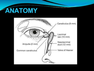
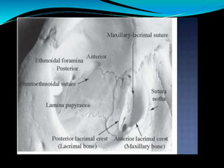
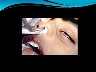
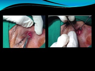
LACRIMAL
SAC SURGERY
-PRANAV KOHLI
ANATOMY
SURGICAL INDICATIONS
Persistent congenital lacrimal duct obstructions
unresponsive to previoustherapies.
Congenital lacrimal duct obstructions associated with
mucocele, dacryocystitis, and not responsive to other
treatments.
Primary acquired nasolacrimal duct obstructions
(PANDO).
Secondary acquired nasolacrimal duct obstructions
(SALDO).
Preoperative requisites
Confirmation of the diagnosis and clinical findings.
Hemoglobin levels.
Bleeding and clotting times.
Blood pressure measurement.
Random blood sugars.
ENT evaluation
Additional general anesthesia investigations when
required.
Nasal decongestant such as otrivin drops should be
given to reduce nasalcongestion
Patient is kept nil by mouth for ease of sedation
STEPS
NASAL PACKING
It is done to keep the mucosa taut and reducebleeding.
Nasal packing should be explained to the patient.
Few drops of 4% topical lignocaine should be instilled first
in the ipsilateral nostril, then nasal pack (roller gauze
soaked in 2% lignocaine-adrenaline jelly) inserted in the
ipsilateral nostril with the help of nasal packing forceps in
the direction of middle meatus, insinuated and negotiated
as deep aspossible
The direction of nasal packing is superior, then
posterior, then inferior.
SKIN
Either a curved incision along lacrimal crest or a
straight incision 8-11mm medial to medial canthus is
made
Orbicularis is split in the line of incision and and a
lacrimal retractor inserted so as to retract it withskin
Angular vessels should beavoided
EXPOSURE OF MPL AND
ANTERIOR LACRIMAL CREST
Identification and exposure of MPL is a very important
step in DCR surgery
Once MPL is exposed, the orbicularis fibers are
separated along the entire length of the incision.
Dis-insertion (not dividing) of MPL is done at the
anterior lacrimal crest by cutting on the bone at
insertion with 11number blade.
EXPOSURE OF BONE
Dis-insertion of MPL automatically opens up the
periosteum, which is now separated along the entire
length of the incision with sharp dissector or
periosteum elevator.
Lacrimal sac is retracted with periosteum elevator.
Baring of periosteum is done to decrease pain andto
aid bone punching.
Periosteum is elevated posteriorly till the lamina
papyracea. Lamina papyracea is a thin bone with
consistency and color different from lacrimalbone.
Periosteum also elevated anteriorly, inferiorly and
superiorly as much as reasonablypossible
EXPOSURE OF NASAL
MUCOSA
Bone removal is started with a small punch and then
with a big punch.
The correct method of using bone punch is as follows:
insinuate, engage the bone with the punch, support
with left thumb, hitch back, crush properly and then
gentle rocking movement to remove thebone.
Bone punch should always be perpendicular to the
punching surface. Clear the punch of bone pieceswith
20G needle.
Osteotomy should be as large as possible and should
be of size of thumbnail.
Extent of osteotomy should be asfollows:
Posteriorly: Till lamina papyracea.
Superiorly: At or slightly above level ofMPL.
Anteriorly and inferiorly: As much as possible
PREPARARTION OF FLAPS
OF SAC
A probe is introduced into the sac through thelower
canaliculus and the sac is incised vertically .
Toprepare anterior and posterior flaps the incisionis
converted into H shape
SUTURING OF FLAPS
Posterior flaps are sutured so that the posterior sac
flap does not block common canalicular ostium in sac.
Onesuture usually is sufficient for posterior flap.
Care should be taken to avoid nasal pack in the suture
Anterior nasal flap is now sutured to the anteriorsac
flap with minimum two 6-0 vicryl sutures(sometimes
three). Inserting lacrimal probe helps to confirm
proper flap suturing.
. MPL re-attachment is done with periosteum using deep
down to the bone bite of 6-0 vicryl on the medial incision
edge at MPL level.
Movement of the head when suture is pulled confirms the
firm suture attachment toperiosteum.
Additional 3-4orbicularis closure stitches are taken.
Skin closure can be achieved with either interrupted or
continuous sub-cuticular sutures..
Quarter folded pad on the wound and half-folded pad on
the eye should beapplied.
. Minimum 4-5 micropore tapes in a criss-cross fashion
with one tape to secure the nasal pack in positionshould
be applied
Adjunctive measures (use of
mitomycin C and intubation)
Mitomycin C in a concentration of 0.04% is used if
there are intra-sac synechiae, soft tissue scarringlike
in failed DCR's and in the presence of a complicated
surgery.
Intubation is also advisable for similar indications but
in addition it is also used in the presence of canalicular
problems and inadequate flaps
Post-operative care
Complete bed rest in propped up position and chin
extension is recommended for 24 hours.
Patients should be told to avoid blowing of nose.
Oral antibiotics, non-steroidal anti-inflammatory
drug (NSAID) - should be given routinely for fivedays.
Dressing and nasal pack removal to be done after 24
hours. Local treatment includes otrivin-P nasal drops
twice daily, antibiotic ointment on the wound twice
daily and antibiotic with steroid eye drop four times
daily.
Sac syringing should be done gently once in 2-3days
for the first week or 10days to remove bloodclots.
Suture removal to be done after 1week
YOUTUBE VIDEO LINK FOR BETTER UNDERSTANDING -
https://www.youtube.com/watch?v=mxEvnZvxYyM

lacrimalsacsurgery-PRANAV.pptx

  • 1.
  • 2.
  • 5.
    SURGICAL INDICATIONS Persistent congenitallacrimal duct obstructions unresponsive to previoustherapies. Congenital lacrimal duct obstructions associated with mucocele, dacryocystitis, and not responsive to other treatments. Primary acquired nasolacrimal duct obstructions (PANDO). Secondary acquired nasolacrimal duct obstructions (SALDO).
  • 6.
    Preoperative requisites Confirmation ofthe diagnosis and clinical findings. Hemoglobin levels. Bleeding and clotting times. Blood pressure measurement. Random blood sugars. ENT evaluation Additional general anesthesia investigations when required. Nasal decongestant such as otrivin drops should be given to reduce nasalcongestion Patient is kept nil by mouth for ease of sedation
  • 7.
    STEPS NASAL PACKING It isdone to keep the mucosa taut and reducebleeding. Nasal packing should be explained to the patient. Few drops of 4% topical lignocaine should be instilled first in the ipsilateral nostril, then nasal pack (roller gauze soaked in 2% lignocaine-adrenaline jelly) inserted in the ipsilateral nostril with the help of nasal packing forceps in the direction of middle meatus, insinuated and negotiated as deep aspossible The direction of nasal packing is superior, then posterior, then inferior.
  • 9.
    SKIN Either a curvedincision along lacrimal crest or a straight incision 8-11mm medial to medial canthus is made Orbicularis is split in the line of incision and and a lacrimal retractor inserted so as to retract it withskin Angular vessels should beavoided
  • 12.
    EXPOSURE OF MPLAND ANTERIOR LACRIMAL CREST Identification and exposure of MPL is a very important step in DCR surgery Once MPL is exposed, the orbicularis fibers are separated along the entire length of the incision. Dis-insertion (not dividing) of MPL is done at the anterior lacrimal crest by cutting on the bone at insertion with 11number blade.
  • 14.
    EXPOSURE OF BONE Dis-insertionof MPL automatically opens up the periosteum, which is now separated along the entire length of the incision with sharp dissector or periosteum elevator. Lacrimal sac is retracted with periosteum elevator. Baring of periosteum is done to decrease pain andto aid bone punching. Periosteum is elevated posteriorly till the lamina papyracea. Lamina papyracea is a thin bone with consistency and color different from lacrimalbone. Periosteum also elevated anteriorly, inferiorly and superiorly as much as reasonablypossible
  • 15.
    EXPOSURE OF NASAL MUCOSA Boneremoval is started with a small punch and then with a big punch. The correct method of using bone punch is as follows: insinuate, engage the bone with the punch, support with left thumb, hitch back, crush properly and then gentle rocking movement to remove thebone. Bone punch should always be perpendicular to the punching surface. Clear the punch of bone pieceswith 20G needle. Osteotomy should be as large as possible and should be of size of thumbnail.
  • 16.
    Extent of osteotomyshould be asfollows: Posteriorly: Till lamina papyracea. Superiorly: At or slightly above level ofMPL. Anteriorly and inferiorly: As much as possible
  • 18.
    PREPARARTION OF FLAPS OFSAC A probe is introduced into the sac through thelower canaliculus and the sac is incised vertically . Toprepare anterior and posterior flaps the incisionis converted into H shape
  • 20.
    SUTURING OF FLAPS Posteriorflaps are sutured so that the posterior sac flap does not block common canalicular ostium in sac. Onesuture usually is sufficient for posterior flap. Care should be taken to avoid nasal pack in the suture Anterior nasal flap is now sutured to the anteriorsac flap with minimum two 6-0 vicryl sutures(sometimes three). Inserting lacrimal probe helps to confirm proper flap suturing.
  • 21.
    . MPL re-attachmentis done with periosteum using deep down to the bone bite of 6-0 vicryl on the medial incision edge at MPL level. Movement of the head when suture is pulled confirms the firm suture attachment toperiosteum. Additional 3-4orbicularis closure stitches are taken. Skin closure can be achieved with either interrupted or continuous sub-cuticular sutures.. Quarter folded pad on the wound and half-folded pad on the eye should beapplied. . Minimum 4-5 micropore tapes in a criss-cross fashion with one tape to secure the nasal pack in positionshould be applied
  • 23.
    Adjunctive measures (useof mitomycin C and intubation) Mitomycin C in a concentration of 0.04% is used if there are intra-sac synechiae, soft tissue scarringlike in failed DCR's and in the presence of a complicated surgery. Intubation is also advisable for similar indications but in addition it is also used in the presence of canalicular problems and inadequate flaps
  • 25.
    Post-operative care Complete bedrest in propped up position and chin extension is recommended for 24 hours. Patients should be told to avoid blowing of nose. Oral antibiotics, non-steroidal anti-inflammatory drug (NSAID) - should be given routinely for fivedays.
  • 26.
    Dressing and nasalpack removal to be done after 24 hours. Local treatment includes otrivin-P nasal drops twice daily, antibiotic ointment on the wound twice daily and antibiotic with steroid eye drop four times daily. Sac syringing should be done gently once in 2-3days for the first week or 10days to remove bloodclots. Suture removal to be done after 1week
  • 27.
    YOUTUBE VIDEO LINKFOR BETTER UNDERSTANDING - https://www.youtube.com/watch?v=mxEvnZvxYyM