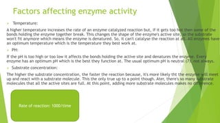
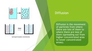
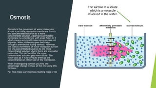
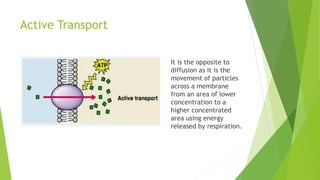
Key concepts in Biology
Cells
All living things are made up of cells and can either be eukaryotic or prokaryotic.
Eukaryotic cells are complex and animal and plant cells are eukaryotic. However,
prokaryotic cells are small and simpler and bacteria cells are prokaryotic.
Plant cell Animal cell
Centriole
Cell wall
Chloroplast
Cell membrane
Ribosomes
Nucleus
Endoplasmic reticulum
Mitochondria
Vacuole
Cytoskeleton
 Bacteria cells don't have a nucleus and have 3 sub-cellular structures:
 1) Chromosomal DNA (one long circular strand of DNA)
 2) Ribosomes
 3) Cell membrane
 4) Plasmid DNA (small loops of extra DNA)
 5) Flagellum (long, hair like structure that helps the cell move.
Specialised cells
Multicellular organisms contain lots of different types of cells. Cells with a structure
that makes them adapted to their function are namd specialised cells.
 Egg cells and sperm cells: specialised for reproduction:
During sexual reproduction a sperm and egg cell combine together which forms a
fertilised egg and then develops into an embryo. The function of the egg cell is to
carry the female DNA and feed the embryo but, the sperm cell function is to carry the
male DNA to the egg.
Egg Cell Sperm Cell
1) An egg cell has nutrients in the cytoplasm to feed
the embryo.
1) It has a tail so, it can swim to the egg.
2) After fertalisation, the cell membrane changes
structure to stop more sperm getting in so, only 1 can
fertilise the egg.
2) Has lots of mitochondria which releases the energy
the sperm needs to swim (faster).
3) Has a haploid nucleus which means that the cells
only have 1/2 the number of chromosomes found in a
normal body cell so, when the sperm and egg cell
combine they have full number of chromosomes.
3) Has an acrosome which stores enzymes that digest
through the membrane of the egg cell.
4) Haploid nucleus ( same as egg cell, 3)
 Ciliated Epithelial cells: Specialised for moving materials.
Ciliated epithelial cells
1) Epithelial cells line the surfaces of organs.
2) Some have cillia on top of the cell (cilliated).
3) Cillia are tiny hair-like structures which moves substances by beating them
along the surface of the tissue.
4) Cilliated epithelial cells in the lining of the airways help to move mucus up
to the throat so it can be swallowed (example).
Microscopy
 Microscopes are used to magnify things using
lenses so, we can identify them clearly.
 Cells were discovered using microscopes as
they're so miniature and only can be seen using
microscopes.
 Light microscopes are used to look at cells as
they show sub cellular structures like chloroplast.
 Electron microscopes were invented after light
microscopes and they make specimens look more
huge as well as revealing more detail than light
microscopes. They give better knowledge of what
sub-cellular structures do.
How to use a microscope
 Preparing your specimen:
1. Take a thin slice of your specimen which is what you're looking at.
2. Grab a clean slide and use a pipette to put one drop of water in the middle of it.
3. Thirdly, use tweezers to place your specimen on the slide.
4. Then you might need a drop of stain to make it easier to see.
5. Cautiously, lower a cover slip onto the slide using a mounted needle and try not to trap any
bubbles under it.
 Viewing your specimen:
1. Clip the slide onto the stage.
2. Choose the objective lens with the lowest magnification.
3. Use the coarse adjustment knob to move the stage up to just underneath the objective lens.
4. Seek down the eyepiece, then move the stage down until the specimen is nearly in focus.
5. Move the fine adjustment knob until you find a clear image.
6. Finally, if you want a bigger image then use an objective lens with a higher magnification.
Maths in Microscopy
Magnification is how many times bigger the
image is and there is a formula to use to find the
total magnification:
 Total magnification= eyepiece lens
magnification x objective lens magnification.
If you don't know what lenses were used then
you can also work out the magnification of an
image. This means you need to know the image
size and the real size of the specimen, this is the
formula:
 Magnification= image size/ real size.
Image size/Real size: mm
Magnification: x (x30)
You might need to convert units.
Enzymes
Enzymes are biological catalysts
(produced by living things) which means
the speed up chemical reactions in living
organisms.
Substrates are molecules that get
changed in a chemical reaction.
An active site is in every enzyme which
is the part where it joins on its substrate
to catalyze the reaction.
Enzymes are substrate specific meaning
they usually only work with one
substrate because, for the enzyme to
function, the substrate has to fit into the
active site.
Proteins, lipids
and some
carbohydrates
are big
molecules and
organisms often
need to
breakdown these
big molecules to
make smaller
ones during
digestion. But,
organisms also
need to
synthesize
(make) big
molecules from
smaller
molecules e.g. to
make new cells.
Enzymes named carbohydrase break
carbohydrates into simple sugars.
Proteases break down proteins into
amino acids. Other enzymes produce
proteins from amino acids.
Lipases break down lipids into glycerol
and fatty acids.
Factors affecting enzyme activity
 Temperature:
A higher temperature increases the rate of an enzyme catalyzed reaction but, if it gets too hot then some of the
bonds holding the enzyme together break. This changes the shape of the enzyme's active site, so the substrate
won't fit anymore which means the enzyme is denatured. So, it can't catalyse the reaction at all. All enzymes have
an optimum temperature which is the temperature they best work at.
 PH:
If the pH is too high or too low it affects the bonds holding the active site and denatures the enzyme. Every
enzyme has an optimum pH which is the best they function at. The usual optimum pH is neutral (7), not always.
 Substrate concentration:
The higher the substrate concentration, the faster the reaction because, it's more likely tht the enzyme will meet
up and react with a substrate molecule. This the only true up to a point though. Ater, there's so many substrate
molecules that all the active sites are full. At this point, adding more substrate molecules makes no difference.
Rate of reaction: 1000/time
Diffusion
Diffusion is the movement
of particles from where
there are lots of them to
where there are less of
them (spreading out from
higher concentrated area
to lower concentrated
areas).
Osmosis
Osmosis is the movement of water molecules
across a partially permeable membrane from a
less concentrated solution to a more
concentrated solution. A partially permeable
membrane is a membrane with small holes in it
which means only small molecules can pass not
big ones. Water molecules pass both ways
through a membrane during osmosis. However,
the overall movement of water molecules is from
the less concentrated solution to the more
concentrated solution where there are less water
molecules. This defines that the more
concentrated solution gets more dilute. The
water acts as if it is trying to even up the
concentration on either side of the membrane.
When investigating osmosis you find the
percentage change in mass at the end using this
formula:
PC: final mass-starting mass/starting mass x 100
The sucrose is a solute
which is a molecule
dissolved in the water.
Active Transport
It is the opposite to
diffusion as it is the
movement of particles
across a membrane
from an area of lower
concentration to a
higher concentrated
area using energy
released by respiration.

Key concepts in biology

  • 1.
  • 2.
    Cells All living thingsare made up of cells and can either be eukaryotic or prokaryotic. Eukaryotic cells are complex and animal and plant cells are eukaryotic. However, prokaryotic cells are small and simpler and bacteria cells are prokaryotic. Plant cell Animal cell Centriole Cell wall Chloroplast Cell membrane Ribosomes Nucleus Endoplasmic reticulum Mitochondria Vacuole Cytoskeleton
  • 3.
     Bacteria cellsdon't have a nucleus and have 3 sub-cellular structures:  1) Chromosomal DNA (one long circular strand of DNA)  2) Ribosomes  3) Cell membrane  4) Plasmid DNA (small loops of extra DNA)  5) Flagellum (long, hair like structure that helps the cell move.
  • 4.
    Specialised cells Multicellular organismscontain lots of different types of cells. Cells with a structure that makes them adapted to their function are namd specialised cells.  Egg cells and sperm cells: specialised for reproduction: During sexual reproduction a sperm and egg cell combine together which forms a fertilised egg and then develops into an embryo. The function of the egg cell is to carry the female DNA and feed the embryo but, the sperm cell function is to carry the male DNA to the egg. Egg Cell Sperm Cell 1) An egg cell has nutrients in the cytoplasm to feed the embryo. 1) It has a tail so, it can swim to the egg. 2) After fertalisation, the cell membrane changes structure to stop more sperm getting in so, only 1 can fertilise the egg. 2) Has lots of mitochondria which releases the energy the sperm needs to swim (faster). 3) Has a haploid nucleus which means that the cells only have 1/2 the number of chromosomes found in a normal body cell so, when the sperm and egg cell combine they have full number of chromosomes. 3) Has an acrosome which stores enzymes that digest through the membrane of the egg cell. 4) Haploid nucleus ( same as egg cell, 3)
  • 5.
     Ciliated Epithelialcells: Specialised for moving materials. Ciliated epithelial cells 1) Epithelial cells line the surfaces of organs. 2) Some have cillia on top of the cell (cilliated). 3) Cillia are tiny hair-like structures which moves substances by beating them along the surface of the tissue. 4) Cilliated epithelial cells in the lining of the airways help to move mucus up to the throat so it can be swallowed (example).
  • 6.
    Microscopy  Microscopes areused to magnify things using lenses so, we can identify them clearly.  Cells were discovered using microscopes as they're so miniature and only can be seen using microscopes.  Light microscopes are used to look at cells as they show sub cellular structures like chloroplast.  Electron microscopes were invented after light microscopes and they make specimens look more huge as well as revealing more detail than light microscopes. They give better knowledge of what sub-cellular structures do.
  • 7.
    How to usea microscope  Preparing your specimen: 1. Take a thin slice of your specimen which is what you're looking at. 2. Grab a clean slide and use a pipette to put one drop of water in the middle of it. 3. Thirdly, use tweezers to place your specimen on the slide. 4. Then you might need a drop of stain to make it easier to see. 5. Cautiously, lower a cover slip onto the slide using a mounted needle and try not to trap any bubbles under it.  Viewing your specimen: 1. Clip the slide onto the stage. 2. Choose the objective lens with the lowest magnification. 3. Use the coarse adjustment knob to move the stage up to just underneath the objective lens. 4. Seek down the eyepiece, then move the stage down until the specimen is nearly in focus. 5. Move the fine adjustment knob until you find a clear image. 6. Finally, if you want a bigger image then use an objective lens with a higher magnification.
  • 8.
    Maths in Microscopy Magnificationis how many times bigger the image is and there is a formula to use to find the total magnification:  Total magnification= eyepiece lens magnification x objective lens magnification. If you don't know what lenses were used then you can also work out the magnification of an image. This means you need to know the image size and the real size of the specimen, this is the formula:  Magnification= image size/ real size. Image size/Real size: mm Magnification: x (x30) You might need to convert units.
  • 9.
    Enzymes Enzymes are biologicalcatalysts (produced by living things) which means the speed up chemical reactions in living organisms. Substrates are molecules that get changed in a chemical reaction. An active site is in every enzyme which is the part where it joins on its substrate to catalyze the reaction. Enzymes are substrate specific meaning they usually only work with one substrate because, for the enzyme to function, the substrate has to fit into the active site.
  • 10.
    Proteins, lipids and some carbohydrates arebig molecules and organisms often need to breakdown these big molecules to make smaller ones during digestion. But, organisms also need to synthesize (make) big molecules from smaller molecules e.g. to make new cells. Enzymes named carbohydrase break carbohydrates into simple sugars. Proteases break down proteins into amino acids. Other enzymes produce proteins from amino acids. Lipases break down lipids into glycerol and fatty acids.
  • 11.
    Factors affecting enzymeactivity  Temperature: A higher temperature increases the rate of an enzyme catalyzed reaction but, if it gets too hot then some of the bonds holding the enzyme together break. This changes the shape of the enzyme's active site, so the substrate won't fit anymore which means the enzyme is denatured. So, it can't catalyse the reaction at all. All enzymes have an optimum temperature which is the temperature they best work at.  PH: If the pH is too high or too low it affects the bonds holding the active site and denatures the enzyme. Every enzyme has an optimum pH which is the best they function at. The usual optimum pH is neutral (7), not always.  Substrate concentration: The higher the substrate concentration, the faster the reaction because, it's more likely tht the enzyme will meet up and react with a substrate molecule. This the only true up to a point though. Ater, there's so many substrate molecules that all the active sites are full. At this point, adding more substrate molecules makes no difference. Rate of reaction: 1000/time
  • 12.
    Diffusion Diffusion is themovement of particles from where there are lots of them to where there are less of them (spreading out from higher concentrated area to lower concentrated areas).
  • 13.
    Osmosis Osmosis is themovement of water molecules across a partially permeable membrane from a less concentrated solution to a more concentrated solution. A partially permeable membrane is a membrane with small holes in it which means only small molecules can pass not big ones. Water molecules pass both ways through a membrane during osmosis. However, the overall movement of water molecules is from the less concentrated solution to the more concentrated solution where there are less water molecules. This defines that the more concentrated solution gets more dilute. The water acts as if it is trying to even up the concentration on either side of the membrane. When investigating osmosis you find the percentage change in mass at the end using this formula: PC: final mass-starting mass/starting mass x 100 The sucrose is a solute which is a molecule dissolved in the water.
  • 14.
    Active Transport It isthe opposite to diffusion as it is the movement of particles across a membrane from an area of lower concentration to a higher concentrated area using energy released by respiration.