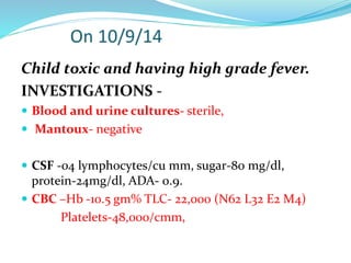
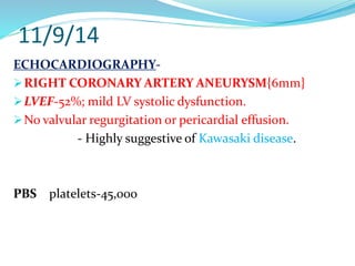
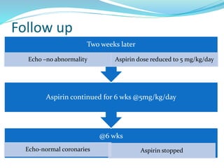
AN UNUSUAL CASE OF
PROLONGED FEVER
SPEAKER - Dr. GNANDAS BARMAN
Pritwish
Mondal
1 year old
Male/Hindu
child from
Bongaon
Admitted on
07/09/2014
•Fever and excessive irritability for -10 days
H/O Present illness
Patient developed high grade continous fever 1o days
prior to admission.
Associated redness of both eyes.
Fever was not associated with rigor, cough, rash,
dysuria, altered sensorium, convulsion.
The child was extremely irritable since the onset of
fever.
On Examination:
 Alert and irritable.
 Vitals- PR-140/min,
BP-80/44mm of Hg,
RR-36/min,
Temp-104.2⁰F,
CRT-2 sec
SpO2-97% in room air.
 AF- closed
 Bilateral bulbar conjunctival congestion.
 Pallor present
On Examination:
 Multiple right posterior cervical
lymphadenopathy-
1.5cm, firm, discrete, mobile , nontender.
 No rash /desquamation.
On Examination:-
 Anthropometry: -
Wt- 10.5 Kg (between 50th - 85th percentile)
Length - 78cm (50th - 85th percentile)
Head circumference-45 cm (15th - 50th
percentile)
G.I. SYSTEM-
 UPPER G.I.- normal
 Liver -4 cms along Rt MCL , liver span-
10 cms, firm, sharp margin, non tender,
left lobe not palpable.
 Spleen-3 cms
 Other systems- WNL
Differential Diagnosis
 VIRAL FEVER.
 HEMATOLOGICAL MALIGNANCIES.
 KAWASAKI DISEASE
INVESTIGATIONS (7/9/14):
 CBC-
Hb: 6 gm%
TLC: 20,000/ cu mm, (N60/L36/M2/E2)
Platelet: 50,000/cumm
ESR- 25 mm,
no abnormal cells in peripheral smear.
 LFT: TSB-0.6,
ALT/AST-80/68,
Alb-2.6,Glb-2.8
Urea/Creatinine-22/0.8
Serum electrolytes-Na/K- 142/4.7
Urine RE/ME: 5-6 pus cells.
MPslide, MPDA- Negative
DengueIgM-NR
Widal- Negative.
 Urine and blood c/s sent.
 Mantoux testdone.
 Chest Xray-WNL
 USG W/A-Hepatosplenomegaly
Treatment:-
 IV Fluids
 Inj Ceftriaxone
 Syr Paracetamol
 PRBC transfusion
Child toxic and having high grade fever.
INVESTIGATIONS -
 Blood and urine cultures- sterile,
 Mantoux- negative
 CSF -04 lymphocytes/cu mm, sugar-80 mg/dl,
protein-24mg/dl, ADA- 0.9.
 CBC –Hb -10.5 gm% TLC- 22,000 (N62 L32 E2 M4)
Platelets-48,000/cmm,
 Bone marrow aspiration study done.
11/9/14
ECHOCARDIOGRAPHY-
RIGHT CORONARY ARTERY ANEURYSM{6mm}
LVEF-52%; mild LV systolic dysfunction.
No valvular regurgitation or pericardial effusion.
- Highly suggestive of Kawasaki disease.
PBS platelets-45,000
ON 12/09/14
 Serum Ferritin-1886 ng/ml(>500)
 Triglyceride-442mg/dl(>265 )
 Fibrinogen-119.6mg/dl(<150)
BONE MARROW STUDY-
Bone marrow examination
showing
Hemophagocytosis
PROPOSED HLH DIAGNOSTIC
CRITERIA,2009:
[1] Molecular diagnosis of hemophagocytic
lymphohistiocytosis(HLH) or X-linked lymphoproliferative
syndrome (XLP).
[2] Or at least 3 of 4:
 a. Fever
 b. Splenomegaly
 c. Cytopenia (minimum 2 cell lines reduced)
 d. Hepatitis
PROPOSED HLH DIAGNOSTIC
CRITERIA,2009:
[3]. And at least 1 of 4:
 a. Hemophagocytosis
 b. ↑ Ferritin
 c. ↑ sIL2Rα (CD25)
 d. Absent or very decreased NK function
[4]. Other results supportive of HLH diagnosis:
 a. Hypertriglyceridemia
 b. Hypofibrinogenemia
 c. Hyponatremia
Filipovich A et al[ASH Education Book Jan 1,2009 vol.2009 no.
1 127-131]
DIAGNOSIS
ATYPICAL KAWASAKI DISEASE WITH SECONDARY
HEMOPHAGOCYTIC LYMPHOHISTIOCYTOSIS[HLH]
TREATMENT
 IV IMMUNOGLOBULIN- 2gms/kg transfused over 24
hours on 13/9/14.
 Platelet count-80,000 on 14/9/14
 CBC on 15/9/14 : Hb-11, Tc-12,000,N52L42B5E1
platelets-1.5 lakh
 Aspirin 80 mg/kg started from 15/9/14.
 Patient became afebrile 36 hrs after IVIG transfusion.
 Hepatosplenomegaly started to regress within 3 days
of IVIG transfusion
Follow up
@6 wks
Echo-normal coronaries Aspirin stopped
Aspirin continued for 6 wks @5mg/kg/day
Two weeks later
Echo –no abnormality Aspirin dose reduced to 5 mg/kg/day
DISCUSSION
1.9% of children with acute kawasaki disease are
reported to develop secondary HLH.†
It result from cytotoxic dysfunction leading to
persistent expansion of T cells and Macrophages ,
escalating production of proinflammatory
cytokines.
†Latino et al[ J Pediatr Hemato Oncol 2000 oct;32(7):527-31]
Present with prolonged and persisting fever beyond initial
IVIG treatment ; KD complicated with secondary HLH is
difficult to distinguish from refractory KD/ recurrent KD.
Onset of secondary HLH or MAS with mean of 13.3 days
[range 3-22 days];
However recurrent KD typically occurred much later at a
mean of 17.9 months[range 1-60 months]‡
Treatment includes Pulse Methyl Prednisolone, Anakinra,
Etoposide.
‡ Kang et al[Blood Res. 2013 Dec; 48(4): 254–257]
CASE
SERIES
PATIENTS OF KD
COMPLICATED WITH
HLH
RESPONSIVE
TO IVIG
ADDITIONAL
IMMUNOMODULATOR
REQUIRED
1. 12 1 11
2. 8 1 7
Kawasaki with hlh,an unusual case of fever

Kawasaki with hlh,an unusual case of fever

  • 1.
    AN UNUSUAL CASEOF PROLONGED FEVER SPEAKER - Dr. GNANDAS BARMAN
  • 2.
    Pritwish Mondal 1 year old Male/Hindu childfrom Bongaon Admitted on 07/09/2014
  • 3.
    •Fever and excessiveirritability for -10 days
  • 4.
    H/O Present illness Patientdeveloped high grade continous fever 1o days prior to admission. Associated redness of both eyes. Fever was not associated with rigor, cough, rash, dysuria, altered sensorium, convulsion. The child was extremely irritable since the onset of fever.
  • 5.
    On Examination:  Alertand irritable.  Vitals- PR-140/min, BP-80/44mm of Hg, RR-36/min, Temp-104.2⁰F, CRT-2 sec SpO2-97% in room air.  AF- closed  Bilateral bulbar conjunctival congestion.  Pallor present
  • 6.
    On Examination:  Multipleright posterior cervical lymphadenopathy- 1.5cm, firm, discrete, mobile , nontender.  No rash /desquamation.
  • 7.
    On Examination:-  Anthropometry:- Wt- 10.5 Kg (between 50th - 85th percentile) Length - 78cm (50th - 85th percentile) Head circumference-45 cm (15th - 50th percentile)
  • 8.
    G.I. SYSTEM-  UPPERG.I.- normal  Liver -4 cms along Rt MCL , liver span- 10 cms, firm, sharp margin, non tender, left lobe not palpable.  Spleen-3 cms  Other systems- WNL
  • 9.
    Differential Diagnosis  VIRALFEVER.  HEMATOLOGICAL MALIGNANCIES.  KAWASAKI DISEASE
  • 10.
    INVESTIGATIONS (7/9/14):  CBC- Hb:6 gm% TLC: 20,000/ cu mm, (N60/L36/M2/E2) Platelet: 50,000/cumm ESR- 25 mm, no abnormal cells in peripheral smear.
  • 11.
     LFT: TSB-0.6, ALT/AST-80/68, Alb-2.6,Glb-2.8 Urea/Creatinine-22/0.8 Serumelectrolytes-Na/K- 142/4.7 Urine RE/ME: 5-6 pus cells.
  • 12.
  • 13.
     Urine andblood c/s sent.  Mantoux testdone.  Chest Xray-WNL  USG W/A-Hepatosplenomegaly
  • 14.
    Treatment:-  IV Fluids Inj Ceftriaxone  Syr Paracetamol  PRBC transfusion
  • 15.
    Child toxic andhaving high grade fever. INVESTIGATIONS -  Blood and urine cultures- sterile,  Mantoux- negative  CSF -04 lymphocytes/cu mm, sugar-80 mg/dl, protein-24mg/dl, ADA- 0.9.  CBC –Hb -10.5 gm% TLC- 22,000 (N62 L32 E2 M4) Platelets-48,000/cmm,
  • 16.
     Bone marrowaspiration study done.
  • 17.
    11/9/14 ECHOCARDIOGRAPHY- RIGHT CORONARY ARTERYANEURYSM{6mm} LVEF-52%; mild LV systolic dysfunction. No valvular regurgitation or pericardial effusion. - Highly suggestive of Kawasaki disease. PBS platelets-45,000
  • 18.
    ON 12/09/14  SerumFerritin-1886 ng/ml(>500)  Triglyceride-442mg/dl(>265 )  Fibrinogen-119.6mg/dl(<150)
  • 19.
    BONE MARROW STUDY- Bonemarrow examination showing Hemophagocytosis
  • 20.
    PROPOSED HLH DIAGNOSTIC CRITERIA,2009: [1]Molecular diagnosis of hemophagocytic lymphohistiocytosis(HLH) or X-linked lymphoproliferative syndrome (XLP). [2] Or at least 3 of 4:  a. Fever  b. Splenomegaly  c. Cytopenia (minimum 2 cell lines reduced)  d. Hepatitis
  • 21.
    PROPOSED HLH DIAGNOSTIC CRITERIA,2009: [3].And at least 1 of 4:  a. Hemophagocytosis  b. ↑ Ferritin  c. ↑ sIL2Rα (CD25)  d. Absent or very decreased NK function [4]. Other results supportive of HLH diagnosis:  a. Hypertriglyceridemia  b. Hypofibrinogenemia  c. Hyponatremia Filipovich A et al[ASH Education Book Jan 1,2009 vol.2009 no. 1 127-131]
  • 22.
    DIAGNOSIS ATYPICAL KAWASAKI DISEASEWITH SECONDARY HEMOPHAGOCYTIC LYMPHOHISTIOCYTOSIS[HLH]
  • 23.
    TREATMENT  IV IMMUNOGLOBULIN-2gms/kg transfused over 24 hours on 13/9/14.  Platelet count-80,000 on 14/9/14  CBC on 15/9/14 : Hb-11, Tc-12,000,N52L42B5E1 platelets-1.5 lakh  Aspirin 80 mg/kg started from 15/9/14.
  • 24.
     Patient becameafebrile 36 hrs after IVIG transfusion.  Hepatosplenomegaly started to regress within 3 days of IVIG transfusion
  • 25.
    Follow up @6 wks Echo-normalcoronaries Aspirin stopped Aspirin continued for 6 wks @5mg/kg/day Two weeks later Echo –no abnormality Aspirin dose reduced to 5 mg/kg/day
  • 26.
    DISCUSSION 1.9% of childrenwith acute kawasaki disease are reported to develop secondary HLH.† It result from cytotoxic dysfunction leading to persistent expansion of T cells and Macrophages , escalating production of proinflammatory cytokines. †Latino et al[ J Pediatr Hemato Oncol 2000 oct;32(7):527-31]
  • 27.
    Present with prolongedand persisting fever beyond initial IVIG treatment ; KD complicated with secondary HLH is difficult to distinguish from refractory KD/ recurrent KD. Onset of secondary HLH or MAS with mean of 13.3 days [range 3-22 days]; However recurrent KD typically occurred much later at a mean of 17.9 months[range 1-60 months]‡ Treatment includes Pulse Methyl Prednisolone, Anakinra, Etoposide. ‡ Kang et al[Blood Res. 2013 Dec; 48(4): 254–257]
  • 28.
    CASE SERIES PATIENTS OF KD COMPLICATEDWITH HLH RESPONSIVE TO IVIG ADDITIONAL IMMUNOMODULATOR REQUIRED 1. 12 1 11 2. 8 1 7