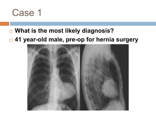
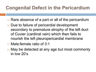
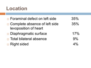
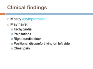
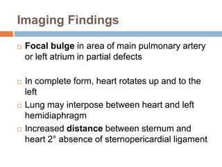
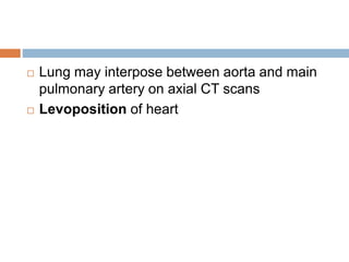
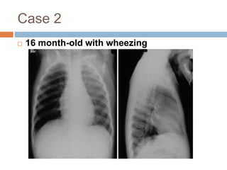
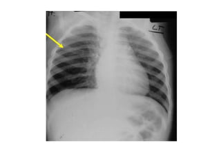
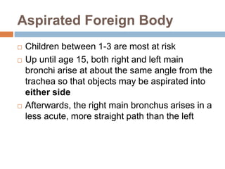
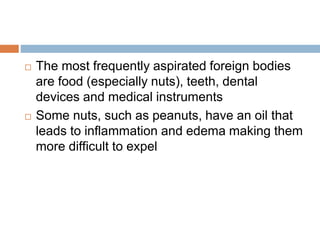
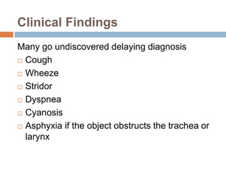
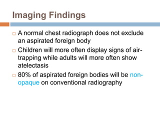
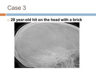
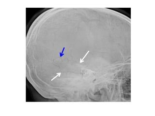
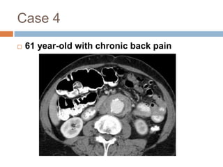
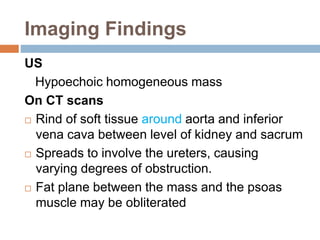
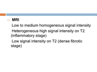
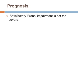
This document discusses congenital heart disease (CHD), including atrial septal defects (ASD) and tetralogy of Fallot (TOF). It defines the conditions, describes their signs and symptoms, and outlines their treatment and nursing care. Key points include the definition of ASD as an abnormal opening between the atria, and TOF as having four structural defects including pulmonary stenosis and a ventricular septal defect. Nursing focuses on decreasing cardiac workload, preventing infection, and supporting nutrition and development.
Congenital heart defects are abnormalities in the heart's structure that are present at birth. They can affect the heart's chambers, valves, or blood vessels. Common defects include ventricular septal defects (holes in the wall between the ventricles), atrial septal defects, patent ductus arteriosus, tetralogy of Fallot, and transposition of the great arteries. Many defects cause no symptoms, but some can lead to heart failure, lung problems, or other serious issues if not treated with medication, catheterization, or surgery. Diagnosis involves medical history, physical exam, chest X-ray, electrocardiogram, and echocardiogram.
The document provides information on congenital heart defects, including their causes, types, signs and symptoms, diagnosis, and treatment. It discusses several specific defects in detail, including aortic stenosis, coarctation of the aorta, pulmonary stenosis, and patent ductus arteriosus. The key points are:
1. Congenital heart defects can involve the heart's chambers, valves, or vessels and have various causes including genetic syndromes.
2. Specific defects like aortic stenosis and pulmonary stenosis can cause obstruction to blood flow while others like patent ductus arteriosus allow extra blood flow to the lungs.
3. Symptoms depend on the severity of the defect but may include heart failure,
This document discusses congenital heart defects present at birth, classified as cyanotic or acyanotic. Acyanotic defects include left-to-right shunts which allow oxygenated blood to pass from the left side of the heart to the right side, and obstructive lesions which obstruct blood flow. Cyanotic defects allow deoxygenated blood to pass from the right side of the heart to the left, reducing oxygen in the blood. Common signs and symptoms, methods of diagnosis, and treatments for heart failure are also outlined.
This document discusses several congenital heart diseases including their incidence, etiology, pathophysiology, clinical manifestations, diagnosis, and management. It provides details on ventricular septal defect (VSD), atrial septal defect (ASD), patent ductus arteriosus (PDA), tetralogy of Fallot, transposition of the great arteries, coarctation of the aorta, and pulmonary stenosis. The overall incidence of congenital heart diseases is about 8-10 per 1000 live births with VSD being the most common type, accounting for 25-30% of cases. Etiologies may include hereditary factors, infections, chromosomal or genetic abnormalities. Clinical exams, imaging tests, and cardiac catheterization
Management of congenital heart disease in infantsSMSRAZA
- Congenital heart disease is the most common birth defect, affecting around 8 per 1000 births. Common defects include ventricular septal defects, atrial septal defects, and tetralogy of Fallot.
- Diagnosis involves a detailed family history, physical exam for murmurs or cyanosis, chest x-ray, electrocardiogram, and echocardiogram. Treatment depends on the specific defect but may include medications, closure devices, surgery, or lifestyle changes.
- Managing congenital heart disease requires a multidisciplinary approach including cardiologists, cardiac surgeons, nutritionists, and mental health professionals to address both physical and psychological needs.
The document discusses various congenital heart defects including their definition, etiology, classification as acyanotic or cyanotic, signs and symptoms, diagnosis, and treatment. Specific defects covered include atrial septal defects, ventricular septal defects, patent ductus arteriosus, and atrioventricular septal defects. The treatment sections provide guidelines for managing each defect medically or surgically depending on its size and severity.
The document discusses several types of congenital heart disease including their causes, clinical features, investigations, and treatments. It begins by describing the fetal circulation and how defects can arise from incomplete changes at birth. It then examines several specific defects in more detail, including atrial septal defect (ASD), ventricular septal defect (VSD), patent ductus arteriosus (PDA), coarctation of the aorta, tetralogy of Fallot, pulmonary stenosis, Ebstein's anomaly, and pulmonary hypertension/Eisenmenger's syndrome. For each condition, it outlines the pathophysiology, typical signs and symptoms, relevant test findings, and current management approaches.
This document discusses congenital heart disease (CHD), which affects nearly 1% of births annually in the United States. It outlines the epidemiology, anatomy, types, clinical approach, history taking, physical exams, investigations and general management of CHD. The most common type is ventricular septal defect. While about 95% of babies with non-critical CHD are expected to survive to 18 years, only 69% of those with critical CHD are expected to survive. The document describes several specific types of CHDs in detail, including their typical features, exams, treatments and management approaches.
This document provides information on several types of congenital and rheumatic heart disease, as well as surgical correction of cardiovascular lesions. It discusses congenital heart defects such as atrial and ventricular septal defects, tetralogy of Fallot, coarctation of the aorta, and patent ductus arteriosus. For each condition, it describes the etiology, clinical features, investigations, and management approaches. The document emphasizes that while many congenital heart defects used to be fatal in childhood, surgery can now correct or partially repair many conditions, allowing survival into adulthood.
This document presents information from a presentation on acyanotic congenital heart disease. It begins with objectives that cover fetal circulation, defining CHD and risk factors, classifying CHD, explaining acyanotic heart disease and specific defects. It then provides detailed information on ventricular septal defect, atrial septal defect, patent ductus arteriosus, aortic stenosis, pulmonary stenosis, and coarctation of aorta. For each defect, it discusses clinical manifestation, diagnostic criteria, management, and complications. It also includes summaries of two research papers on neurodevelopmental outcomes after surgery for acyanotic CHD and a comparison of renal function between cyanotic and acyanotic CHD in children.
CONGENITAL HEART DISEASE: APPROACH TO DIAGNOSISNizam Uddin
This document discusses the diagnosis and classification of congenital heart disease. It begins by classifying CHDs into acyanotic or cyanotic types based on whether pulmonary blood flow is increased, normal, decreased, or increased. It emphasizes the importance of using a pulse oximeter to detect cyanosis, as the human eye cannot reliably detect low oxygen saturation above 85-93%. The document then discusses specific CHD types like atrial and ventricular septal defects, patent ductus arteriosus, tetralogy of Fallot, and transposition of the great arteries. It provides guidance on when surgical intervention is indicated for different CHDs. Overall, the document provides an overview of evaluating and classifying CHDs, with a focus on
Congenital heart disease refers to heart defects that are present at birth. Some defects may not cause issues, but complex defects can be life-threatening. Advances in diagnosis and treatment have allowed babies with congenital heart disease to survive into adulthood. There are many types of congenital heart defects, ranging from holes in the heart to problems with heart valves or blood vessels. Causes may include genetic factors, viral infections during pregnancy, or certain medications or behaviors. Treatment options depend on the type and severity of defect and may include medications, surgery, cardiac devices, or even heart transplant.
This document provides an overview of congenital heart disease (CHD). It begins with objectives and an outline of the topics to be covered, including a case presentation of a 4 day old infant with a patent ductus arteriosus. Key points include: the causes of CHD can be genetic or environmental; CHD is classified into categories like septal defects, cyanotic defects, and obstructive defects; signs and symptoms vary depending on the specific defect but may include murmurs, breathing difficulties, and cyanosis; and management involves treatments like medication or surgery depending on the severity and type of defect.
Congenital heart disease has a prevalence of 0.5-0.8% of live births. It can be caused by genetic and environmental factors. Defects are classified as acyanotic if there is increased blood flow to the lungs, or cyanotic if there is decreased pulmonary blood flow. Common defects include atrial septal defects, ventricular septal defects, and patent ductus arteriosus. Diagnosis involves tests like echocardiogram and chest x-ray. Treatment options include procedures to close defects, heart transplantation, open heart surgery, and medications to manage symptoms.
Congenital heart diseases are abnormalities present at birth that are caused by defects in heart development during weeks 3-8 of gestation. They range from mild to severe. The most common types are atrial septal defect (ASD), ventricular septal defect (VSD), patent ductus arteriosus (PDA), tetralogy of Fallot, transposition of the great arteries, and coarctation of the aorta. Left-to-right shunts cause increased pulmonary blood flow while right-to-left shunts allow deoxygenated blood to bypass the lungs, causing cyanosis. Most defects are now surgically corrected.
The lecture is for medical student. It is from Dr RUSINGIZA Emmanuel, MD, senior lecture at UR( UNIVERSITY OF RWANDA) .
It will help to understand heart diseases in newborn, infants and children.
This document discusses pulmonary embolism (PE) and the use of CT angiography (CTA) for diagnosis. It provides information on the epidemiology and risk factors for PE. It describes the signs and symptoms of PE and clinical diagnostic criteria. The document outlines the technique for CTA for PE diagnosis including contrast injection parameters and common pitfalls. It details CT findings of acute, chronic and evolving PE and discusses prognostic factors seen on CTA such as right ventricular enlargement.
A "hole" in the wall that separates the top two chambers of the heart.
This defect allows oxygen-rich blood to leak into the oxygen-poor blood chambers in the heart. ASD is a defect in the septum between the heart's two upper chambers (atria). The septum is a wall that separates the heart's left and right sides
Congenital heart disease for undergraduates student uod 2015Azad Haleem
The document provides an overview of pediatric cardiology including:
1. The anatomy and physiology of the heart is described along with embryological development and how circulation changes with age.
2. Congenital heart diseases are discussed including prevalence, etiology, evaluation, diagnosis, and classifications.
3. Key aspects of history taking and physical examination for congenital heart disease are outlined along with important investigations like ECG, chest x-ray, and echocardiography.
This document discusses primary cardiac tumors, including their presentation, diagnosis, and types. It covers the following key points:
- The majority (75%) of primary cardiac tumors are benign neoplasms. Myxomas are the most common benign tumor, usually originating in the left atrium.
- Clinical presentation varies depending on tumor location and can include embolic phenomena, cardiac manifestations like obstruction, or symptoms of metastatic disease. Echocardiography is the primary diagnostic tool.
- Other common benign primary tumors include papillary fibroelastomas (found on heart valves) and lipomas. Rhabdomyomas are most often seen in children with tuberous sclerosis and can cause arrhythmias
This document provides a historical overview of the development of congenital heart disease (CHD) diagnosis and treatment. It discusses early discoveries and innovations such as the first closure of a stab wound in the right ventricle in 1896. It then outlines major advances including cardiac catheterization in 1929, the development of pediatric cardiology in the 1940s, and the first successful open-heart surgery using hypothermia and cross-circulation in 1954. The document concludes with a discussion of modern CHD management and new frontiers in causation, gene-based treatments, and imaging.
This document discusses congenital heart defects, specifically patent ductus arteriosus (PDA). It provides that PDA is one of the most common congenital heart defects, occurring in 1 in 2,500-5,000 infants and affecting females twice as often as males. PDA is caused by factors like prematurity, prostaglandin E, and other congenital defects. Clinical manifestations of PDA include respiratory distress, heart failure, low immune system, and frequent respiratory infections. The document lists nursing diagnoses related to PDA such as decreased cardiac output, impaired gas exchange, and risk for infection.
The document describes various congenital heart diseases classified into 3 groups: left to right shunts, right to left shunts, and obstructive lesions. Specific conditions are described in detail, including tetralogy of Fallot, transposition of the great arteries, truncus arteriosus, tricuspid atresia, and total anomalous pulmonary venous return. For each condition, typical presentations, physical exam findings, investigations, management, and surgical procedures are summarized.
Mrs. K, a 55-year-old woman, was admitted with recurrent vomiting for the past 5 months and generalized weakness. She had lost significant weight and could no longer perform daily activities. On examination, she was emaciated and had pigmentation on her skin. Tests found low sodium and potassium levels with high renin and aldosterone levels, consistent with adrenal insufficiency. The provisional diagnosis was hypertension and Addison's disease. Further tests were needed to confirm whether the condition was primary or secondary adrenal insufficiency.
The Indian Dental Academy is the Leader in continuing dental education , training dentists in all aspects of dentistry and
offering a wide range of dental certified courses in different formats.for more details please visit
www.indiandentalacademy.com
A 14-year-old boy presented with short stature and failure to develop secondary sexual characteristics. His height was significantly below average and he showed no signs of puberty. Tests found low levels of growth hormone, testosterone, and a relatively smaller pituitary gland on MRI. He was diagnosed with short stature and delayed puberty due to partial empty sella syndrome, where the pituitary gland is smaller and sits in an empty sella in the skull. The causes and further management plan were noted as problems to address.
This document discusses the workup and evaluation of hemolytic anemia. Common tests include a complete blood count, peripheral smear, LDH, haptoglobin, and indirect bilirubin. Changes in LDH and low haptoglobin are sensitive indicators of hemolysis. Hemolytic anemias can be hereditary or acquired. The workup depends on the suspected cause and may include hemoglobin electrophoresis, Coombs testing, and flow cytometry to identify the specific type of hemolytic anemia.
This document discusses shock, including its definition, pathophysiology, stages, types (hypovolemic, distributive, cardiogenic), and management. Shock is defined as inadequate tissue perfusion with oxygenated blood. It outlines the initial, compensatory, progressive, and irreversible stages of shock. Hypovolemic shock is the most common type in trauma patients and results from blood or fluid loss. Initial fluid resuscitation for trauma patients in hemorrhagic shock consists of 2 L of isotonic saline as rapidly as possible. Ongoing fluid resuscitation is guided by monitoring the patient's response and signs of end organ perfusion. Blood transfusion may be needed for patients who are transient or non
The osmotic fragility test is used to diagnose different types of anemia by examining how red blood cells react in solutions of different tonicity. Red blood cells will swell and rupture in hypotonic solutions like distilled water due to excess water entering the cell. They will shrink and rupture in hypertonic solutions with high salt content due to water leaving the cell. In a normal saline solution that is isotonic, the red blood cells will remain intact. The test involves adding blood to solutions of varying tonicity, then centrifuging and observing if the fluid portion becomes colored, indicating red blood cell rupture.
Panhypopituitarism is a condition where the pituitary gland produces insufficient amounts of hormones. This can lead to a variety of symptoms depending on which hormones are deficient. Symptoms may include blurred vision, infertility, stunted growth, hypothyroidism, and fatigue. The condition is usually caused by damage to the pituitary gland from issues like tumors, infections, strokes, or genetic factors. Treatment involves hormone replacement therapy to restore normal hormone levels.
This document provides information on reticluocyte count, which assesses bone marrow erythropoietic activity. It describes how a reticluocyte is an immature red blood cell that contains RNA aggregates. The routine reticluocyte count involves staining a blood sample and counting the number of reticluocytes per 1000 red blood cells under a microscope. Quality control measures and different counting methods using a calibrated disk are discussed. The reticluocyte percentage, absolute count, corrected count, and production index are defined and their clinical significance and reference ranges are explained.
Hypopituitarism is a rare disorder where the pituitary gland fails to secrete hormones that regulate important body functions. It can affect one or multiple hormones. Common causes include tumors, injuries, infections, or genetic defects. Symptoms vary depending on which hormones are deficient but may include fatigue, weight changes, and fertility issues. Treatment involves lifelong hormone replacement therapy. Prognosis is generally good if treated, though the condition itself is permanent.
Eczema - A Case Presentation (by Dr. Julius King Kwedhi)Dr. Julius Kwedhi
Eczema: Come from the Greek name for boiling, a reference to the tiny vesicles (bubbles) that are commonly seen in the early acute stage of the disease
An immune-mediated inflammation of the skin arising from an interaction between genetic (e.g. epidermal barrier function, immune system) and environmental factors (foods, airborne allergens, Staphylococcus aureus colonization on skin due to deficiencies in endogenous antimicrobial peptides, topical products)
The eczemas are a disparate group of diseases, but unified by the presence of itch and, in the acute stages, of oedema (spongiosis) in the epidermis
Hemolytic anemia, Hereditary spherocytosis and G6PD deficiencyThe Medical Post
This document discusses hereditary spherocytosis and G6PD deficiency, two causes of hemolytic anemia. Hereditary spherocytosis is caused by a defect in the red blood cell membrane that results in spherical shaped red blood cells. G6PD deficiency results in hemolytic anemia during times of oxidative stress due to the lack of an enzyme, glucose-6-phosphate dehydrogenase, that protects red blood cells. The document describes the clinical presentations, treatments, and diagnostic testing for each condition.
Osmotic fragility & rbc membrane defects 050916Anwar Siddiqui
This document discusses red blood cell membrane defects and osmotic fragility testing. It begins by introducing the structure and components of the red blood cell membrane, including integral proteins, lipids, and peripheral proteins that make up the cytoskeleton. Key membrane defects are then described, such as hereditary spherocytosis caused by weakened interactions between membrane proteins, and hereditary elliptocytosis caused by defects in spectrin. The document concludes by explaining how the osmotic fragility test measures red blood cell resistance to lysis in saline solutions of varying concentrations to evaluate membrane stability and defects.
1) Adrenal insufficiency is characterized by inadequate cortisol secretion that cannot meet the body's stress requirements. It can be primary, secondary, or tertiary depending on whether the problem originates in the adrenals, pituitary, or hypothalamus.
2) Primary adrenal failure has causes including congenital defects, autoimmune destruction, infections, hemorrhage, or infiltration of the adrenal glands. Secondary adrenal failure is due to pituitary problems like tumors or trauma.
3) Diagnosis involves checking for low blood sugar, high potassium, symptoms like weakness, and confirming a lack of cortisol response to ACTH stimulation.
4) Treatment focuses on stabilizing the patient
This document presents a case of a 59-year-old man with COPD and a history of smoking who is experiencing increased shortness of breath. After assessing the patient according to GOLD 2017 guidelines and categorizing him as GOLD stage 2B, a pharmaceutical care plan is developed that includes stopping his current COPD medications, starting new medications, smoking cessation counseling, and patient education. Newly approved COPD medications including Bevespi Aerosphere, Stiolto Respimat, and Utibron Neohaler are also briefly summarized.
This is a case study on Viral Pneumonia where a patient came with fever, generalised bodyache and fatigue but was undiagnosed , but when she suddenly, developed respiratory distress, desaturated,then the whole story got changed.so, may this study be of some help to you all!
- A 55-year-old male presented with 3 weeks of weakness, fatigue, and weight loss. Physical exam found him to be thin and lean with oral pigmentation.
- Labs found leukopenia, a positive PPD test, low cortisol, increased ACTH, and a normal chest x-ray.
- The final diagnosis was tuberculosis adrenalitis, which had caused primary adrenal insufficiency.
The document discusses cardiovascular disorders in children, including congenital heart diseases like ventricular septal defect (VSD). It provides details on the anatomy and physiology of the cardiovascular system in children, changes after birth, and diagnostic techniques for congenital heart diseases such as echocardiography and catheterization. VSD is described as a hole in the septum between the right and left ventricles allowing left-to-right shunting, which can cause congestive heart failure or pulmonary hypertension.
This document discusses congenital heart diseases (CHD), which are the most common type of heart disease in children. CHD can be caused by genetic factors like chromosomal abnormalities or environmental factors like infections during pregnancy. They are classified as defects causing left-to-right shunts, right-to-left shunts, or obstructions. Common examples of each type are described along with their typical presentations, morphologies, and clinical outcomes. Surgical correction or intervention is often needed for severe defects.
The document discusses congenital heart defects, specifically ventricular septal defects (VSD). A VSD is an opening in the ventricular septum that allows blood to pass between the left and right ventricles. VSDs are the most common type of congenital heart defect. While small VSDs may cause no symptoms, larger VSDs can lead to issues like congestive heart failure in infants. Physical exam may reveal murmurs. Treatment options include medical management or surgical closure of the defect.
This document discusses congenital heart defects, specifically atrial septal defects (ASD). It describes the types of ASDs, including ostium secundum, patent foramen ovale, and ostium primum. Clinical manifestations are typically asymptomatic for infants and children, with possible findings of a heart murmur, EKG changes, and enlarged heart on xray. Echocardiogram can diagnose and show the defect. Small defects may close on their own, while larger defects are treated with devices or surgery if causing heart failure or pulmonary hypertension. Surgical repair involves closing the defect with sutures or a patch.
The document summarizes key information about congenital heart disease presented at a pediatrics conference on pathology of congenital heart disease. Some key points:
- Congenital heart disease occurs in 0.5-0.8% of live births and has various causes including genetic factors, environmental factors, and multifactorial causes.
- Common types of congenital heart disease include VSD (42%), ASD (10%), pulmonary stenosis (8%), PDA (7%), and TOF (5%).
- Evaluation of congenital heart disease involves history, physical exam, chest x-ray, ECG, and may involve other tests. Findings provide clues to the type of defect present.
Congenital Heart Disease is present in approximately 0.8% of children. It is commonly divided into cyanotic and noncyanotic categories based on the direction of blood flow. Tetralogy of Fallot is the most common cyanotic congenital heart defect, characterized by four anatomical abnormalities that result in deoxygenated blood mixing with oxygenated blood. Clinical features include progressive cyanosis after infancy and hypoxic spells in young children. Surgical repair is usually required. Atrial septal defect is a common noncyanotic defect where a hole exists between the upper chambers of the heart. It often causes no symptoms and may close on its own, but larger defects require closure to prevent lung and heart damage.
ACYANOTIC DISEASE- Non cyanotic heart diseasesNelsonNgulube
ETIOLOGY AND EPIDEMIOLOGY
Congenital heart disease occurs in 8 per 1,000 births. The spectrum of lesions ranges from asymptomatic to fatal. Although most cases of congenital heart disease are multifactorial, some lesions are associated with chromosomal disorders, single gene defects, teratogens, or maternal metabolic disease (see Table139-2).
Congenital heart defects can be divided into three pathophysiological groups (Table 143.1).
1. Left-to-right shunts
2. Right-to-left shunts
3. Obstructive, stenotic lesions
Acyanotic congenital heart disease includes left-to-right shunts resulting in an increase in pulmonary blood flow (patent ductus arteriosus [PDA], ventricular septal defect [VSD], atrial septal defect [ASD]) and obstructive lesions (aortic stenosis, pulmonary stenosis, coarctation of the aorta), which usually have normal pulmonary blood flow.
VENTRICULAR SEPTAL DEFECTEtiology and Epidemiology
The ventricular septum is a complex structure that can be divided
into four components. The largest component is the muscular
septum. The inlet or posterior septum comprises endocardial
cushion tissue. The subarterial or supracristal septum com
prises conotruncal tissue. The membranous septum is below
the aortic valve and is relatively small. VSDs occur when any of these components fail to develop normally (Fig. 143.1). VSD,
the most common congenital heart defect, accounts for 25% of all congenital heart disease. Perimembranous VSD
Peripheral arterial disease affects around 12% of the adult population in the US. It most commonly presents as intermittent claudication. This document discusses the diagnosis and management of various vascular conditions. It covers topics like aneurysms, arterial occlusions, and venous diseases. Evaluation involves history, exam, imaging studies like duplex ultrasound and angiography. Treatment depends on the specific condition but may include lifestyle changes, medications, endovascular procedures, or surgery.
Congenital heart disease (CHD) describes abnormalities present at birth that affect the structure and function of the heart. The most common CHDs are ventricular septal defects, atrial septal defects, tetralogy of Fallot, transposition of the great arteries, coarctation of the aorta, and patent ductus arteriosus. CHDs can cause cyanosis, heart failure, or no symptoms depending on the specific defect and severity of shunting.
Antenatal Diagnosis of Fetal Heart DiseaseTarique Ajij
The document discusses several congenital heart defects including atrial septal defects, ventricular septal defects, atrioventricular septal defects, Ebstein's anomaly, hypoplastic left heart syndrome, tetralogy of Fallot, double outlet right ventricle, transposition of the great arteries, and coarctation of the aorta. It provides details on the incidence, types, sonographic criteria for diagnosis, and prognosis for each condition.
Congenital heart disease occurs in approximately 8 per 1000 live births and is the most common type of heart disease in children. The most common types are ventricular septal defects (30.5%), patent ductus arteriosus (9.7%), and atrial septal defects (9.8%). Congenital heart disease is classified as defects causing left-to-right shunts, right-to-left shunts, or obstructions. Common examples of each type include ventricular septal defects, tetralogy of Fallot, and coarctation of the aorta, respectively. Primary cardiac tumors are rare but include myxomas, rhabdomyomas, and lipomas.
Pulmonary embolism (PE) is a common and potentially fatal condition where blood clots block arteries in the lungs. An estimated 5 million venous thromboses occur annually worldwide, with 10-30% of PE cases correctly diagnosed. Risk factors include cancer, obesity, pregnancy, prolonged immobility, and genetic hypercoagulable states. Diagnosis involves assessing clinical probability, d-dimer testing, imaging like CT scans or V/Q scans, and echocardiography. Treatment consists of anticoagulants like heparin or warfarin to prevent further clotting while the body breaks down existing clots.
Pulmonary embolism (PE) is a common and potentially fatal condition where blood clots block arteries in the lungs. An estimated 5 million venous thromboses occur annually worldwide, with 10-30% of cases resulting in PE. Risk factors include immobilization, surgery, cancer, and estrogen use. Diagnosis involves assessing clinical probability based on symptoms and risk factors, followed by tests like D-dimer, chest imaging, ultrasound, V/Q scan, CT, or angiogram. Treatment aims to prevent further clotting with anticoagulants like heparin and warfarin, provide supportive care, and in some severe cases utilize thrombolysis or embolectomy.
Pulmonary embolism (PE) is a common and potentially fatal condition where blood clots block arteries in the lungs. An estimated 5 million venous thromboses occur annually worldwide, with 10-30% of PE cases correctly diagnosed. Risk factors include older age, cancer, obesity, surgery, trauma, and genetic or acquired hypercoagulable states. Diagnosis involves assessing clinical probability, blood tests like D-dimer, imaging like CT scans or ventilation-perfusion scans, and echocardiography. Treatment focuses on anticoagulation to prevent further clotting and allow natural lysis, along with supportive care and thrombolysis or embolectomy in severe cases.
Pulmonary embolism (PE) is a common and potentially fatal condition where blood clots block arteries in the lungs. An estimated 5 million venous thromboses occur annually worldwide, with 10-30% of PE cases correctly diagnosed. Risk factors include cancer, obesity, pregnancy, prolonged immobility, and genetic predispositions. Diagnosis involves evaluating symptoms, physical exam findings, blood tests like D-dimer, imaging like CT scans and V/Q scans, and echocardiograms. Treatment focuses on anticoagulation with heparin or warfarin to prevent further clotting while the body breaks down existing clots.
Pulmonary embolism (PE) is a common and potentially fatal condition where blood clots block arteries in the lungs. An estimated 5 million venous thromboses occur annually worldwide, with 10-30% of cases resulting in PE. Risk factors include cancer, obesity, pregnancy, prolonged immobility, and genetic predispositions. Diagnosis involves evaluating symptoms, medical history, imaging tests like CT scans and ventilation-perfusion scans, and blood tests. Treatment focuses on anticoagulation to prevent further clotting and allow natural dissolution, with thrombolysis or embolectomy for severe cases.
Pulmonary embolism (PE) is a common and potentially fatal condition where blood clots block arteries in the lungs. An estimated 5 million venous thromboses occur annually worldwide, with 10-30% of PE cases correctly diagnosed. Risk factors include older age, cancer, obesity, surgery, trauma, and genetic or acquired hypercoagulable states. Diagnosis involves assessing clinical probability based on symptoms and risk factors, then confirming with tests like D-dimer, chest imaging, ventilation-perfusion scanning, pulmonary angiography, or CT pulmonary angiography. Treatment focuses on anticoagulation to prevent further clotting while allowing natural lysis of existing thrombi.
The document discusses optimizing cardiac health through the role of physiotherapists in treating congenital heart disease. It begins with an introduction to congenital heart disease, including epidemiology and risk factors. It then covers classification of various types of congenital heart defects and their signs and symptoms. The document outlines assessment of congenital heart disease and discusses physiotherapy management and recent advancements in the field. Key congenital heart defects are explained in more detail including their characteristics, causes, and typical presentations.
1) Kaposi's sarcoma, lymphoma, and lymphocytic interstitial pneumonitis are common AIDS-associated malignancies and conditions that can be seen radiographically.
2) Chest x-rays and CT scans of patients with these conditions often show bilateral pulmonary infiltrates, nodules, ground glass opacities, and lymphadenopathy.
3) Specific findings include peribronchovascular thickening, interlobular septal thickening, and poorly defined small nodules in Kaposi's sarcoma and reticulonodular infiltrates without pleural effusions in lymphocytic interstitial pneumonitis.
This document discusses various congenital lung abnormalities including tracheobronchial abnormalities (such as tracheal agenesis, stenosis, and tracheo-esophageal fistula), pulmonary underdevelopment (such as lung agenesis and lobar hypoplasia seen in Scimitar syndrome), bronchopulmonary foregut malformations (including bronchogenic cysts, enteric cysts, and cystic adenomatoid malformation), diaphragmatic abnormalities (congenital diaphragmatic hernia and eventration), and pulmonary arteriovenous malformations. Imaging plays an important role in evaluating these conditions and establishing diagnoses.
This document discusses uterine fibroids and adenomyosis. Uterine fibroids are benign tumors of the myometrium that are common in women of reproductive age. They are responsive to hormones and often cause symptoms like menorrhagia, pain, and infertility. Adenomyosis is a condition where endometrial tissue is present in the myometrium, causing symptoms like menorrhagia and dysmenorrhea. Both conditions are evaluated using ultrasound, CT, and MRI. Ultrasound can detect fibroids and features suggestive of adenomyosis. MRI is best for diagnosing and characterizing adenomyosis by detecting thickening of the junctional zone. Treatment involves managing symptoms, while complications may include
This document discusses various disorders of the scrotum and testes that can be evaluated with ultrasound imaging. It covers congenital anomalies like undescended testes, acquired conditions like hydrocele and epididymitis, and traumatic injuries. Ultrasound is described as the preferred method for diagnosing these conditions by identifying features like cysts, swelling, blood flow changes, and ruptures. Timely ultrasound exams are important for urgent issues like testicular torsion.
This document discusses upper urinary tract trauma including renal and ureteric injuries. It covers the typical causes, imaging findings, and classifications of renal trauma including contusions, lacerations, hematomas, and vascular injuries. CT is usually the preferred initial imaging method. Most minor renal injuries can be managed conservatively but major injuries sometimes require surgery or percutaneous drainage. Ureteric injuries are less common and may appear as extravasation, hydronephrosis, or discontinuity on IVU or CT. Long-term complications can include hypertension, infection, or calculi formation if not properly treated.
The document discusses factors that impact radiographic image quality, including film factors, geometric factors, and subject factors. Key aspects of image quality are spatial resolution, contrast resolution, noise, and artifacts. Spatial resolution refers to the ability to distinguish small structures, while contrast resolution is the ability to distinguish similar densities. Noise comes from film graininess and quantum mottle. The film's characteristic curve shows the relationship between exposure and optical density. Proper processing, including time, temperature and chemicals, is important for achieving good radiographic quality.
This document provides information about intravenous urography (IVU):
- IVU remains the primary modality for visualizing the pelvicalyceal system, though its use has decreased with the rise of CT, US, and MR.
- It is used to evaluate conditions like hematuria, renal colic, urinary tract infections, renal tumors, and kidney stones.
- The procedure involves injecting iodinated contrast intravenously and obtaining x-ray images of the kidneys, ureters, and bladder over time.
- It allows assessment of renal and bladder anatomy and function but carries risks of minor side effects from the contrast or potential complications from the catheterization.
This document summarizes 10 cases involving the genitourinary system. Case 1 is neurogenic bladder resulting from injury to nerves controlling urination. Case 2 is caudal regression syndrome, a spectrum of defects in the caudal region. Case 3 is posterior urethral valves, the most common congenital obstructive urethra lesion. The remaining cases include Hutch diverticulum, sacro-coccygeal teratoma, xanthogranulomatous pyelonephritis, a tumor in a bladder diverticulum, emphysematous pyelonephritis, a Bosniak 2 renal cyst, and a CT classification system for renal trauma.
1. Renal radionuclide imaging uses various radiotracers like DMSA, MAG3, and DTPA with technetium-99m or iodine-131 to evaluate renal perfusion and function, obstruction, renovascular hypertension, infection, and transplant and congenital kidney anomalies.
2. DMSA scintigraphy with technetium-99m is commonly used to assess renal morphology and detect infections, masses, and post-infection scarring by visualizing renal contours and detecting "cold" defects.
3. Diuretic renal scans with MAG3 or DTPA and Lasix are used to evaluate obstruction by assessing tracer washout from the renal pelvises,
This document discusses various types of intestinal obstructions in neonates. It describes high intestinal obstructions, which occur proximal to the ileum such as gastric, duodenal or jejunal obstructions. It also describes low intestinal obstructions, which occur distal to the ileum and in the colon. Specific causes of obstruction discussed include duodenal atresia, intestinal malrotation, necrotizing enterocolitis, meconium ileus and Hirschsprung's disease. Diagnosis involves abdominal x-rays and contrast studies to identify the location and cause of obstruction.
MR spectroscopy is a non-invasive technique that uses MRI to measure brain chemistry. It provides information about metabolites like NAA, creatine, and choline to help characterize lesions and diseases. Single-voxel MRS is less advanced but faster, while multi-voxel MRS examines more areas but takes longer. MRS is an additive test that is interpreted along with conventional MRI images to aid diagnosis.
This document describes schizencephaly, a rare brain malformation characterized by clefts that extend from the ventricles to the brain surface. It is caused by failed neuronal migration during early gestation. On imaging, schizencephaly appears as grey matter-lined clefts and is classified as open or closed lip. It is associated with other cortical abnormalities and presents variably with seizures, weakness, or developmental delays depending on extent of involvement.
The document contains findings from 14 radiology cases summarized in 3 sentences or less:
Case 1 describes bilateral symmetrical bulky hila and a diagnosis of sarcoidosis stage 1. Case 2 finds large rounded nodules with calcification, a diagnosis of pneumoconiosis with progressive massive fibrosis. Case 3 finds left upper lobe hyperinflation and herniation in a young child, diagnosed as congenital lobar emphysema.
This document provides guidance on chest X-ray positioning and interpretation. It outlines different chest X-ray views including PA, lateral, AP, decubitus, and inspiratory-expiratory views. For a PA view, the patient faces the cassette with the tube 6 feet away. Proper inspiration is important, with the diaphragm at the 8th-10th posterior or 5th-6th anterior rib. Key areas to examine include the trachea, heart, diaphragm, lungs, pleural spaces, and bones. Paired inspiratory-expiratory views can demonstrate air trapping and diagnose foreign bodies.
Basic anatomy Views -importance and positioning Interpretation Skull radiographyairwave12
The document provides instructions for various skull and sinus x-ray views including positioning, collimation, and interpretation guidelines. Key views covered include PA, Caldwell, Chamberlain-Townes, lateral, base, Schuller's, Water's, sinus lateral, and basilar views. Proper positioning is emphasized to ensure quality images and evaluation of important anatomical structures like the sinuses, orbits, and temporomandibular joints.
This document appears to be a musculoskeletal assessment containing 10 case studies. Each case study describes a different musculoskeletal condition, including fibrous dysplasia, hemolytic anemia, ankylosing spondylitis with fracture, aneurysmal bone cyst of vertebrae, pedicle metastasis/plasmacytoma, DISH, exostosis, stress fracture, osteomylitis, and radial ray condition. The document concludes by asking how many of the case studies the reader was able to correctly identify.
This document discusses two bone conditions: Ewing's sarcoma and simple bone cyst. Ewing's sarcoma is a cancerous tumor most common in children and adolescents that typically presents with pain and swelling in the long bones or axial skeleton. It is diagnosed through imaging like MRI, CT, bone scan and PET scan. Treatment involves chemotherapy, radiation therapy and surgical resection. Simple bone cyst is a benign bone lesion most common in teenagers that appears as a lytic lesion on x-ray, usually in the long bones. It is typically asymptomatic but can cause pain or fracture. Treatment options include curettage, bone grafting, or injections to promote healing.
Tumors arising from nerve tissue & fat tissue in bonesairwave12
Neurofibromas are benign peripheral nerve sheath tumors that are the most common manifestation of neurofibromatosis type 1 (NF1). They typically present in young adults as slow-growing, solitary masses under 5cm located on superficial cutaneous nerves of the head and neck. Neurofibromas associated with NF1 can be diffuse, involving long nerve segments beyond the epineurium. Imaging shows well-defined hypodense masses on CT without enhancement, and hyperintense lesions on T2-weighted MRI with heterogeneous enhancement. Lipomas are benign fatty tumors that most commonly involve the long bones, presenting as osteolytic lesions on plain films.
10 Benefits an EPCR Software should Bring to EMS Organizations Traumasoft LLC
The benefits of an ePCR solution should extend to the whole EMS organization, not just certain groups of people or certain departments. It should provide more than just a form for entering and a database for storing information. It should also include a workflow of how information is communicated, used and stored across the entire organization.
Promoting Wellbeing - Applied Social Psychology - Psychology SuperNotesPsychoTech Services
A proprietary approach developed by bringing together the best of learning theories from Psychology, design principles from the world of visualization, and pedagogical methods from over a decade of training experience, that enables you to: Learn better, faster!
These lecture slides, by Dr Sidra Arshad, offer a simplified look into the mechanisms involved in the regulation of respiration:
Learning objectives:
1. Describe the organisation of respiratory center
2. Describe the nervous control of inspiration and respiratory rhythm
3. Describe the functions of the dorsal and respiratory groups of neurons
4. Describe the influences of the Pneumotaxic and Apneustic centers
5. Explain the role of Hering-Breur inflation reflex in regulation of inspiration
6. Explain the role of central chemoreceptors in regulation of respiration
7. Explain the role of peripheral chemoreceptors in regulation of respiration
8. Explain the regulation of respiration during exercise
9. Integrate the respiratory regulatory mechanisms
10. Describe the Cheyne-Stokes breathing
Study Resources:
1. Chapter 42, Guyton and Hall Textbook of Medical Physiology, 14th edition
2. Chapter 36, Ganong’s Review of Medical Physiology, 26th edition
3. Chapter 13, Human Physiology by Lauralee Sherwood, 9th edition
Clinic ^%[+27633867063*Abortion Pills For Sale In Tembisa Central19various
Clinic ^%[+27633867063*Abortion Pills For Sale In Tembisa Central Clinic ^%[+27633867063*Abortion Pills For Sale In Tembisa CentralClinic ^%[+27633867063*Abortion Pills For Sale In Tembisa CentralClinic ^%[+27633867063*Abortion Pills For Sale In Tembisa CentralClinic ^%[+27633867063*Abortion Pills For Sale In Tembisa Central
The skin is the largest organ and its health plays a vital role among the other sense organs. The skin concerns like acne breakout, psoriasis, or anything similar along the lines, finding a qualified and experienced dermatologist becomes paramount.
Travel Clinic Cardiff: Health Advice for International TravelersNX Healthcare
Travel Clinic Cardiff offers comprehensive travel health services, including vaccinations, travel advice, and preventive care for international travelers. Our expert team ensures you are well-prepared and protected for your journey, providing personalized consultations tailored to your destination. Conveniently located in Cardiff, we help you travel with confidence and peace of mind. Visit us: www.nxhealthcare.co.uk
Does Over-Masturbation Contribute to Chronic Prostatitis.pptxwalterHu5
In some case, your chronic prostatitis may be related to over-masturbation. Generally, natural medicine Diuretic and Anti-inflammatory Pill can help mee get a cure.
DECLARATION OF HELSINKI - History and principlesanaghabharat01
This SlideShare presentation provides a comprehensive overview of the Declaration of Helsinki, a foundational document outlining ethical guidelines for conducting medical research involving human subjects.
REGULATION FOR COMBINATION PRODUCTS AND MEDICAL DEVICES.pptx
Journal club cases
1. Journal club cases
1.Congenital Defect in the Pericardium
2.Aspirated Foreign Body
3.Wormian Bones
4.Retroperitoneal Fibrosis
2. Case 1
What is the most likely diagnosis?
41 year-old male, pre-op for hernia surgery
3. Frontal and lateral chest radiographs demonstrate an unusually-shaped (yellow arrow)
levopositioned (green arrow) heart. The heart is displaced upward from the left
hemidiaphragm (white arrow) and there is a clear space between the sternum and the
heart (blue arrow) on the lateral image.
4. Congenital Defect in the Pericardium
Rare absence of a part or all of the pericardium
Due to failure of pericardial development
secondary to premature atrophy of the left duct
of Cuvier (cardinal vein) which then fails to
nourish the left pleuropericardial membrane
Male:female ratio of 3:1
May be detected at any age but most commonly
in low 20’s
5. Location
Foraminal defect on left side 35%
Complete absence of left side 35%
levoposition of heart
Diaphragmatic surface 17%
Total bilateral absence 9%
Right sided 4%
6. Clinical findings
Mostly asymptomatic
May have:
Tachycardia
Palpitations
Right bundle block
Positional discomfort lying on left side
Chest pain
7. Imaging Findings
Focal bulge in area of main pulmonary artery
or left atrium in partial defects
In complete form, heart rotates up and to the
left
Lung may interpose between heart and left
hemidiaphragm
Increased distance between sternum and
heart 2° absence of sternopericardial ligament
8. Lung may interpose between aorta and main
pulmonary artery on axial CT scans
Levoposition of heart
9. Treatment
Asymptomatic, complete absence of the
pericardium and very small defects present no
danger to the patient and require no intervention
Symptomatic foraminal defect may require
surgery because of herniation and strangulation
of left atrial appendage or herniation of LA/LV
Surgery can be enlargement of the defect to prevent
strangulation or closure of the defect
10. Complications and associations
Associated congenital anomalies occur in about
30 per cent of the reported cases
Bronchogenic cysts
ASD, VSD, PDA, Tetralogy of Fallot, Mitral
stenosis
Diaphragmatic hernia
Pulmonary sequestration
13. Aspirated Foreign Body
Children between 1-3 are most at risk
Up until age 15, both right and left main
bronchi arise at about the same angle from the
trachea so that objects may be aspirated into
either side
Afterwards, the right main bronchus arises in a
less acute, more straight path than the left
14. The most frequently aspirated foreign bodies
are food (especially nuts), teeth, dental
devices and medical instruments
Some nuts, such as peanuts, have an oil that
leads to inflammation and edema making them
more difficult to expel
15. Clinical Findings
Many go undiscovered delaying diagnosis
Cough
Wheeze
Stridor
Dyspnea
Cyanosis
Asphyxia if the object obstructs the trachea or
larynx
16. Imaging Findings
A normal chest radiograph does not exclude
an aspirated foreign body
Children will more often display signs of air-trapping
while adults will more often show
atelectasis
80% of aspirated foreign bodies will be non-opaque
on conventional radiography
17. Hyperinflation of one lung or lobe may occur
(obstructive emphysema)
Lobar or segmental atelectasis
Mediastinal shift
Pneumomediastinum
CT may demonstrate the foreign body or better
show the narrowing of the bronchus
18. Differential Diagnosis
Asthma
In an adult, a large pulmonary embolus may
appear to cause increased lucency of one
hemithorax
Swyer-James syndrome
Lack of soft tissue on one side, such as from a
mastectomy or Poland Syndrome
19. complications
Mediastinitis or tracheoesophageal fistulas
Air trapping leading
Obstructive emphysema
Atelectasis
Post-obstructive pneumonia
Abscess
Bronchiectasis
21. Case 3
28 year-old hit on the head with a brick
22.
23. Wormian Bones
Accessory bones within a suture of the skull
Most often the lambdoid suture
Usually a normal variant occurring in as many
as 80% of Asian population
Males more frequently affected than females
Pathological only when greater than 10 in
number or large
24. A larger, single, centrally located intrasutural
bone at the junction of the lambdoid suture
and sagittal suture is called the os inca
27. Retroperitoneal Fibrosis
Relatively uncommon
More common in males than in females
Predominantly patients aged 40-60 years
In almost 70% of patients, no cause is found
28. Primary or Idiopathic Retroperitoneal Fibrosis
(RPF)
May be an autoimmune response.
Primary biliary cirrhosis
Fibrosing mediastinitis
Glomerulonephritis
Panhypopituitarism
31. Desmoplastic response to malignancy
Lymphoma
Carcinoid
Retroperitoneal metastases (breast, lung,
thyroid, GI tract, GU organs)
32. Retroperitoneal fluid collection as in trauma,
surgery or infection
Aneurysm of the aorta or iliac arteries
(desmoplastic response)
Radiation therapy
33. Clinical findings
Most common presentation is flank, back,
scrotal or lower abdominal pain
Fever
Weight loss
Nausea and vomiting
Symptoms relating to renal impairment and
hypertension are common clinical features
34. Imaging Findings
US
Hypoechoic homogeneous mass
On CT scans
Rind of soft tissue around aorta and inferior
vena cava between level of kidney and sacrum
Spreads to involve the ureters, causing
varying degrees of obstruction.
Fat plane between the mass and the psoas
muscle may be obliterated
35. MRI
Low to medium homogeneous signal intensity
Heterogeneous high signal intensity on T2
(inflammatory stage)
Low signal intensity on T2 (dense fibrotic
stage)