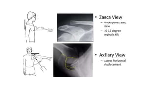
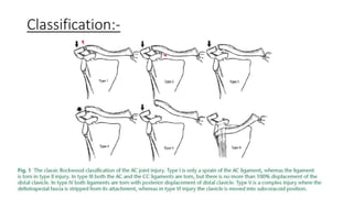
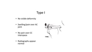
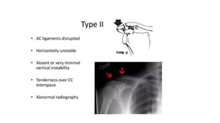
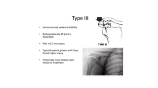
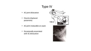
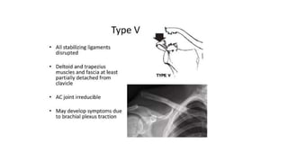
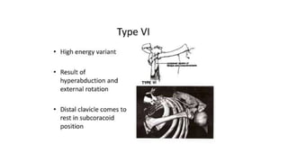
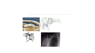
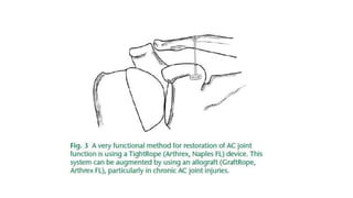
Acromioclavicular joint injuries: diagnosis,
classification and ligamentoplasty
procedures
July 2018
Evrim Sirin
Nuri Aydin
Osman Mert Topkar
Presenter:- Dr. Niraj Raj
Guide - Dr. John Sir
Introductıon:-
• AC joint injury often found among athletes and people involved in contact
sports after an acute shoulder trauma .
• 5 times common in men than in women, with the highest incidence in the
20- to 30-year old age group.
• MOI :- falling on an outstretched arm or direct trauma to the apex of the
shoulder with the arm in adducted position.
• C/O pain and tenderness over the shoulder, particularly over the AC joint.
Anatomy :-
• The AC joint is a diarthrodial joint that serves as a primary link between the
axial skeleton and the upper extremity.
• The joint has dynamic and static stabilizers and it is movable in all planes so
it is not a rigid structure.
• The AC and CC (coracoclavicular) ligaments are the static stabilizers
whereas the deltoid and trapezoid muscles are the dynamic stabilizers.
• The normal AC joint is capable of translating 4 to 6 mm in the
anterior, posterior and superior planes under 70-N loads.
• The joint also accommodates rotary motion of 5° to 8° during
scapulothoracic motion and 40° to 45° with shoulder abduction and
elevation.
Clinical presentation
• Tenderness over AC joint & limited shoulder range of motion.
• Provocative tests for AC joint pathology (cross-arm adduction and
loading of the AC joint) to localize shoulder pain to the AC joint.
Imaging
• Standard AP, lateral and axillary views.
• The axillary view to visualize the amount of posterior displacement of
the clavicle.
• Zanca view is often helpful and is obtained by tilting the radiograph
beam 10° to 15° cephalad compared with a standard shoulder
radiograph.
• Weighted stress views of the clavicle to distinguish type II and III
injuries.
• MRI is not a routine imaging method, done to assess AC and CC
ligamentous disruptions.
Classification:-
Management:-
• The management of AC injuries is still a controversial.
• Type I and type II injuries are the result of low energy forces and they
are treated by conservative methods using either a harness or a sling.
• On the other hand, type IV, V and VI injuries all result from high-
energy forces and they are generally treated by operative methods.
• The treatment of type III injuries is still a matter of debate.
• The main principle of surgical therapy is accurate reduction of the AC joint
in both coronal and sagittal planes.
• This is achieved either by primary repair or by reconstruction of injured
ligaments and maintaining stability to protect this repair or reconstruction.
• The traditional Weaver-Dunn CA ligament transfer procedure has largely
fallen into disfavour today.
• Surgical procedures have been describe are screws, plates, muscle
transfers, ligamentoplasty procedures and ligament reconstruction using
either autograft or allografts.
• Anatomical ligament reconstruction using tendon graft can be performed
by open and arthroscopic methods.
• The hook plate, AC joint transfixion with K wires (Phemister technique) and
CC fixation with a screw (Bosworth technique) are recognized as non-
anatomical procedures which reduce mobility and are related to high rates
of fixation failure and complications.
• Recent treatment modalities for AC joint dislocation have focused on either
CC ligament reconstruction or CC interval fixation.
• Many devices such as screws, plates, suture anchors or synthetic tapes
have been used as fixation material;
• however, none of these methods are free of complications such as implant
failure or implant migration, bony erosions and fractures of the clavicle,
and recurrent dislocation.
• Numerous open or mini-open surgical fixations are currently performed,
but they are associated with certain complications such as infection, failure
of fixation or implant fixation.
• Recently there has been increasing interest in arthroscopic management of
these injuries.
• Management requires reconstruction of the AC ligament as well as the
superior joint capsule.
Arthroscopic technique
• Arthroscopy allows a better, greater and clearer visibility around the
coracoid, and extensive dissection of the deltotrapezial fascia is not
required.
• 3 portals are mainly used: namely a posterior portal; an anterolateral
portal for the optical device; and an operative anterosuperior portal.
• The patient is placed in the ‘beach-chair’ position and a 30° arthroscope is
used to visualize the glenohumeral joint through a posterior portal.
• Arthroscopic techniques usually rely on bony tunnels and suture-graft fixation.
• Use of single tunnel versus double,suture versus graft, autograft versus
allograft and adjustable-length suture versus non-adjustable suture are all
different variables.
• A/C to Scheibel et al, drilling two tunnels for the reconstruction of the CC
complex and using an adjustable-length suture is a more anatomical
method with good to excellent results.
• Today the most commonly used arthroscopic systems are the
TightRope system, suture-shuttle device and all arthroscopic
systems.
TightRope:-
• A very functional method for restoration of AC joint function is using
a TightRope® (Arthrex, Naples, FL, USA) device, where a single or
double drill hole through the clavicle is made.
• contains two titanium buttons
• One button is for the clavicle and the other is for the coracoid
process; they are joined by a loop of No. 5 fiberwire suture.
• This TightRope device can either be used by itself or with the
augmentation of tendon allograft particularly for chronic AC joint
dislocations (GraftRope, Arthrex, FL, USA).
• Augmentation with tendon allograft reduces the stress-rising effects
of titanium buttons around the clavicle and the coracoid, so the risk
of failure by suture cut-out is minimized.
Suture-shuttle (coracoid cerclage technique)
• Suture-shuttle device augmented by a tendon graft, especially in
chronic AC injuries.
All-arthroscopic method
• The other arthroscopic procedure is the all-arthroscopic technique
that is best reserved for acute cases, particularly for acute grade 4
and 5 lesions.
• This procedure consists of two stages.
• In the first stage, the reconstruction of CC ligaments is performed,
• And in the second stage, the reconstruction of superior AC ligaments
is performed.
• This system relies on restoration of the AC joint on both the
horizontal and coronal planes,
Take home message
• There is no current gold standard for the treatment of AC joint
injuries.
• With recent advances in arthroscopic techniques and introduction of
new equipment, the treatment modalities are shifted towards less
invasive and more arthroscopically-assisted methods.

JC Acromioclavicular joint injuries.pptx

  • 1.
    Acromioclavicular joint injuries:diagnosis, classification and ligamentoplasty procedures July 2018 Evrim Sirin Nuri Aydin Osman Mert Topkar Presenter:- Dr. Niraj Raj Guide - Dr. John Sir
  • 2.
    Introductıon:- • AC jointinjury often found among athletes and people involved in contact sports after an acute shoulder trauma . • 5 times common in men than in women, with the highest incidence in the 20- to 30-year old age group. • MOI :- falling on an outstretched arm or direct trauma to the apex of the shoulder with the arm in adducted position. • C/O pain and tenderness over the shoulder, particularly over the AC joint.
  • 3.
    Anatomy :- • TheAC joint is a diarthrodial joint that serves as a primary link between the axial skeleton and the upper extremity. • The joint has dynamic and static stabilizers and it is movable in all planes so it is not a rigid structure. • The AC and CC (coracoclavicular) ligaments are the static stabilizers whereas the deltoid and trapezoid muscles are the dynamic stabilizers.
  • 4.
    • The normalAC joint is capable of translating 4 to 6 mm in the anterior, posterior and superior planes under 70-N loads. • The joint also accommodates rotary motion of 5° to 8° during scapulothoracic motion and 40° to 45° with shoulder abduction and elevation.
  • 5.
    Clinical presentation • Tendernessover AC joint & limited shoulder range of motion. • Provocative tests for AC joint pathology (cross-arm adduction and loading of the AC joint) to localize shoulder pain to the AC joint.
  • 6.
    Imaging • Standard AP,lateral and axillary views. • The axillary view to visualize the amount of posterior displacement of the clavicle. • Zanca view is often helpful and is obtained by tilting the radiograph beam 10° to 15° cephalad compared with a standard shoulder radiograph. • Weighted stress views of the clavicle to distinguish type II and III injuries. • MRI is not a routine imaging method, done to assess AC and CC ligamentous disruptions.
  • 8.
  • 15.
    Management:- • The managementof AC injuries is still a controversial. • Type I and type II injuries are the result of low energy forces and they are treated by conservative methods using either a harness or a sling. • On the other hand, type IV, V and VI injuries all result from high- energy forces and they are generally treated by operative methods. • The treatment of type III injuries is still a matter of debate.
  • 17.
    • The mainprinciple of surgical therapy is accurate reduction of the AC joint in both coronal and sagittal planes. • This is achieved either by primary repair or by reconstruction of injured ligaments and maintaining stability to protect this repair or reconstruction. • The traditional Weaver-Dunn CA ligament transfer procedure has largely fallen into disfavour today.
  • 18.
    • Surgical procedureshave been describe are screws, plates, muscle transfers, ligamentoplasty procedures and ligament reconstruction using either autograft or allografts. • Anatomical ligament reconstruction using tendon graft can be performed by open and arthroscopic methods. • The hook plate, AC joint transfixion with K wires (Phemister technique) and CC fixation with a screw (Bosworth technique) are recognized as non- anatomical procedures which reduce mobility and are related to high rates of fixation failure and complications.
  • 20.
    • Recent treatmentmodalities for AC joint dislocation have focused on either CC ligament reconstruction or CC interval fixation. • Many devices such as screws, plates, suture anchors or synthetic tapes have been used as fixation material; • however, none of these methods are free of complications such as implant failure or implant migration, bony erosions and fractures of the clavicle, and recurrent dislocation.
  • 21.
    • Numerous openor mini-open surgical fixations are currently performed, but they are associated with certain complications such as infection, failure of fixation or implant fixation. • Recently there has been increasing interest in arthroscopic management of these injuries. • Management requires reconstruction of the AC ligament as well as the superior joint capsule.
  • 22.
    Arthroscopic technique • Arthroscopyallows a better, greater and clearer visibility around the coracoid, and extensive dissection of the deltotrapezial fascia is not required. • 3 portals are mainly used: namely a posterior portal; an anterolateral portal for the optical device; and an operative anterosuperior portal. • The patient is placed in the ‘beach-chair’ position and a 30° arthroscope is used to visualize the glenohumeral joint through a posterior portal.
  • 23.
    • Arthroscopic techniquesusually rely on bony tunnels and suture-graft fixation. • Use of single tunnel versus double,suture versus graft, autograft versus allograft and adjustable-length suture versus non-adjustable suture are all different variables. • A/C to Scheibel et al, drilling two tunnels for the reconstruction of the CC complex and using an adjustable-length suture is a more anatomical method with good to excellent results.
  • 24.
    • Today themost commonly used arthroscopic systems are the TightRope system, suture-shuttle device and all arthroscopic systems.
  • 25.
    TightRope:- • A veryfunctional method for restoration of AC joint function is using a TightRope® (Arthrex, Naples, FL, USA) device, where a single or double drill hole through the clavicle is made. • contains two titanium buttons • One button is for the clavicle and the other is for the coracoid process; they are joined by a loop of No. 5 fiberwire suture.
  • 26.
    • This TightRopedevice can either be used by itself or with the augmentation of tendon allograft particularly for chronic AC joint dislocations (GraftRope, Arthrex, FL, USA). • Augmentation with tendon allograft reduces the stress-rising effects of titanium buttons around the clavicle and the coracoid, so the risk of failure by suture cut-out is minimized.
  • 28.
    Suture-shuttle (coracoid cerclagetechnique) • Suture-shuttle device augmented by a tendon graft, especially in chronic AC injuries.
  • 29.
    All-arthroscopic method • Theother arthroscopic procedure is the all-arthroscopic technique that is best reserved for acute cases, particularly for acute grade 4 and 5 lesions. • This procedure consists of two stages. • In the first stage, the reconstruction of CC ligaments is performed, • And in the second stage, the reconstruction of superior AC ligaments is performed. • This system relies on restoration of the AC joint on both the horizontal and coronal planes,
  • 30.
    Take home message •There is no current gold standard for the treatment of AC joint injuries. • With recent advances in arthroscopic techniques and introduction of new equipment, the treatment modalities are shifted towards less invasive and more arthroscopically-assisted methods.