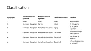
AC Joint Dislocation
• Acromioclavicular (AC) dislocation is a common injury especially among
sportsmen.
• There is still a lack of consensus on whether to conserve or operate
type III AC joint dislocations.
• There are a plethora of choices between mechanical fixation or
synthetic materials or biologic anatomic reconstructions.
• Even among surgeons, there is a choice between open repairs and the
latest—arthroscopic reconstructions.
INTRODUCTION
• The treatment for AC joint dislocations has not been uniform and the
results also vary, based on type of treatment.
• Strapping is restrictive and often nonproductive and a variety of
techniques, from K-wire fixation to synthetic grafts have led to lack of
confidence in choosing the correct treatment.
• AC joint dislocation has an overall incidence of 3 to 4 per 100 000 in
the general population, with 25 % to 52 % occurring during sporting
activities [2, 3].
2. Allman FL., Jr Fractures and ligamentous injuries of the clavicle and its articulation. J Bone Joint Surg Am. 1967;49:774–84.
3. Dias JJ, Gregg PJ. Acromioclavicular joint injuries in sport: recommendations for treatment. Sports Med. 1991;11:125–32. doi: 10.2165/00007256-199111020-00004.
ANATOMY
• The AC joint is a diarthrodial joint formed by the distal clavicle and the medial facet of
the acromion.
• Interposed in the joint is a fibrocartilaginous disk.
• The acromioclavicular capsule is reinforced by anterior, superior, posterior, and inferior
ligaments.
• The posterior and superior ligaments are the strongest and are invested by the
deltotrapezial fascia.
• Biomechanical studies have shown that the AC ligaments and capsule are important in
providing anterior-posterior stability to the AC joint.
• Coracoclavicular ligaments are the laterally located trapezoid ligament and the more
medial conoid ligament.
• These are thought to be the primary suspensory ligaments of the shoulder.
• The coracoclavicular (CC) ligaments prevent superior-inferior displacement of the clavicle
Mechanism of injury
• The mechanism of injury usually involves a direct blow to the lateral
aspect of the shoulder with the arm in an adducted position, leading
to downward displacement of the scapula opposed by impaction of
the clavicle onto the first rib [3].
• The CC ligament is one of the strongest ligaments in the body.
• As the force perpetuates, further energy is transmitted to the
coracoclavicular ligaments, resulting in greater displacement of the
clavicle with reference to the acromion.
• A major injury will lead to further transmission of force and disruption
of the deltoid and trapezius muscles, as the lateral end of clavicle
herniates through it [4].
3. Dias JJ, Gregg PJ. Acromioclavicular joint injuries in sport: recommendations for treatment. Sports Med. 1991;11:125–32. doi: 10.2165/00007256-199111020-00004
4. Lemos MJ. The evaluation and treatment of the injured acromioclavicular joint in athletes. Am J Sports Med. 1998;1:137–44.
Classification
Injury type
Acromioclavicular
ligament
Coracoclavicular
ligament
Deltotrapezial fascia Direction
I Sprain Intact Intact Nondisplaced
II Complete disruption Sprain Intact 25 % Superior
III Complete disruption Complete disruption Injury
25 %–100 %
Superior
IV Complete disruption Complete disruption Detached
Posterior through
the trapezius
V Complete disruption Complete disruption Detached
100 %–300 %
Superior
VI Complete disruption Complete disruption Detached
Inferior to acromion
or coracoid
Clinical features
• Local tenderness and excruciating pain is expected in the acute setting.
• In a type IV and V dislocation injury, gross deformity and tenting of
Trapezius and deltoid is significant along with displacement of skin.
• The ranges are often unaffected, though 20 % of our patients turn up with
a stiff shoulder, which is possibly due to collateral injury to rotator cuff or
surrounding muscles.
• The most common complaint in the late setting is a nagging medial
scapular pain.
• Piano sign may be elicited with ballottement of the lateral end of clavicle.
• The O’Brien test may be particularly helpful when attempting to
differentiate symptoms of AC joint arthroses from intra-articular lesions,
especially with lesions of the superior glenoid labrum.
Radiographic features
• Anteroposterior, lateral, and axial views are standard views taken for
the shoulder; however, a Zanca view [12] is the most accurate view to
look at the AC joint.
• This view is performed by tilting the X-ray beam 10°–15° toward the
cephalic direction.
• To assess AC joint instability, it is desirable to use a 5 kg weight in
each hand and perform bilateral comparative radiographs.
• Weighted X-rays can help differentiate type I from type II injuries and
more importantly type II from occult type III injuries.
• The axial view of the shoulder is important in differentiating a type III
AC joint injury from a type IV injury.
• Visualization of the acromion anterior to the clavicle will indicate a
type IV lesion.
• An increase of more than 25 %–50 % distance in CC interval by
comparing the unaffected side is suggestive of complete disruption of
CC ligament as per [13].
Nonoperative management
• Grade I and II dislocations can be treated nonoperatively with an
appropriate sling such as a RJ bandage or Kenny Brace.
• Three to 6 weeks immobilization followed by rehab should restore
function between 2–3 months after injury.
• In our practice we have extended the conservative program even for
elderly patients with type III and type IV patients, when cosmetic
issues are not of concern and functional restoration is possible due to
the lower level of activities required then.
Operative management
• Early fixation techniques described were using K-wires, Steinman pins
and cerclage wires.
• The use of hardware across the AC joint may worsen the intra-
articular injury and may hasten the onset of joint arthrosis.
• Since the CC ligament is ignored, fixation failure is common with K-
wire migration, also a possibility.
Hook plate
• The Hook Plate was originally designed for lateral end clavicle
fractures.
• This application has been extended to AC joint dislocations.
• It has been associated with numerous complications including
Acromial Fractures, plate bending, and AC arthritis as high as 41 %,
and a definite second surgery for hardware removal [14].
• Hook plate eroding through acromion has been noted as early as 32
days after surgery.
Bosworth screw
• Stabilization of AC joint with a screw between clavicle and coracoid.
• A rigid fixation between Coracoid and clavicle has been an appealing
prospect in the form of screw.
• Due to motion between the coracoid and the clavicle, fatigue of the
implant occurs over time.
• Biomechanical studies in cadaveric models showed that the use of a
Coraco-Clavicular screw, reduced joint motion, and significantly increased
joint contact pressures, which could have implications for early joint
degeneration when this technique is used [15].
• Failure could present as Lateral end clavicle osteolysis, hardware failure, or
even fracture of coracoid or clavicle [16–19].
• There have been reports of high failure of mechanical devices [20].
AC Joint Dislocation .pptx

AC Joint Dislocation .pptx

  • 1.
  • 2.
    • Acromioclavicular (AC)dislocation is a common injury especially among sportsmen. • There is still a lack of consensus on whether to conserve or operate type III AC joint dislocations. • There are a plethora of choices between mechanical fixation or synthetic materials or biologic anatomic reconstructions. • Even among surgeons, there is a choice between open repairs and the latest—arthroscopic reconstructions.
  • 3.
    INTRODUCTION • The treatmentfor AC joint dislocations has not been uniform and the results also vary, based on type of treatment. • Strapping is restrictive and often nonproductive and a variety of techniques, from K-wire fixation to synthetic grafts have led to lack of confidence in choosing the correct treatment. • AC joint dislocation has an overall incidence of 3 to 4 per 100 000 in the general population, with 25 % to 52 % occurring during sporting activities [2, 3]. 2. Allman FL., Jr Fractures and ligamentous injuries of the clavicle and its articulation. J Bone Joint Surg Am. 1967;49:774–84. 3. Dias JJ, Gregg PJ. Acromioclavicular joint injuries in sport: recommendations for treatment. Sports Med. 1991;11:125–32. doi: 10.2165/00007256-199111020-00004.
  • 4.
    ANATOMY • The ACjoint is a diarthrodial joint formed by the distal clavicle and the medial facet of the acromion. • Interposed in the joint is a fibrocartilaginous disk. • The acromioclavicular capsule is reinforced by anterior, superior, posterior, and inferior ligaments. • The posterior and superior ligaments are the strongest and are invested by the deltotrapezial fascia. • Biomechanical studies have shown that the AC ligaments and capsule are important in providing anterior-posterior stability to the AC joint. • Coracoclavicular ligaments are the laterally located trapezoid ligament and the more medial conoid ligament. • These are thought to be the primary suspensory ligaments of the shoulder. • The coracoclavicular (CC) ligaments prevent superior-inferior displacement of the clavicle
  • 5.
    Mechanism of injury •The mechanism of injury usually involves a direct blow to the lateral aspect of the shoulder with the arm in an adducted position, leading to downward displacement of the scapula opposed by impaction of the clavicle onto the first rib [3]. • The CC ligament is one of the strongest ligaments in the body. • As the force perpetuates, further energy is transmitted to the coracoclavicular ligaments, resulting in greater displacement of the clavicle with reference to the acromion. • A major injury will lead to further transmission of force and disruption of the deltoid and trapezius muscles, as the lateral end of clavicle herniates through it [4]. 3. Dias JJ, Gregg PJ. Acromioclavicular joint injuries in sport: recommendations for treatment. Sports Med. 1991;11:125–32. doi: 10.2165/00007256-199111020-00004 4. Lemos MJ. The evaluation and treatment of the injured acromioclavicular joint in athletes. Am J Sports Med. 1998;1:137–44.
  • 6.
    Classification Injury type Acromioclavicular ligament Coracoclavicular ligament Deltotrapezial fasciaDirection I Sprain Intact Intact Nondisplaced II Complete disruption Sprain Intact 25 % Superior III Complete disruption Complete disruption Injury 25 %–100 % Superior IV Complete disruption Complete disruption Detached Posterior through the trapezius V Complete disruption Complete disruption Detached 100 %–300 % Superior VI Complete disruption Complete disruption Detached Inferior to acromion or coracoid
  • 7.
    Clinical features • Localtenderness and excruciating pain is expected in the acute setting. • In a type IV and V dislocation injury, gross deformity and tenting of Trapezius and deltoid is significant along with displacement of skin. • The ranges are often unaffected, though 20 % of our patients turn up with a stiff shoulder, which is possibly due to collateral injury to rotator cuff or surrounding muscles. • The most common complaint in the late setting is a nagging medial scapular pain. • Piano sign may be elicited with ballottement of the lateral end of clavicle. • The O’Brien test may be particularly helpful when attempting to differentiate symptoms of AC joint arthroses from intra-articular lesions, especially with lesions of the superior glenoid labrum.
  • 8.
    Radiographic features • Anteroposterior,lateral, and axial views are standard views taken for the shoulder; however, a Zanca view [12] is the most accurate view to look at the AC joint. • This view is performed by tilting the X-ray beam 10°–15° toward the cephalic direction. • To assess AC joint instability, it is desirable to use a 5 kg weight in each hand and perform bilateral comparative radiographs. • Weighted X-rays can help differentiate type I from type II injuries and more importantly type II from occult type III injuries.
  • 9.
    • The axialview of the shoulder is important in differentiating a type III AC joint injury from a type IV injury. • Visualization of the acromion anterior to the clavicle will indicate a type IV lesion. • An increase of more than 25 %–50 % distance in CC interval by comparing the unaffected side is suggestive of complete disruption of CC ligament as per [13].
  • 10.
    Nonoperative management • GradeI and II dislocations can be treated nonoperatively with an appropriate sling such as a RJ bandage or Kenny Brace. • Three to 6 weeks immobilization followed by rehab should restore function between 2–3 months after injury. • In our practice we have extended the conservative program even for elderly patients with type III and type IV patients, when cosmetic issues are not of concern and functional restoration is possible due to the lower level of activities required then.
  • 11.
    Operative management • Earlyfixation techniques described were using K-wires, Steinman pins and cerclage wires. • The use of hardware across the AC joint may worsen the intra- articular injury and may hasten the onset of joint arthrosis. • Since the CC ligament is ignored, fixation failure is common with K- wire migration, also a possibility.
  • 12.
    Hook plate • TheHook Plate was originally designed for lateral end clavicle fractures. • This application has been extended to AC joint dislocations. • It has been associated with numerous complications including Acromial Fractures, plate bending, and AC arthritis as high as 41 %, and a definite second surgery for hardware removal [14]. • Hook plate eroding through acromion has been noted as early as 32 days after surgery.
  • 13.
    Bosworth screw • Stabilizationof AC joint with a screw between clavicle and coracoid. • A rigid fixation between Coracoid and clavicle has been an appealing prospect in the form of screw. • Due to motion between the coracoid and the clavicle, fatigue of the implant occurs over time. • Biomechanical studies in cadaveric models showed that the use of a Coraco-Clavicular screw, reduced joint motion, and significantly increased joint contact pressures, which could have implications for early joint degeneration when this technique is used [15]. • Failure could present as Lateral end clavicle osteolysis, hardware failure, or even fracture of coracoid or clavicle [16–19]. • There have been reports of high failure of mechanical devices [20].