SlideShare a Scribd company logo
1 of 10
Download to read offline
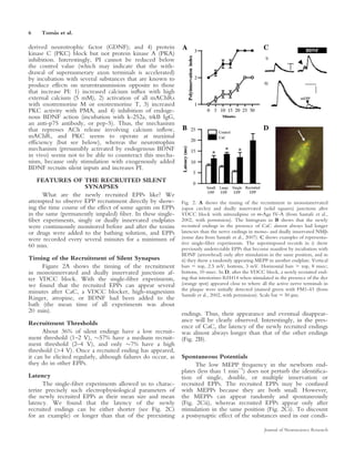
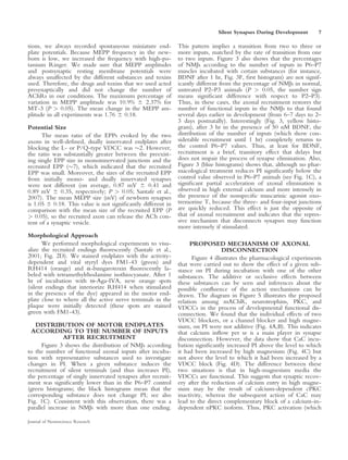
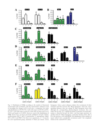
Journal of Neuroscience Research 89:3–12 (2011)


Mini-Review
Silent Synapses in Neuromuscular Junction
Development
Josep Tomas,* Manel M. Santafe, Maria A. Lanuza, Neus Garcıa, Nuria Besalduch,
          `                  ´                            ´
              `
and Marta Tomas
                                                                    `
Unitat d’Histologia i Neurobiologia (UHN), Facultat de Medicina i Ciencies de la Salut,
Universitat Rovira i Virgili, Reus, Spain

In the last few years, evidence has been found to suggest            SYNAPSE LOSS IN THE NEUROMUSCULAR
that some synaptic contacts become silent but can be                                         JUNCTION
functionally recruited before they completely retract during               The neuromuscular junction (NMJ) is a classic model
postnatal synapse elimination in muscle. The physiological         from which much of our understanding of synaptic transmis-
mechanism of developmental synapse elimination may be              sion has emerged (Katz, 1996), including the calcium-depend-
better understood by studying this synapse recruitment.            ent quantal nature of transmitter release and the response of the
This Mini-Review collects previously published data and            postsynaptic receptors. This is also true for the different forms
new results to propose a molecular mechanism for axonal            of nervous system plasticity. In general, the basic NMJ
disconnection. The mechanism is based on protein kinase            mechanisms operate throughout the nervous system. Synapse
C (PKC)-dependent inhibition of acetylcholine (ACh)                elimination during initial synaptogenesis occurs in the NMJs
release. PKC activity may be stimulated by a methoctr-             (Thompson, 1985), as it does throughout the nervous struc-
amine-sensitive M2-type muscarinic receptor and by cal-            tures (Bourgeois and Rakic, 1993). The skeletal muscle cells in
cium inflow though P/Q- and L-type voltage-dependent                newborn vertebrates are transiently polyinnervated at a single
calcium channels. In addition, tropomyosin-related tyro-           synaptic site by several motor axons (Redfern, 1970; Brown
sine kinase B (trkB) receptor-mediated brain-derived neu-          et al., 1976; Ribchester and Barry, 1994). Figure 1A shows a
rotrophic factor (BDNF) activity may oppose the PKC-               pool of polyinnervated NMJ from a P6 rodent levator auris
mediated ACh release depression. Thus, a balance                   longus (LAL) muscle. Most evidence shows that, in the first
between trkB and muscarinic pathways may contribute to             postnatal days, differential activity among the axons determines
the final functional suppression of some neuromuscular              which endings are lost because relatively inactive synapses are
synapses during development. V 2010 Wiley-Liss, Inc.
                                   C
                                                                   permanently removed by the activity elicited by more active
                                                                   inputs (Jansen and Fladby, 1990; Sanes and Lichtman, 1999;
Key words: postnatal synapse elimination; voltage-
                                                                   however, see Callaway et al., 1987). Synapse elimination is ac-
dependent calcium channels; muscarinic acetylcholine
                                                                   tivity dependent, because it slows down or speeds up when
receptors; protein kinases; neurotrophins
                                                                   total neuromuscular activity decreases or increases, respectively.
                                                                   In addition, the axon terminals that fire coordinately with the
                                                                   postsynaptic cells can be enlarged and strengthened, whereas
  DEVELOPMENTAL SYNAPSE ELIMINATION                                asynchronous synapses can be weakened (Bi and Poo, 2001;
                                                                   Favero et al., 2010). Finally, in the adult, endplates are inner-
      The development of the nervous system involves               vated by a single axon (Benoit and Changeux, 1975; O’Brien
an initially exuberant production of neurons that estab-           et al., 1978). Thus, in a nonrandom, active process, strong syn-
lish excessive synaptic contacts and the subsequent
reduction in both neurons and synapses. This develop-
mental process consists of an initial synapse overproduc-          Contract grant sponsor: MEC; Contract grant number: SAF 2008-02836;
tion to promote broad connectivity and a subsequent ac-            Contract grant sponsor: Catalan Government; Contract grant number:
                                                                   2009SGR01248.
tivity-dependent reduction in synapse number. This
allows connectivity to be refined and specificity gained.                                              `
                                                                   *Correspondence to: Dr. J. Tomas, Unitat d’Histologia i Neurobiologia
Hebbian competition between the nerve terminals of                                                     `
                                                                   (UHN), Facultat de Medicina i Ciencies de la Salut, Universitat Rovira i
axons with different activities seems to be the fundamen-          Virgili, Carrer St. Llorenc num 21, 43201-Reus, Spain.
                                                                                             ¸
                                                                   E-mail: jmtf@fmcs.urv.es
tal characteristic of this process of synapse elimination,
which leads to the loss of roughly half of the overpro-            Received 22 April 2010; Revised 23 June 2010; Accepted 11 July 2010
duced elements (Fields and Nelson, 1992; Sanes and                 Published online 20 September 2010 in Wiley Online Library
Lichtman, 1999; Mennerick and Zorumski, 2000).                     (wileyonlinelibrary.com). DOI: 10.1002/jnr.22494

' 2010 Wiley-Liss, Inc.
4        `
      Tomas et al.

aptic connections decrease the effectiveness of other inputs and      Most studies have found a progressive diminution of the neu-
weak inputs can neither support themselves nor eliminate other        rotransmitter release capacity of the nerve terminals that are dis-
inputs. A genetic method of selectively inhibiting neurotrans-        connected. At the same time, the postsynaptic receptors are
mission from one of two inputs to a single target cell shows          reorganized. However, the causal relation between axonal re-
that more powerful inputs are strongly favored competitors            traction and postsynaptic receptor changes are not fully under-
during synapse elimination (Jia et al., 1999; Buffelli et al.,        stood (Colman et al., 1997; Lanuza et al., 2002).
2003). However, local differential effectiveness rather than (or
in addition to) differential activity should be the key determi-           OCCURRENCE OF SILENT SYNAPSES
nant of eventual success, because an axon that fails at one syn-        DURING DEVELOPMENTAL ELIMINATION
apse can be successful at another (Keller-Peck et al., 2001),                             IN MUSCLE
which suggests very local involvement of the postsynaptic mus-             We know that, in polyinnervated synapses, quantal
cle cell and postsynaptic (and glial cell)-derived trophic factors.   responses clearly decrease in both size and number


                                                                Figure 1.

                                                                                                            Journal of Neuroscience Research
Silent Synapses During Development             5

before axonal withdrawal is completed (Dunia and                             functional inputs for a large number of P6–P7 NMJs
Herrera, 1993; Colman et al., 1997). Neurotransmitter                        from rat LAL muscle. Then, we calculated the mean
release from the axon that survives is characterized by a                    value, defined as the polyinnervation index of the
greater quantal content, whereas the efficiency of the                        muscle (PI). The nerve was stimulated with increasing
input(s) that is removed decreases progressively, because                    intensity from zero until an endplate potential (EPP)
a small quantal content is associated with reduced postsy-                   was observed. If the size and latency of the EPP
naptic receptor density (Colman et al., 1997; Culican                        remained constant as the stimulus was increased, we
et al., 1998). The competitive interactions may affect                       concluded that the endplate was monoinnervated. In
neurotransmitter release first, and axonal detachment                         endplates with polyneuronal innervation, increasing the
may occur after the neurotransmitter release has been                        stimulus amplitude recruited one or more axons,
totally abolished. Does the release machinery still operate                  which produced a stepwise increment in the EPP
for some time after the cessation of neurotransmission?                      (Redfern, 1970). This compound EPP is built by
With this question in mind, we test the hypothesis that                      recruiting two or more axons. In many polyinnervated
some nerve terminals become silent before they com-                          synapses, the endings are separated not by the thresh-
pletely retract (and shed membrane-bound remnants;                           old but by the latency. To study the PI, we consider
Bishop et al., 2004), and before the complete end of the                     only those synapses in which the different inputs are
repression period but that they retain certain capabilities                  clearly separated, on successive stimulations (10–20
for evoked release that can be recovered. Understanding                      EPPs at 0.5 Hz), by the excitability threshold, the la-
the role that important molecules play in ACh release at                     tency, or both. In this way, reproducibility is guaran-
the time of synapse elimination may help in understand-                      teed and the individual inputs can be accurately iden-
ing this important mechanism.                                                tified (see Fig. 1B).
                                                                                    In P6–P7 muscles, the PI was 1.63 6 0.14 with
   FUNCTIONAL RECRUITMENT OF SILENT                                          47.92% 6 2.08% of monoinnervated junctions (Lanuza
                     SYNAPSES                                                et al., 2001; Santafe et al., 2001). Figure 1C shows the
      Imposed changes in synaptic activity can acceler-                      effect on PI of blocking or activating several key mole-
ate or delay the developmental synapse elimination                           cules involved in ACh release. An increase in PI can be
process (Jansen and Fladby, 1990). In most cases, devi-                      observed in the following circumstances: 1) specific
ations from the normal physiological tempo can occur                         block of calcium entry through L- and P/Q-type (but
for several hours or even days (Nelson et al., 2005).                        not N-type) voltage-dependent calcium channels
Here, we investigate the almost immediate response                           (VDCC) or high magnesium-mediated nonspecific cal-
(1 hr) of some motor nerve terminals that recover ac-                        cium inflow reduction; 2) M2-type muscarinic acetyl-
etylcholine (ACh) release by acute exposure to modu-                         choline autoreceptor (mAChR) block (but not M1, M3,
lators of the molecular pathways involved in neuro-                          and M4 subtypes block); 3) brain-derived neurotrophic
transmission. We used intracellular recording of the                         factor (BDNF) incubation but not stimulation with neu-
evoked synaptic potentials to observe the number of                          rotrophin-3 (NT-3), neurotrophin-4 (NT-4), or glial-
3
Fig. 1. A shows examples of polyinnervated NMJ from a P6 rodent              that blocking the M2 mAChR, the L-type or the P/Q-type VDCC, or
levator auris longus (LAL) muscle. The electrophysiological raw data in      the PKC and stimulating the neuromuscular preparation with exoge-
B shows two doubly innervated endplates (left; vertical bars 5 2 mV,         nous BDNF show similar (increasing) PI response patterns. Calcium
horizontal bars 5 10 msec) and a polyinnervated NMJ (right; vertical         inflow agents: Mg21, 5 mM; the N-type VDCC blocker x-conotoxin
bar 5 4 mV, horizontal bar 5 10 msec) in a P6 LAL muscle. The                GVI-A (x-CgTx GVI-A), 1 lM; the L-type VDCC blocker Nitrendi-
superimposed traces show synapses with inputs that, on successive            pine, 1 lM; the P/Q-type VDCC blocker x-agatoxin IV-A (x-Aga
stimulations, are clearly separated by the excitability threshold (V,        IV-A), 100 nM; Ca21, 5 mM. Muscarinic agents: The nonspecific
graded nerve stimulation) or the latency, so the individual inputs can       muscarinic blocker atropine (AT), 2 lM; the M2 blocker methoctr-
be accurately identified. In C, the diagrams are a graphic representation     amine (METHOC), 1 lM; the M1 muscarinic blockers pirenzepine
that collectively show the pattern of action of the different agents used    (PIR), 10 lM; muscarinic toxin 7 (MT-7), 100 nM; the M4 blockers
on changing polyinnervation index (PI). These substances block or            tropicamide (TPC), 1 lM; muscarinic toxin 3 (MT-3), 100 nM; the
activate different molecules related to ACh release and PI can be            M3 blocker 4-DAMP, 1 lM; the muscarinic agonists oxotremorine M
observed to increase in some circumstances. Some data are from Santafe       (OXO-M), 1 lM; and oxotremorine T (OXO-T), 1 lM. Serine
et al. (2009) and Garcia et al. (2010). However, most values of the          kinase agents: The PKC blockers calphostin C (CaC), 1 lM, stauro-
polyinnervation index have not been previously published: 4-DAMP,            sporine (STP), 200 nM, and chelerythrine (CEL), 1 lM; the PKC
oxotremorine M, oxotremorine T, staurosporine, PMA, H-89, Sp-8-              stimulator phorbol ester (PMA), 10 nM; the PKA blocker H-89,
BrcAMPs, K252-A, TrkB-IgG, Ac anti p75, PEP-5, NT-4, NT-3,                   5 lM; the PKA stimulator Sp-8-BrcAMPs, 10 lM. Neurotrophin
GDNF. For these new experiments, n 5 5 muscles for each experi-              agents: The tyrosine kinase blocker K252-A, 200 nM; the BDNF
mental group, minimum 15 fibers per muscle. The thick black line              neutralizing fusion protein TrkB-IgG (TrkB-IgG), 1 lg/ml; the neu-
means ratio 1 (experimental PI/control PI) or ‘‘no effect.’’ The experi-     tralizing antibody against p75NTR (Ac anti-p75), 5 lg/ml; the p75NTR
mental effect of the different substances is shown by the thick blue line.   pathway blocker PEP-5, 1 lM; brain-derived neurotrophic factor
Green circles mean that the experimental PI is significantly different        (BDNF), 50 nM; neurotrophin 4 (NT-4), 50 ng/ml; neurotrophin
from the control PI (P < 0.05), and blue squares mean that there is no       3 (NT-3), 200 ng/ml; glial-derived neurotrophic factor (GDNF),
difference (P > 0.05). SEMs are eliminated for clarity. It can be noted      200 ng/ml. Scale bar 5 10 lm.

Journal of Neuroscience Research
6       `
     Tomas et al.

derived neurotrophic factor (GDNF); and 4) protein
kinase C (PKC) block but not protein kinase A (PKA)
inhibition. Interestingly, PI cannot be reduced below
the control value (which may indicate that the with-
drawal of supernumerary axon terminals is accelerated)
by incubation with several substances that are known to
produce effects on neurotransmission opposite to those
that increase PI: 1) increased calcium influx with high
external calcium (5 mM), 2) activation of all mAChRs
with oxotremorine M or oxotremorine T, 3) increased
PKC activity with PMA, and 4) inhibition of endoge-
nous BDNF action (incubation with k-252a, trkB IgG,
an anti-p75 antibody, or pep-5). Thus, the mechanism
that represses ACh release involving calcium inflow,
mAChR, and PKC seems to operate at maximal
efficiency (but see below), whereas the neurotrophin
mechanism (presumibly activated by endogenous BDNF
in vivo) seems not to be able to counteract this mecha-
nism, because only stimulation with exogenously added
BDNF recruits silent inputs and increases PI.
    FEATURES OF THE RECRUITED SILENT
                      SYNAPSES
      What are the newly recruited EPPs like? We
attempted to observe EPP recruitment directly by show-         Fig. 2. A shows the timing of the recruitment in monoinnervated
ing the time course of the effect of some agents on EPPs       (open circles) and dually innervated (solid squares) junctions after
in the same (permanently impaled) fiber. In these single-       VDCC block with nitrendipine or x-Aga IV-A (from Santafe et al.,
fiber experiments, singly or dually innervated endplates        2002, with permission). The histogram in B shows that the newly
were continuously monitored before and after the toxins        recruited endings in the presence of CaC almost always had longer
or drugs were added to the bathing solution, and EPPs          latencies than the nerve endings in mono- and dually innervated NMJs
were recorded every several minutes for a minimum of           (some data from Santafe et al., 2007). C shows examples of representa-
                                                               tive single-fiber experiments. The superimposed records in i) show
60 min.                                                        previously undetectable EPPs that become manifest by incubation with
                                                               BDNF (arrowhead) only after stimulation in the same position, and in
Timing of the Recruitment of Silent Synapses                   ii) they show a randomly appearing MEPP in another endplate. Vertical
     Figure 2A shows the timing of the recruitment             bars 5 top, 2.5 mV; bottom, 3 mV. Horizontal bars 5 top, 8 msec;
in monoinnervated and dually innervated junctions af-          bottom, 10 msec. In D, after the VDCC block, a newly recruited end-
ter VDCC block. With the single-fiber experiments,              ing that interiorizes RH414 when stimulated in the presence of the dye
we found that the recruited EPPs can appear several            (orange spot) appeared close to where all the active nerve terminals in
                                                               the plaque were initially detected (stained green with FM1-43 (from
minutes after CaC, a VDCC blocker, high-magnesium              Santafe et al., 2002, with permission). Scale bar 5 50 lm.
Ringer, atropine, or BDNF had been added to the
bath (the mean time of all experiments was about
20 min).                                                       endings. Thus, their appearance and eventual disappear-
Recruitment Thresholds                                         ance will be clearly observed. Interestingly, in the pres-
                                                               ence of CaC, the latency of the newly recruited endings
      About 36% of silent endings have a low recruit-          was almost always longer than that of the other endings
ment threshold (1–2 V), 57% have a medium recruit-            (Fig. 2B).
ment threshold (2–4 V), and only 7% have a high
threshold (4 V). Once a recruited ending has appeared,
it can be elicited regularly, although failures do occur, as   Spontaneous Potentials
they do in other EPPs.                                               The low MEPP frequency in the newborn end-
                                                               plates (less than 1 min–1) does not perturb the identifica-
Latency                                                        tion of single, double, or multiple innervation or
      The single-fiber experiments allowed us to charac-        recruited EPPs. The recruited EPPs may be confused
terize precisely such electrophysiological parameters of       with MEPPs because they are both small. However,
the newly recruited EPPs as their mean size and mean           the MEPPs can appear randomly and spontaneously
latency. We found that the latency of the newly                (Fig. 2Cii), whereas recruited EPPs appear only after
recruited endings can be either shorter (see Fig. 2Ci          stimulation in the same position (Fig. 2Ci). To discount
for an example) or longer than that of the preexisting         a postsynaptic effect of the substances used in our condi-

                                                                                                       Journal of Neuroscience Research
Silent Synapses During Development       7

tions, we always recorded spontaneous miniature end-          This pattern implies a transition from two to three or
plate potentials. Because MEPP frequency in the new-          more inputs, matched by the rate of transition from one
born is low, we increased the frequency with high-po-         to two inputs. Figure 3 also shows that the percentages
tassium Ringer. We made sure that MEPP amplitudes             of NMJs according to the number of inputs in P6–P7
and postsynaptic resting membrane potentials were             muscles incubated with certain substances (for instance,
always unaffected by the different substances and toxins      BDNF after 1 hr, Fig. 3F, first histogram) are not signif-
used. Therefore, the drugs and toxins that we used acted      icantly different from the percentage of NMJs in normal,
presynaptically and did not change the number of              untreated P2–P3 animals (P  0.05, the number sign
AChRs in our conditions. The maximum percentage of            means significant difference with respect to P2–P3).
variation in MEPP amplitude was 10.9% 6 2.37% for             Thus, in these cases, the axonal recruitment restores the
MT-3 (P  0.05). The mean change in the MEPP am-              number of functional inputs in the NMJs to that found
plitude in all experiments was 1.76 6 0.18.                   several days earlier in development (from 6–7 days to 2–
                                                              3 days postnatally). Interestingly (Fig. 3, yellow histo-
Potential Size                                                gram), after 3 hr in the presence of 50 nM BDNF, the
      The mean ratio of the EPPs evoked by the two            distribution of the number of inputs (which show con-
axons in well-defined, dually innervated endplates after       siderable recruitment until 1 hr) completely returns to
blocking the L- or P/Q-type VDCC was 2. However,             the control P6–P7 values. Thus, at least for BDNF,
the ratio was substantially greater between the preexist-     recruitment is a brief, transitory effect that delays but
ing single EPP size in monoinnervated junctions and the       does not impair the process of synapse elimination. Also,
recruited EPP (7), which indicated that the recruited        Figure 3 (blue histograms) shows that, although no phar-
EPP was small. Moreover, the sizes of the recruited EPP       macological treatment reduces PI significantly below the
from initially mono- and dually innervated synapses           control value observed in P6–P7 animals (see Fig. 1C), a
were not different (on average, 0.87 mV 6 0.41 and            significant partial acceleration of axonal elimination is
0.89 mV 6 0.35, respectively; P  0.05; Santafe et al.,       observed in high external calcium and more intensely in
2007). The mean MEPP size (mV) of newborn synapses            the presence of the nonspecific muscarinic agonist oxo-
is 1.05 6 0.18. This value is not significantly different in   tremorine T, because the three- and four-input junctions
comparison with the mean size of the recruited EPP (P         are quickly reduced. This effect is just the opposite of
 0.05), so the recruited axon can release the ACh con-       that of axonal recruitment and indicates that the repres-
tent of a synaptic vesicle.                                   sive mechanism that disconnects synapses may function
                                                              more intensely if stimulated.
Morphological Approach
      We performed morphological experiments to visu-               PROPOSED MECHANISM OF AXONAL
alize the recruited endings fluorescently (Santafe et al.,                         DISCONNECTION
2001; Fig. 2D). We stained endplates with the activity-              Figure 4 illustrates the pharmacological experiments
dependent and vital styryl dyes FM1-43 (green) and            that were carried out to show the effect of a given sub-
RH414 (orange) and a-bungarotoxin fluorescently la-            stance on PI during incubation with one of the other
beled with tetramethylrhodamine isothiocyanate. After 1       substances. The additive or occlusive effects between
hr of incubation with x-Aga-IVA, new orange spots             these substances can be seen and inferences about the
(silent endings that interiorize RH414 when stimulated        possible confluence of the action mechanisms can be
in the presence of the dye) appeared in the motor end-        drawn. The diagram in Figure 5 illustrates the proposed
plate close to where all the active nerve terminals in the    relation among mAChR, neurotrophins, PKC, and
plaque were initially detected (these spots are stained       VDCCs in the process of developmental functional dis-
green with FM1-43).                                           connection. We found that the individual effects of two
                                                              VDCC blockers, or a channel blocker and high magne-
   DISTRIBUTION OF MOTOR ENDPLATES                            sium, on PI were not additive (Fig. 4A,B). This indicates
  ACCORDING TO THE NUMBER OF INPUTS                           that calcium inflow per se is a main player in synapse
               AFTER RECRUITMENT                              disconnection. However, the data show that CaC incu-
      Figure 3 shows the distribution of NMJs according       bation significantly increased PI above the level to which
to the number of functional axonal inputs after incuba-       it had been increased by high magnesium (Fig. 4C) but
tion with representative substances used to investigate       not above the level to which it had been increased by a
changes in PI. When a given substance induces the             VDCC block (Fig. 4D). The difference between these
recruitment of silent terminals (and thus increases PI),      two situations is that in high-magnesium media the
the percentage of singly innervated synapses after recruit-   VDCCs are functional. This suggests that synaptic recov-
ment was significantly lower than in the P6–P7 control         ery after the reduction of calcium entry in high magne-
(green histograms; the black histograms mean that the         sium may be the result of calcium-dependent cPKC
corresponding substance does not change PI; see also          inactivity, whereas the subsequent action of CaC may
Fig. 1C). Consistent with this observation, there was a       lead to the direct complementary block of a calcium-in-
parallel increase in NMJs with more than one ending.          dependent nPKC isoform. Thus, PKC activation (which

Journal of Neuroscience Research
Fig. 3. Distribution of NMJs according to the number of functional        histograms: when a given substance induces the recruitment of silent
axonal inputs after incubation with the representative substances used    terminals (and thus increases PI). Black histograms: when the corre-
to investigate the changes in PI. In all cases, a comparison was made     sponding substance does not change PI. Blue histograms shows that
with distributions at P2–P3 (#P  0.05 with respect to P2–P3) and P6–     although the corresponding pharmacological treatments does not
P7 (*P  0.05 with respect to P6–P7). Some data are from Garcia et al.    reduce PI (see Fig. 1C), a significant partial acceleration of axonal elim-
(2010) and Santafe et al. (2009). However, most of the distributions of   ination is observed because the three- and four-input junctions are
NMJs shown have not been previously published: x-CgTx GVI-A,              quickly reduced. Yellow histogram: a particular situation when, after 3
methoctramine MT-7, MT-3, oxotremorine T, staurosporine, PMA,             hr in the presence of 50 nM BDNF, the distribution of the number of
NT-4, NT-3 and PEP-5. For these new experiments, n 5 5 muscles            inputs (which show considerable recruitment until 1 hr) completely
for each experimental group, minimum 15 fibers per muscle. Green           returns to the control P6–P7 values.
Silent Synapses During Development             9


Fig. 4. Pharmacological experiments designed to show the effect of a      ment, n 5 5 muscles, minimum 15 fibers per muscle. Values are
given substance on PI during incubation with one of the other sub-        expressed as a percentage (mean 6 SEM) of EPP amplitude at time 0
stances. Thus, the additive or occlusive effects between these substan-   (control). *P  0.05 with respect to control values (T 5 0 min). #P
ces can be studied. Most data are from Santafe et al. (2009). The data     0.05 with respect to T 5 60 min. Each line represents three to six
shown in F have not been previously published. For this new experi-       single-fiber experiments obtained from different muscles.


was finally blocked by CaC) may be partially calcium in-                         Which extracellular signals from the local context
dependent. Nevertheless, the calcium channels them-                       of the activity-dependent axonal competition affect the
selves seem to be an important PKC target, because,                       PKC-VDCC mechanism in the synaptic contacts that
when they are blocked (see P/Q in Fig. 4D), CaC-                          are disconnected? Our experiments indicate that the
induced PKC inhibition no longer has any nerve ending                     BDNF-trkB (Fig. 5, point 5) and the ACh-mAChRs
recruiting activity. Thus, we hypothesize that PKC con-                   (Fig. 5, point 6) pathways may have a leading role.
tributes at least partially to synapse disconnection by
modulating VDCC operation (Fig. 5, point 1). Calcium                      Involvement of BDNF
entry also modulates PKC involvement in the pathway                             As with CaC, Figure 4G shows that BDNF can
(Fig. 5, point 2). In this regard, it is known that the                   extend the effect of high magnesium and also consider-
VDCCs are targets for PKCs that may regulate the effi-                     ably increase functional polyinnervation (mainly the tri-
cacy of calcium permeation. The influence of both PKC                      ple synapses). However, previous incubation with
and PKA on ACh release depends on the P/Q-type                            nitrendipine or CaC (Fig. 4H,I, respectively) completely
VDCC (Santafe et al., 2006), and PKC can regulate N-                      impairs any additional effect of BDNF. Thus, only when
type (Yokoyama et al., 2005) and L-type (Arenson and                      the first incubation is performed in high magnesium can
Evans, 2001) channels. Finally, calcium entry may have a                  both CaC and BDNF extend recruitment above that
direct influence on synapse disconnection, probably                        observed in high magnesium. This suggests that the
through a calcium-dependent protease system (Fig. 5,                      pathway turned on by BDNF may inhibit the PKC
point 3), and/or indirectly through a cPKC-mediated                       mechanism or at least nPKC, because CaC seems to do
phosphorylation in another intermediary step leading to                   in high magnesium media. These results indicate that
synaptic withdrawal (Fig. 5, point 4).                                    incubation with exogenous BDNF (but not any other

Journal of Neuroscience Research
10        `
       Tomas et al.

                                                                         sall, 1998; Nathanson, 2000). Both intracellular pathways
                                                                         can modulate the VDCC.
                                                                                We believe that the involvement of presynaptic
                                                                         mAChRs in the elimination process may allow direct
                                                                         interaction between nerve endings through differential
                                                                         activity-dependent ACh release. Thus, the most active
                                                                         ending may directly punish endings that are less active
                                                                         (ACh-filled arrow in Figure 5 indicates an ACh stream
                                                                         coming from a strong nerve terminal). There is some
                                                                         evidence to suggest that, in synapse elimination, active
                                                                         synapses prosper by punishing their inactive neighbors
                                                                         (see, e.g., Thompson, 1985). Axons may compete by
                                                                         generating activity-mediated signals to destabilize directly
                                                                         synaptic sites associated with other inputs. The with-
Fig. 5. Proposed relation among M2-type mAChR, the BDNF-trkB
                                                                         drawal process is spatially regulated so that the branches
pathway, PKC and L- and P/Q-type VDCCs in the control of the             of an axon that are nearest (50 lm) to the competitor’s
ACh release mechanism, and neurotransmission from the axonal             territory are removed before the more distant branches
inputs in the process of developmental functional disconnection.         undergo retraction (Gan and Lichtman, 1998). This short
Taking all the findings into consideration, we believe that the best      distance is compatible with a local diffusion of ACh
interpretation of the results is as illustrated here. The green arrows   within the common synaptic gutter between competing
indicate a stimulating action, and the red arrows indicate an inhibi-    endings. We have observed that the different axonal
tory one. See text for explanation.                                      inputs to a given NMJ in the LAL muscle are generally
                                                                         intermingled (indeed, they share the same postsynaptic
                                                                         gutter of 10 lm) in the same endplate site, especially
neurotrophin or the neurotrophic cytokine GDNF, in                       at the beginning and in the first half of the elimination
gray in Fig. 5) has the same effect as PKC and L-type                    process. However, although one neuron propagates the
VDCC block. Thus, BDNF may modulate the PKC-                             same activity pattern and level to all its axonal branches,
VDCC intracellular cascade that, when fully active in                    the competitive success of these branches can vary
certain motor nerve terminals, leads to synapse discon-                  greatly in different polyinnervated junctions (Keller-Peck
nection. We observed that the blockade of the trkB re-                   et al., 2001). This suggests local interactions that involve
ceptor (but not of p75NTR signalling) prevents exoge-                    the postsynaptic muscle cell. A target-derived neurotro-
nous BDNF-induced recruitment (Garcia et al., 2010).                     phic factor may oppose the M2 mAChR and PKC-
It seems as if the silent endings have lost the p75NTR                   mediated depressing action on release. Thus, we
signalling but not the trkB pathway, which may remain                    hypothesize that there is a displacement of the balance
precariously coupled to release, because resources for                   between both BDNF-trkB and muscarinic pathways that
neurotransmission progressively decrease as synapse elim-                might contribute to the final functional suppression of
ination progresses. However, as previously stated, only                  some neuromuscular synapses during development.
added BDNF recruits silent inputs; preventing the action
of endogenous BDNF in vivo (by blocking the trkB re-
ceptor or with the trkB-IgG chimera) does not change
PI (see Fig. 1C). These observations suggest that exoge-                                     CONCLUSIONS
nous BDNF only shows the existence of an ineffective                           Our results suggest that there is a functional dis-
pathway, because the low activity-dependent production                   connection period before the complete morphological
of BDNF near poorly active nerve endings could not                       disconnection of synapses during development. We
counteract the PKC-VDCC inhibitory mechanism.                            identified a small number of important molecules among
                                                                         those that may potentially modulate the functional syn-
                                                                         aptic disconnection. We found that blocking the M2
Involvement of mAChR                                                     mAChR signaling cascade, the L-type or the P/Q-type
      The specific block of P- or L-type VDCC or a                        VDCC, or the PKC or stimulating the neuromuscular
simple reduction in calcium inflow, PKC block, or stim-                   preparation with exogenous BDNF increases ACh
ulation with BDNF results in the same synapse recruit-                   release (just enough to be detected) in functionally silent
ment as M2-type mAChR block. However, the M2                             axon terminals. These endings may become silent before
blocker methoctramine recovers no silent endings when                    they are completely (anatomically) retracted and before
it acts after VDCC block (the L-type in Fig. 4F), which                  synapse elimination. The calcium ions, VDCC,
indicates that the M2-mediated effect shares the pro-                    mAChR, and PKC may act through a common active
posed VDCC-PKC mechanism. Muscarinic receptors                           synapse elimination mechanism. BDNF separately may
are known to couple to G-proteins to stimulate phos-                     stimulate synapse generation, for instance, as it does in
pholipase C (and PKC) or inhibit adenylyl cyclase and                    the visual cortex (Cabelli et al., 1995). The low activity-
PKA (Caulfield, 1993; Felder, 1995; Caulfield and Bird-                    dependent production of endogenous BDNF near

                                                                                                           Journal of Neuroscience Research
Silent Synapses During Development                   11

poorly active nerve endings could not counteract the                          disassembly of the postsynaptic specialization and withdrawal of
mAChR-PKC-VDCC inhibitory mechanism.                                          Schwann cell processes. J Neurosci 18:4953–4965.
      We did not identify true silent synapses, structural                  Dunia R, Herrera AA. 1993. Synapse formation and elimination during
specializations for neurotransmission that do not produce                     growth of the pectoral muscle in Xenopus laevis. J Physiol 469:501–509.
                                                                            Favero M, Buffelli M, Cangiano A, Busetto G. 2010. The timing of
a physiological response in the receiving cell although                       impulse activity shapes the process of synaptic competition at the neu-
they become fully functional upon the induction of ac-                        romuscular junction. Neuroscience 167:343–353.
tivity-dependent processes such as long-term potentia-                      Felder CC. 1995. Muscarinic acetylcholine receptors: signal transduction
tion (Atwood and Wojtowicz, 1999). We did, however,                           through multiple effectors. FASEB J 9:619–625.
observe functional synapses that became silent during a                     Fields RD, Nelson PG. 1992. Activity-dependent development of the
stereotyped developmental process that eliminates some                        vertebrate nervous system. Int Rev Neurobiol 34:133–214.
nerve endings before they completely retract. One prop-                     Gan WB, Lichtman JW. 1998. Synaptic segregation at the developing
erty of the silent synapses that we have described is the                     neuromuscular junction. Science 282:1508–1511.
rapid restoration (in minutes) of the release capacity after                Garcia N, Santafe MM, Tomas M, Lanuza A, Besalduch B, Tomas J.
interfering with disconnection. Mammalian synapses                            2010. Involvement of brain-derived neurotrophic factor (BDNF) in the
have been observed to appear and be turned over rapidly                       functional elimination of synaptic contacts at polyinnervated neuromus-
in both physiological and ultrastructural studies in tissue                   cular synapses during development. J Neurosci Res (in press).
                                                                            Gasparini S, Saviane C, Voronin LL, Cherubini E. 2000. Silent synapses
culture (Haydon and Drapeau, 1995) and brain slices                           in the developing hippocampus: lack of functional AMPA receptors or
(Isaac et al., 1997; Rumpel et al., 1998; Choi et al.,                        low probability of glutamate release? Proc Natl Acad Sci U S A
2000; Gasparini et al., 2000).                                                97:9741–9746.
                                                                            Haydon PG, Drapeau P. 1995. From contact to connection: early events
                         REFERENCES                                           during synaptogenesis. Trends Neurosci 18:196–201.
                                                                            Isaac JT, Crair MC, Nicoll RA, Malenka RC. 1997. Silent synapses dur-
Arenson MS, Evans SC. 2001. Activation of protein kinase C increases
                                                                              ing development of thalamocortical inputs. Neuron 18:269–280.
 acetylcholine release from frog motor nerves by a direct action on L-
 type Ca21 channels and apparently not by depolarisation of the termi-      Jansen JK, Fladby T. 1990. The perinatal reorganization of the innerva-
 nal. Neuroscience 104:1157–1164.                                             tion of skeletal muscle in mammals. Prog Neurobiol 34:39–90.
Atwood HL, Wojtowicz JM. 1999. Silent synapses in neural plasticity:        Jia M, Li M, Dunlap V, Nelson PG. 1999. The thrombin receptor medi-
 current evidence. Learn Mem 6:542–571.                                       ates functional activity-dependent neuromuscular synapse reduction via
Benoit P, Changeux JP. 1975. Consequences of tenotomy on the evolu-           protein kinase C activation in vitro. J Neurobiol 38:369–381.
 tion of multiinnervation in developing rat soleus muscle. Brain Res        Katz B. 1996. Neural transmitter release: from quantal secretion to exo-
 99:354–358.                                                                  cytosis and beyond. The Fenn Lecture. J Neurocytol 25:677–686.
Bi G, Poo M. 2001. Synaptic modification by correlated activity: Hebb’s      Keller-Peck CR, Walsh MK, Gan WB, Feng G, Sanes JR, Lichtman
 postulate revisited. Annu Rev Neurosci 24:139–166.                           JW. 2001. Asynchronous synapse elimination in neonatal motor units:
Bishop DL, Misgeld T, Walsh MK, Gan WB, Lichtman JW. 2004. Axon               studies using GFP transgenic mice. Neuron 31:381–394.
 branch removal at developing synapses by axosome shedding. Neuron          Lanuza MA, Garcia N, Santafe M, Nelson PG, Fenoll-Brunet MR,
 44:651–661.                                                                  Tomas J. 2001. Pertussis toxin-sensitive G-protein and protein kinase C
Bourgeois JP, Rakic P. 1993. Changes of synaptic density in the primary       activity are involved in normal synapse elimination in the neonatal rat
 visual cortex of the macaque monkey from fetal to adult stage. J Neu-        muscle. J Neurosci Res 63:330–340.
 rosci 13:2801–2820.                                                        Lanuza MA, Garcia N, Santafe M, Gonzalez CM, Alonso I, Nelson PG,
Brown MC, Jansen JK, Van Essen D. 1976. Polyneuronal innervation of           Tomas J. 2002. Pre- and postsynaptic maturation of the neuromuscular
 skeletal muscle in new-born rats and its elimination during maturation.      junction during neonatal synapse elimination depends on protein kinase
 J Physiol 261:387–422.                                                       C. J Neurosci Res 67:607–617.
Buffelli M, Burgess RW, Feng G, Lobe CG, Lichtman JW, Sanes JR.             Mennerick S, Zorumski CF. 2000. Neural activity and survival in the
 2003. Genetic evidence that relative synaptic efficacy biases the out-        developing nervous system. Mol Neurobiol 22:41–54.
 come of synaptic competition. Nature 424:430–434.                          Nathanson NM. 2000. A multiplicity of muscarinic mechanisms: enough
Cabelli RJ, Hohn A, Shatz CJ. 1995. Inhibition of ocular dominance            signaling pathways to take your breath away. Proc Natl Acad Sci U S
 column formation by infusion of NT-4/5 or BDNF. Science                      A 97:6245–6247.
 267:1662–1666.                                                             Nelson PG, Jia M, Li M-X, Gizaw R, Lanuza MA, Tomas J. 2005.
Callaway EM, Soha JM, Van Essen DC. 1987. Competition favouring               Coordinated pre- and postsynaptic changes involved in developmental
 inactive over active motor neurons during synapse elimination. Nature        activity-dependent synapse elimination. In: Stanton P, Brauham C,
 328:422–426.                                                                 Scharfman H, editors. The transsynaptic dialogue in synaptic plasticity.
Caulfield MP. 1993. Muscarinic receptors—characterization, coupling            New York: Springer. p 441–456.
 and function. Pharmacol Ther 58:319–379.                                   O’Brien RA, Ostberg AJ, Vrbova G. 1978. Observations on the elimina-
Caulfield MP, Birdsall NJ. 1998. International Union of Pharmacology.          tion of polyneuronal innervation in developing mammalian skeletal
 XVII. Classification of muscarinic acetylcholine receptors. Pharmacol         muscle. J Physiol 282:571–582.
 Rev 50:279–290.                                                            Redfern PA. 1970. Neuromuscular transmission in new-born rats. J
Choi S, Klingauf J, Tsien RW. 2000. Postfusional regulation of cleft glu-     Physiol 209:701–709.
 tamate concentration during LTP at ‘‘silent synapses.’’ Nat Neurosci       Ribchester RR, Barry JA. 1994. Spatial versus consumptive competition
 3:330–336.                                                                   at polyneuronally innervated neuromuscular junctions. Exp Physiol
Colman H, Nabekura J, Lichtman JW. 1997. Alterations in synaptic              79:465–494.
 strength preceding axon withdrawal. Science 275:356–361.                   Rumpel S, Hatt H, Gottmann K. 1998. Silent synapses in the developing
Culican SM, Nelson CC, Lichtman JW. 1998. Axon withdrawal during              rat visual cortex: evidence for postsynaptic expression of synaptic plas-
 synapse elimination at the neuromuscular junction is accompanied by          ticity. J Neurosci 18:8863–8874.

Journal of Neuroscience Research
12         `
        Tomas et al.

Sanes JR, Lichtman JW. 1999. Development of the vertebrate neuromus-            and high release conditions on the rat motor nerve terminal. Neuro-
  cular junction. Annu Rev Neurosci 22:389–442.                                 science 148:432–440.
Santafe MM, Garcia N, Lanuza MA, Uchitel OD, Tomas J. 2001. Calcium            Santafe MM, Garcia N, Lanuza MA, Tomas M, Besalduch N, Tomas J.
  channels coupled to neurotransmitter release at dually innervated neuro-       2009. Presynaptic muscarinic receptors, calcium channels, and protein
  muscular junctions in the newborn rat. Neuroscience 102:697–708.               kinase C modulate the functional disconnection of weak inputs at poly-
Santafe MM, Garcia N, Lanuza MA, Uchitel OD, Salon I, Tomas J.                   innervated neonatal neuromuscular synapses. J Neurosci Res 87:1195–
  2002. Decreased calcium influx into the neonatal rat motor nerve ter-           1206.
  minals can recruit additional neuromuscular junctions during the syn-
                                                                               Thompson WJ. 1985. Activity and synapse elimination at the neuromus-
  apse elimination period. Neuroscience 110:147–154.
                                                                                cular junction. Cell Mol Neurobiol 5:167–182.
Santafe MM, Lanuza MA, Garcia N, Tomas J. 2006. Muscarinic autorecep-
  tors modulate transmitter release through protein kinase C and protein ki-   Yokoyama CT, Myers SJ, Fu J, Mockus SM, Scheuer T, Catterall WA.
  nase A in the rat motor nerve terminal. Eur J Neurosci 23:2048–2056.          2005. Mechanism of SNARE protein binding and regulation of Cav2
Santafe MM, Lanuza MA, Garcia N, Tomas M, Tomas J. 2007. Cou-                   channels by phosphorylation of the synaptic protein interaction site.
  pling of presynaptic muscarinic autoreceptors to serine kinases in low        Mol Cell Neurosci 28:1–17.


                                                                                                                       Journal of Neuroscience Research

More Related Content

What's hot

Molecular Neurobiology Overview Presentation
Molecular Neurobiology Overview PresentationMolecular Neurobiology Overview Presentation
Molecular Neurobiology Overview PresentationRita Barakat
 
Neural Hypothesis Model
Neural Hypothesis ModelNeural Hypothesis Model
Neural Hypothesis ModelDru Sin
 
The Collaborative Mind: Neuroplasticity and Cybernetic Social Cognition
The Collaborative Mind: Neuroplasticity and Cybernetic Social CognitionThe Collaborative Mind: Neuroplasticity and Cybernetic Social Cognition
The Collaborative Mind: Neuroplasticity and Cybernetic Social CognitionMicah Allen
 
Dendritic excitability and synaptic plasticity
Dendritic excitability and synaptic plasticityDendritic excitability and synaptic plasticity
Dendritic excitability and synaptic plasticityMasuma Sani
 
Neuroplasticity of brain
Neuroplasticity of brainNeuroplasticity of brain
Neuroplasticity of brainDiptanshu Das
 
Issues in brainmapping...EEG and brainmap spectral profiles in cortical lesio...
Issues in brainmapping...EEG and brainmap spectral profiles in cortical lesio...Issues in brainmapping...EEG and brainmap spectral profiles in cortical lesio...
Issues in brainmapping...EEG and brainmap spectral profiles in cortical lesio...Professor Yasser Metwally
 
Anatomical plasticity of adult brain is titrated by nogo receptor 1 (06 March...
Anatomical plasticity of adult brain is titrated by nogo receptor 1 (06 March...Anatomical plasticity of adult brain is titrated by nogo receptor 1 (06 March...
Anatomical plasticity of adult brain is titrated by nogo receptor 1 (06 March...Loki Stormbringer
 
Ikrar Et Al (Cell Type Specific Regulation Of Cortical Excitability Through T...
Ikrar Et Al (Cell Type Specific Regulation Of Cortical Excitability Through T...Ikrar Et Al (Cell Type Specific Regulation Of Cortical Excitability Through T...
Ikrar Et Al (Cell Type Specific Regulation Of Cortical Excitability Through T...Taruna Ikrar
 
Kimpo et al_JNsci 2003
Kimpo et al_JNsci 2003Kimpo et al_JNsci 2003
Kimpo et al_JNsci 2003Rhea Kimpo
 
A Neurovascular Niche for Neurogenesis after Stroke
A Neurovascular Niche for Neurogenesis after StrokeA Neurovascular Niche for Neurogenesis after Stroke
A Neurovascular Niche for Neurogenesis after Strokejohnohab
 
Brain metabolism, eeg, 2012 case study from madrid spain
Brain metabolism, eeg, 2012 case study from madrid spainBrain metabolism, eeg, 2012 case study from madrid spain
Brain metabolism, eeg, 2012 case study from madrid spainRalph Sherman
 

What's hot (18)

Molecular Neurobiology Overview Presentation
Molecular Neurobiology Overview PresentationMolecular Neurobiology Overview Presentation
Molecular Neurobiology Overview Presentation
 
Developmental neuroanatomy and neurophysiology of pain
Developmental neuroanatomy and neurophysiology of painDevelopmental neuroanatomy and neurophysiology of pain
Developmental neuroanatomy and neurophysiology of pain
 
Neural Hypothesis Model
Neural Hypothesis ModelNeural Hypothesis Model
Neural Hypothesis Model
 
The Collaborative Mind: Neuroplasticity and Cybernetic Social Cognition
The Collaborative Mind: Neuroplasticity and Cybernetic Social CognitionThe Collaborative Mind: Neuroplasticity and Cybernetic Social Cognition
The Collaborative Mind: Neuroplasticity and Cybernetic Social Cognition
 
Dendritic excitability and synaptic plasticity
Dendritic excitability and synaptic plasticityDendritic excitability and synaptic plasticity
Dendritic excitability and synaptic plasticity
 
Neuroplasticity of brain
Neuroplasticity of brainNeuroplasticity of brain
Neuroplasticity of brain
 
Issues in brainmapping...EEG and brainmap spectral profiles in cortical lesio...
Issues in brainmapping...EEG and brainmap spectral profiles in cortical lesio...Issues in brainmapping...EEG and brainmap spectral profiles in cortical lesio...
Issues in brainmapping...EEG and brainmap spectral profiles in cortical lesio...
 
Anatomical plasticity of adult brain is titrated by nogo receptor 1 (06 March...
Anatomical plasticity of adult brain is titrated by nogo receptor 1 (06 March...Anatomical plasticity of adult brain is titrated by nogo receptor 1 (06 March...
Anatomical plasticity of adult brain is titrated by nogo receptor 1 (06 March...
 
Neuroplasticity and anesthesia
Neuroplasticity and anesthesia Neuroplasticity and anesthesia
Neuroplasticity and anesthesia
 
Sleep and anesthesia
Sleep and anesthesiaSleep and anesthesia
Sleep and anesthesia
 
The neuroanatomy, neurophysiology, and neurochemistry of pain, stress, and an...
The neuroanatomy, neurophysiology, and neurochemistry of pain, stress, and an...The neuroanatomy, neurophysiology, and neurochemistry of pain, stress, and an...
The neuroanatomy, neurophysiology, and neurochemistry of pain, stress, and an...
 
Ikrar Et Al (Cell Type Specific Regulation Of Cortical Excitability Through T...
Ikrar Et Al (Cell Type Specific Regulation Of Cortical Excitability Through T...Ikrar Et Al (Cell Type Specific Regulation Of Cortical Excitability Through T...
Ikrar Et Al (Cell Type Specific Regulation Of Cortical Excitability Through T...
 
Kimpo et al_JNsci 2003
Kimpo et al_JNsci 2003Kimpo et al_JNsci 2003
Kimpo et al_JNsci 2003
 
Neuroplasticity
NeuroplasticityNeuroplasticity
Neuroplasticity
 
A Neurovascular Niche for Neurogenesis after Stroke
A Neurovascular Niche for Neurogenesis after StrokeA Neurovascular Niche for Neurogenesis after Stroke
A Neurovascular Niche for Neurogenesis after Stroke
 
Brain metabolism, eeg, 2012 case study from madrid spain
Brain metabolism, eeg, 2012 case study from madrid spainBrain metabolism, eeg, 2012 case study from madrid spain
Brain metabolism, eeg, 2012 case study from madrid spain
 
Genes,brain & behavior1
Genes,brain & behavior1Genes,brain & behavior1
Genes,brain & behavior1
 
The brain and energy
The brain and energyThe brain and energy
The brain and energy
 

Viewers also liked (20)

Trabajo de biologia
Trabajo de biologiaTrabajo de biologia
Trabajo de biologia
 
HISTORIA DEL SIDA
HISTORIA DEL SIDAHISTORIA DEL SIDA
HISTORIA DEL SIDA
 
Presentación vih-sida
Presentación vih-sidaPresentación vih-sida
Presentación vih-sida
 
Vih infeccion-tx
Vih infeccion-txVih infeccion-tx
Vih infeccion-tx
 
Conferencia: VIH/SIDA día 1
Conferencia: VIH/SIDA día 1Conferencia: VIH/SIDA día 1
Conferencia: VIH/SIDA día 1
 
Virus del SIDA: VIH
Virus del SIDA: VIHVirus del SIDA: VIH
Virus del SIDA: VIH
 
VIH campaña gobierno de Chile (resumen)
VIH campaña gobierno de Chile (resumen)VIH campaña gobierno de Chile (resumen)
VIH campaña gobierno de Chile (resumen)
 
El sida
El sida El sida
El sida
 
Historia natural de vih , periodo latente , periodo de ventana y pruba ...
Historia  natural de  vih , periodo  latente , periodo de  ventana y  pruba  ...Historia  natural de  vih , periodo  latente , periodo de  ventana y  pruba  ...
Historia natural de vih , periodo latente , periodo de ventana y pruba ...
 
VIH SIDA
VIH SIDAVIH SIDA
VIH SIDA
 
Historia natural y_niveles_de_prevencion_2007
Historia natural y_niveles_de_prevencion_2007Historia natural y_niveles_de_prevencion_2007
Historia natural y_niveles_de_prevencion_2007
 
Historia vih
Historia vihHistoria vih
Historia vih
 
Infección por VIH
Infección por VIHInfección por VIH
Infección por VIH
 
Historia natural de la infección por el vih
Historia natural de la infección por el vihHistoria natural de la infección por el vih
Historia natural de la infección por el vih
 
Historia Del V I H - SIDA
Historia Del V I H - SIDAHistoria Del V I H - SIDA
Historia Del V I H - SIDA
 
VIH/SIDA
VIH/SIDAVIH/SIDA
VIH/SIDA
 
Vih sida odontologia
Vih sida odontologiaVih sida odontologia
Vih sida odontologia
 
VIH SIDA
VIH SIDAVIH SIDA
VIH SIDA
 
Historia natural de la enfermedad vih
Historia natural de la enfermedad vihHistoria natural de la enfermedad vih
Historia natural de la enfermedad vih
 
VIH - SIDA
VIH - SIDAVIH - SIDA
VIH - SIDA
 

Similar to 22494 ftp

Potencial de Acción_nihms97019.pdf
Potencial de Acción_nihms97019.pdfPotencial de Acción_nihms97019.pdf
Potencial de Acción_nihms97019.pdfJuanHernandez790853
 
Pten Deletion in Adult Neural Stem/Progenitor Cells Enhances Constitutive Neu...
Pten Deletion in Adult Neural Stem/Progenitor Cells Enhances Constitutive Neu...Pten Deletion in Adult Neural Stem/Progenitor Cells Enhances Constitutive Neu...
Pten Deletion in Adult Neural Stem/Progenitor Cells Enhances Constitutive Neu...johnohab
 
Neurotransmission and neuromuscular junction
Neurotransmission and neuromuscular junctionNeurotransmission and neuromuscular junction
Neurotransmission and neuromuscular junctionInbarajAnandan
 
Introduction to Neuroplasticity & its application in neuro rehabilitation
Introduction to Neuroplasticity & its application in neuro rehabilitationIntroduction to Neuroplasticity & its application in neuro rehabilitation
Introduction to Neuroplasticity & its application in neuro rehabilitationPhinoj K Abraham
 
What is different about activities on the two sides of the synapse?
What is different about activities on the two sides of the synapse?What is different about activities on the two sides of the synapse?
What is different about activities on the two sides of the synapse?Salman Ul Islam
 
Neuroscience: Transforming Visual Percepts into Memories
Neuroscience: Transforming Visual Percepts into MemoriesNeuroscience: Transforming Visual Percepts into Memories
Neuroscience: Transforming Visual Percepts into Memoriesmustafa sarac
 
Neuroplasticity ma
Neuroplasticity maNeuroplasticity ma
Neuroplasticity maishasonkar2
 
Beyond faithful conduction short term dynamics
Beyond faithful conduction short term dynamicsBeyond faithful conduction short term dynamics
Beyond faithful conduction short term dynamicsRebeka09
 
neuromuscular junction physiology
neuromuscular junction physiologyneuromuscular junction physiology
neuromuscular junction physiologyGowri Shankar
 
Nerve1
Nerve1Nerve1
Nerve1HQAED
 
The Nervous System
The Nervous SystemThe Nervous System
The Nervous SystemVijay A Raj
 
Molecular neuropharma
Molecular neuropharmaMolecular neuropharma
Molecular neuropharmaFred Ecaldre
 

Similar to 22494 ftp (20)

Potencial de Acción_nihms97019.pdf
Potencial de Acción_nihms97019.pdfPotencial de Acción_nihms97019.pdf
Potencial de Acción_nihms97019.pdf
 
nervous system
 nervous system nervous system
nervous system
 
Pten Deletion in Adult Neural Stem/Progenitor Cells Enhances Constitutive Neu...
Pten Deletion in Adult Neural Stem/Progenitor Cells Enhances Constitutive Neu...Pten Deletion in Adult Neural Stem/Progenitor Cells Enhances Constitutive Neu...
Pten Deletion in Adult Neural Stem/Progenitor Cells Enhances Constitutive Neu...
 
Cognitive neuroscience synapses
Cognitive neuroscience   synapsesCognitive neuroscience   synapses
Cognitive neuroscience synapses
 
Neurotransmission and neuromuscular junction
Neurotransmission and neuromuscular junctionNeurotransmission and neuromuscular junction
Neurotransmission and neuromuscular junction
 
Neuro humoral transmission
Neuro humoral transmissionNeuro humoral transmission
Neuro humoral transmission
 
TBI feature
TBI featureTBI feature
TBI feature
 
Introduction to Neuroplasticity & its application in neuro rehabilitation
Introduction to Neuroplasticity & its application in neuro rehabilitationIntroduction to Neuroplasticity & its application in neuro rehabilitation
Introduction to Neuroplasticity & its application in neuro rehabilitation
 
What is different about activities on the two sides of the synapse?
What is different about activities on the two sides of the synapse?What is different about activities on the two sides of the synapse?
What is different about activities on the two sides of the synapse?
 
Neuroscience: Transforming Visual Percepts into Memories
Neuroscience: Transforming Visual Percepts into MemoriesNeuroscience: Transforming Visual Percepts into Memories
Neuroscience: Transforming Visual Percepts into Memories
 
Neuroplasticity ma
Neuroplasticity maNeuroplasticity ma
Neuroplasticity ma
 
Anatomy
AnatomyAnatomy
Anatomy
 
Beyond faithful conduction short term dynamics
Beyond faithful conduction short term dynamicsBeyond faithful conduction short term dynamics
Beyond faithful conduction short term dynamics
 
neuromuscular junction physiology
neuromuscular junction physiologyneuromuscular junction physiology
neuromuscular junction physiology
 
lab 5 final
lab 5 finallab 5 final
lab 5 final
 
Neurotransmittors
Neurotransmittors Neurotransmittors
Neurotransmittors
 
Banin munir
Banin munirBanin munir
Banin munir
 
Nerve1
Nerve1Nerve1
Nerve1
 
The Nervous System
The Nervous SystemThe Nervous System
The Nervous System
 
Molecular neuropharma
Molecular neuropharmaMolecular neuropharma
Molecular neuropharma
 

More from Christopher Cazares Castro (16)

Fetal dev report_es (3)
Fetal dev report_es (3)Fetal dev report_es (3)
Fetal dev report_es (3)
 
4 fb6s ey m-amb
4 fb6s ey m-amb4 fb6s ey m-amb
4 fb6s ey m-amb
 
Tectónica de placas
Tectónica de placasTectónica de placas
Tectónica de placas
 
Problemasresueltosdealgebra
ProblemasresueltosdealgebraProblemasresueltosdealgebra
Problemasresueltosdealgebra
 
Sistema locomotor
Sistema locomotorSistema locomotor
Sistema locomotor
 
Sistema inmunologico
Sistema inmunologicoSistema inmunologico
Sistema inmunologico
 
Presentacion sist resp
Presentacion sist respPresentacion sist resp
Presentacion sist resp
 
Agua y equilibrio_hidirco
Agua y equilibrio_hidircoAgua y equilibrio_hidirco
Agua y equilibrio_hidirco
 
Sistema linfatico presentacion
Sistema linfatico presentacionSistema linfatico presentacion
Sistema linfatico presentacion
 
Lípidos (1)
Lípidos (1)Lípidos (1)
Lípidos (1)
 
Enzimas
EnzimasEnzimas
Enzimas
 
Presentation1
Presentation1Presentation1
Presentation1
 
2 tipos de celulas
2 tipos de celulas2 tipos de celulas
2 tipos de celulas
 
Poblaciones
PoblacionesPoblaciones
Poblaciones
 
Pares craneales
Pares cranealesPares craneales
Pares craneales
 
Las estadísticas en la geografía ccc
Las estadísticas en la geografía cccLas estadísticas en la geografía ccc
Las estadísticas en la geografía ccc
 

22494 ftp

  • 1. Journal of Neuroscience Research 89:3–12 (2011) Mini-Review Silent Synapses in Neuromuscular Junction Development Josep Tomas,* Manel M. Santafe, Maria A. Lanuza, Neus Garcıa, Nuria Besalduch, ` ´ ´ ` and Marta Tomas ` Unitat d’Histologia i Neurobiologia (UHN), Facultat de Medicina i Ciencies de la Salut, Universitat Rovira i Virgili, Reus, Spain In the last few years, evidence has been found to suggest SYNAPSE LOSS IN THE NEUROMUSCULAR that some synaptic contacts become silent but can be JUNCTION functionally recruited before they completely retract during The neuromuscular junction (NMJ) is a classic model postnatal synapse elimination in muscle. The physiological from which much of our understanding of synaptic transmis- mechanism of developmental synapse elimination may be sion has emerged (Katz, 1996), including the calcium-depend- better understood by studying this synapse recruitment. ent quantal nature of transmitter release and the response of the This Mini-Review collects previously published data and postsynaptic receptors. This is also true for the different forms new results to propose a molecular mechanism for axonal of nervous system plasticity. In general, the basic NMJ disconnection. The mechanism is based on protein kinase mechanisms operate throughout the nervous system. Synapse C (PKC)-dependent inhibition of acetylcholine (ACh) elimination during initial synaptogenesis occurs in the NMJs release. PKC activity may be stimulated by a methoctr- (Thompson, 1985), as it does throughout the nervous struc- amine-sensitive M2-type muscarinic receptor and by cal- tures (Bourgeois and Rakic, 1993). The skeletal muscle cells in cium inflow though P/Q- and L-type voltage-dependent newborn vertebrates are transiently polyinnervated at a single calcium channels. In addition, tropomyosin-related tyro- synaptic site by several motor axons (Redfern, 1970; Brown sine kinase B (trkB) receptor-mediated brain-derived neu- et al., 1976; Ribchester and Barry, 1994). Figure 1A shows a rotrophic factor (BDNF) activity may oppose the PKC- pool of polyinnervated NMJ from a P6 rodent levator auris mediated ACh release depression. Thus, a balance longus (LAL) muscle. Most evidence shows that, in the first between trkB and muscarinic pathways may contribute to postnatal days, differential activity among the axons determines the final functional suppression of some neuromuscular which endings are lost because relatively inactive synapses are synapses during development. V 2010 Wiley-Liss, Inc. C permanently removed by the activity elicited by more active inputs (Jansen and Fladby, 1990; Sanes and Lichtman, 1999; Key words: postnatal synapse elimination; voltage- however, see Callaway et al., 1987). Synapse elimination is ac- dependent calcium channels; muscarinic acetylcholine tivity dependent, because it slows down or speeds up when receptors; protein kinases; neurotrophins total neuromuscular activity decreases or increases, respectively. In addition, the axon terminals that fire coordinately with the postsynaptic cells can be enlarged and strengthened, whereas DEVELOPMENTAL SYNAPSE ELIMINATION asynchronous synapses can be weakened (Bi and Poo, 2001; Favero et al., 2010). Finally, in the adult, endplates are inner- The development of the nervous system involves vated by a single axon (Benoit and Changeux, 1975; O’Brien an initially exuberant production of neurons that estab- et al., 1978). Thus, in a nonrandom, active process, strong syn- lish excessive synaptic contacts and the subsequent reduction in both neurons and synapses. This develop- mental process consists of an initial synapse overproduc- Contract grant sponsor: MEC; Contract grant number: SAF 2008-02836; tion to promote broad connectivity and a subsequent ac- Contract grant sponsor: Catalan Government; Contract grant number: 2009SGR01248. tivity-dependent reduction in synapse number. This allows connectivity to be refined and specificity gained. ` *Correspondence to: Dr. J. Tomas, Unitat d’Histologia i Neurobiologia Hebbian competition between the nerve terminals of ` (UHN), Facultat de Medicina i Ciencies de la Salut, Universitat Rovira i axons with different activities seems to be the fundamen- Virgili, Carrer St. Llorenc num 21, 43201-Reus, Spain. ¸ E-mail: jmtf@fmcs.urv.es tal characteristic of this process of synapse elimination, which leads to the loss of roughly half of the overpro- Received 22 April 2010; Revised 23 June 2010; Accepted 11 July 2010 duced elements (Fields and Nelson, 1992; Sanes and Published online 20 September 2010 in Wiley Online Library Lichtman, 1999; Mennerick and Zorumski, 2000). (wileyonlinelibrary.com). DOI: 10.1002/jnr.22494 ' 2010 Wiley-Liss, Inc.
  • 2. 4 ` Tomas et al. aptic connections decrease the effectiveness of other inputs and Most studies have found a progressive diminution of the neu- weak inputs can neither support themselves nor eliminate other rotransmitter release capacity of the nerve terminals that are dis- inputs. A genetic method of selectively inhibiting neurotrans- connected. At the same time, the postsynaptic receptors are mission from one of two inputs to a single target cell shows reorganized. However, the causal relation between axonal re- that more powerful inputs are strongly favored competitors traction and postsynaptic receptor changes are not fully under- during synapse elimination (Jia et al., 1999; Buffelli et al., stood (Colman et al., 1997; Lanuza et al., 2002). 2003). However, local differential effectiveness rather than (or in addition to) differential activity should be the key determi- OCCURRENCE OF SILENT SYNAPSES nant of eventual success, because an axon that fails at one syn- DURING DEVELOPMENTAL ELIMINATION apse can be successful at another (Keller-Peck et al., 2001), IN MUSCLE which suggests very local involvement of the postsynaptic mus- We know that, in polyinnervated synapses, quantal cle cell and postsynaptic (and glial cell)-derived trophic factors. responses clearly decrease in both size and number Figure 1. Journal of Neuroscience Research
  • 3. Silent Synapses During Development 5 before axonal withdrawal is completed (Dunia and functional inputs for a large number of P6–P7 NMJs Herrera, 1993; Colman et al., 1997). Neurotransmitter from rat LAL muscle. Then, we calculated the mean release from the axon that survives is characterized by a value, defined as the polyinnervation index of the greater quantal content, whereas the efficiency of the muscle (PI). The nerve was stimulated with increasing input(s) that is removed decreases progressively, because intensity from zero until an endplate potential (EPP) a small quantal content is associated with reduced postsy- was observed. If the size and latency of the EPP naptic receptor density (Colman et al., 1997; Culican remained constant as the stimulus was increased, we et al., 1998). The competitive interactions may affect concluded that the endplate was monoinnervated. In neurotransmitter release first, and axonal detachment endplates with polyneuronal innervation, increasing the may occur after the neurotransmitter release has been stimulus amplitude recruited one or more axons, totally abolished. Does the release machinery still operate which produced a stepwise increment in the EPP for some time after the cessation of neurotransmission? (Redfern, 1970). This compound EPP is built by With this question in mind, we test the hypothesis that recruiting two or more axons. In many polyinnervated some nerve terminals become silent before they com- synapses, the endings are separated not by the thresh- pletely retract (and shed membrane-bound remnants; old but by the latency. To study the PI, we consider Bishop et al., 2004), and before the complete end of the only those synapses in which the different inputs are repression period but that they retain certain capabilities clearly separated, on successive stimulations (10–20 for evoked release that can be recovered. Understanding EPPs at 0.5 Hz), by the excitability threshold, the la- the role that important molecules play in ACh release at tency, or both. In this way, reproducibility is guaran- the time of synapse elimination may help in understand- teed and the individual inputs can be accurately iden- ing this important mechanism. tified (see Fig. 1B). In P6–P7 muscles, the PI was 1.63 6 0.14 with FUNCTIONAL RECRUITMENT OF SILENT 47.92% 6 2.08% of monoinnervated junctions (Lanuza SYNAPSES et al., 2001; Santafe et al., 2001). Figure 1C shows the Imposed changes in synaptic activity can acceler- effect on PI of blocking or activating several key mole- ate or delay the developmental synapse elimination cules involved in ACh release. An increase in PI can be process (Jansen and Fladby, 1990). In most cases, devi- observed in the following circumstances: 1) specific ations from the normal physiological tempo can occur block of calcium entry through L- and P/Q-type (but for several hours or even days (Nelson et al., 2005). not N-type) voltage-dependent calcium channels Here, we investigate the almost immediate response (VDCC) or high magnesium-mediated nonspecific cal- (1 hr) of some motor nerve terminals that recover ac- cium inflow reduction; 2) M2-type muscarinic acetyl- etylcholine (ACh) release by acute exposure to modu- choline autoreceptor (mAChR) block (but not M1, M3, lators of the molecular pathways involved in neuro- and M4 subtypes block); 3) brain-derived neurotrophic transmission. We used intracellular recording of the factor (BDNF) incubation but not stimulation with neu- evoked synaptic potentials to observe the number of rotrophin-3 (NT-3), neurotrophin-4 (NT-4), or glial- 3 Fig. 1. A shows examples of polyinnervated NMJ from a P6 rodent that blocking the M2 mAChR, the L-type or the P/Q-type VDCC, or levator auris longus (LAL) muscle. The electrophysiological raw data in the PKC and stimulating the neuromuscular preparation with exoge- B shows two doubly innervated endplates (left; vertical bars 5 2 mV, nous BDNF show similar (increasing) PI response patterns. Calcium horizontal bars 5 10 msec) and a polyinnervated NMJ (right; vertical inflow agents: Mg21, 5 mM; the N-type VDCC blocker x-conotoxin bar 5 4 mV, horizontal bar 5 10 msec) in a P6 LAL muscle. The GVI-A (x-CgTx GVI-A), 1 lM; the L-type VDCC blocker Nitrendi- superimposed traces show synapses with inputs that, on successive pine, 1 lM; the P/Q-type VDCC blocker x-agatoxin IV-A (x-Aga stimulations, are clearly separated by the excitability threshold (V, IV-A), 100 nM; Ca21, 5 mM. Muscarinic agents: The nonspecific graded nerve stimulation) or the latency, so the individual inputs can muscarinic blocker atropine (AT), 2 lM; the M2 blocker methoctr- be accurately identified. In C, the diagrams are a graphic representation amine (METHOC), 1 lM; the M1 muscarinic blockers pirenzepine that collectively show the pattern of action of the different agents used (PIR), 10 lM; muscarinic toxin 7 (MT-7), 100 nM; the M4 blockers on changing polyinnervation index (PI). These substances block or tropicamide (TPC), 1 lM; muscarinic toxin 3 (MT-3), 100 nM; the activate different molecules related to ACh release and PI can be M3 blocker 4-DAMP, 1 lM; the muscarinic agonists oxotremorine M observed to increase in some circumstances. Some data are from Santafe (OXO-M), 1 lM; and oxotremorine T (OXO-T), 1 lM. Serine et al. (2009) and Garcia et al. (2010). However, most values of the kinase agents: The PKC blockers calphostin C (CaC), 1 lM, stauro- polyinnervation index have not been previously published: 4-DAMP, sporine (STP), 200 nM, and chelerythrine (CEL), 1 lM; the PKC oxotremorine M, oxotremorine T, staurosporine, PMA, H-89, Sp-8- stimulator phorbol ester (PMA), 10 nM; the PKA blocker H-89, BrcAMPs, K252-A, TrkB-IgG, Ac anti p75, PEP-5, NT-4, NT-3, 5 lM; the PKA stimulator Sp-8-BrcAMPs, 10 lM. Neurotrophin GDNF. For these new experiments, n 5 5 muscles for each experi- agents: The tyrosine kinase blocker K252-A, 200 nM; the BDNF mental group, minimum 15 fibers per muscle. The thick black line neutralizing fusion protein TrkB-IgG (TrkB-IgG), 1 lg/ml; the neu- means ratio 1 (experimental PI/control PI) or ‘‘no effect.’’ The experi- tralizing antibody against p75NTR (Ac anti-p75), 5 lg/ml; the p75NTR mental effect of the different substances is shown by the thick blue line. pathway blocker PEP-5, 1 lM; brain-derived neurotrophic factor Green circles mean that the experimental PI is significantly different (BDNF), 50 nM; neurotrophin 4 (NT-4), 50 ng/ml; neurotrophin from the control PI (P < 0.05), and blue squares mean that there is no 3 (NT-3), 200 ng/ml; glial-derived neurotrophic factor (GDNF), difference (P > 0.05). SEMs are eliminated for clarity. It can be noted 200 ng/ml. Scale bar 5 10 lm. Journal of Neuroscience Research
  • 4. 6 ` Tomas et al. derived neurotrophic factor (GDNF); and 4) protein kinase C (PKC) block but not protein kinase A (PKA) inhibition. Interestingly, PI cannot be reduced below the control value (which may indicate that the with- drawal of supernumerary axon terminals is accelerated) by incubation with several substances that are known to produce effects on neurotransmission opposite to those that increase PI: 1) increased calcium influx with high external calcium (5 mM), 2) activation of all mAChRs with oxotremorine M or oxotremorine T, 3) increased PKC activity with PMA, and 4) inhibition of endoge- nous BDNF action (incubation with k-252a, trkB IgG, an anti-p75 antibody, or pep-5). Thus, the mechanism that represses ACh release involving calcium inflow, mAChR, and PKC seems to operate at maximal efficiency (but see below), whereas the neurotrophin mechanism (presumibly activated by endogenous BDNF in vivo) seems not to be able to counteract this mecha- nism, because only stimulation with exogenously added BDNF recruits silent inputs and increases PI. FEATURES OF THE RECRUITED SILENT SYNAPSES What are the newly recruited EPPs like? We attempted to observe EPP recruitment directly by show- Fig. 2. A shows the timing of the recruitment in monoinnervated ing the time course of the effect of some agents on EPPs (open circles) and dually innervated (solid squares) junctions after in the same (permanently impaled) fiber. In these single- VDCC block with nitrendipine or x-Aga IV-A (from Santafe et al., fiber experiments, singly or dually innervated endplates 2002, with permission). The histogram in B shows that the newly were continuously monitored before and after the toxins recruited endings in the presence of CaC almost always had longer or drugs were added to the bathing solution, and EPPs latencies than the nerve endings in mono- and dually innervated NMJs were recorded every several minutes for a minimum of (some data from Santafe et al., 2007). C shows examples of representa- tive single-fiber experiments. The superimposed records in i) show 60 min. previously undetectable EPPs that become manifest by incubation with BDNF (arrowhead) only after stimulation in the same position, and in Timing of the Recruitment of Silent Synapses ii) they show a randomly appearing MEPP in another endplate. Vertical Figure 2A shows the timing of the recruitment bars 5 top, 2.5 mV; bottom, 3 mV. Horizontal bars 5 top, 8 msec; in monoinnervated and dually innervated junctions af- bottom, 10 msec. In D, after the VDCC block, a newly recruited end- ter VDCC block. With the single-fiber experiments, ing that interiorizes RH414 when stimulated in the presence of the dye we found that the recruited EPPs can appear several (orange spot) appeared close to where all the active nerve terminals in the plaque were initially detected (stained green with FM1-43 (from minutes after CaC, a VDCC blocker, high-magnesium Santafe et al., 2002, with permission). Scale bar 5 50 lm. Ringer, atropine, or BDNF had been added to the bath (the mean time of all experiments was about 20 min). endings. Thus, their appearance and eventual disappear- Recruitment Thresholds ance will be clearly observed. Interestingly, in the pres- ence of CaC, the latency of the newly recruited endings About 36% of silent endings have a low recruit- was almost always longer than that of the other endings ment threshold (1–2 V), 57% have a medium recruit- (Fig. 2B). ment threshold (2–4 V), and only 7% have a high threshold (4 V). Once a recruited ending has appeared, it can be elicited regularly, although failures do occur, as Spontaneous Potentials they do in other EPPs. The low MEPP frequency in the newborn end- plates (less than 1 min–1) does not perturb the identifica- Latency tion of single, double, or multiple innervation or The single-fiber experiments allowed us to charac- recruited EPPs. The recruited EPPs may be confused terize precisely such electrophysiological parameters of with MEPPs because they are both small. However, the newly recruited EPPs as their mean size and mean the MEPPs can appear randomly and spontaneously latency. We found that the latency of the newly (Fig. 2Cii), whereas recruited EPPs appear only after recruited endings can be either shorter (see Fig. 2Ci stimulation in the same position (Fig. 2Ci). To discount for an example) or longer than that of the preexisting a postsynaptic effect of the substances used in our condi- Journal of Neuroscience Research
  • 5. Silent Synapses During Development 7 tions, we always recorded spontaneous miniature end- This pattern implies a transition from two to three or plate potentials. Because MEPP frequency in the new- more inputs, matched by the rate of transition from one born is low, we increased the frequency with high-po- to two inputs. Figure 3 also shows that the percentages tassium Ringer. We made sure that MEPP amplitudes of NMJs according to the number of inputs in P6–P7 and postsynaptic resting membrane potentials were muscles incubated with certain substances (for instance, always unaffected by the different substances and toxins BDNF after 1 hr, Fig. 3F, first histogram) are not signif- used. Therefore, the drugs and toxins that we used acted icantly different from the percentage of NMJs in normal, presynaptically and did not change the number of untreated P2–P3 animals (P 0.05, the number sign AChRs in our conditions. The maximum percentage of means significant difference with respect to P2–P3). variation in MEPP amplitude was 10.9% 6 2.37% for Thus, in these cases, the axonal recruitment restores the MT-3 (P 0.05). The mean change in the MEPP am- number of functional inputs in the NMJs to that found plitude in all experiments was 1.76 6 0.18. several days earlier in development (from 6–7 days to 2– 3 days postnatally). Interestingly (Fig. 3, yellow histo- Potential Size gram), after 3 hr in the presence of 50 nM BDNF, the The mean ratio of the EPPs evoked by the two distribution of the number of inputs (which show con- axons in well-defined, dually innervated endplates after siderable recruitment until 1 hr) completely returns to blocking the L- or P/Q-type VDCC was 2. However, the control P6–P7 values. Thus, at least for BDNF, the ratio was substantially greater between the preexist- recruitment is a brief, transitory effect that delays but ing single EPP size in monoinnervated junctions and the does not impair the process of synapse elimination. Also, recruited EPP (7), which indicated that the recruited Figure 3 (blue histograms) shows that, although no phar- EPP was small. Moreover, the sizes of the recruited EPP macological treatment reduces PI significantly below the from initially mono- and dually innervated synapses control value observed in P6–P7 animals (see Fig. 1C), a were not different (on average, 0.87 mV 6 0.41 and significant partial acceleration of axonal elimination is 0.89 mV 6 0.35, respectively; P 0.05; Santafe et al., observed in high external calcium and more intensely in 2007). The mean MEPP size (mV) of newborn synapses the presence of the nonspecific muscarinic agonist oxo- is 1.05 6 0.18. This value is not significantly different in tremorine T, because the three- and four-input junctions comparison with the mean size of the recruited EPP (P are quickly reduced. This effect is just the opposite of 0.05), so the recruited axon can release the ACh con- that of axonal recruitment and indicates that the repres- tent of a synaptic vesicle. sive mechanism that disconnects synapses may function more intensely if stimulated. Morphological Approach We performed morphological experiments to visu- PROPOSED MECHANISM OF AXONAL alize the recruited endings fluorescently (Santafe et al., DISCONNECTION 2001; Fig. 2D). We stained endplates with the activity- Figure 4 illustrates the pharmacological experiments dependent and vital styryl dyes FM1-43 (green) and that were carried out to show the effect of a given sub- RH414 (orange) and a-bungarotoxin fluorescently la- stance on PI during incubation with one of the other beled with tetramethylrhodamine isothiocyanate. After 1 substances. The additive or occlusive effects between hr of incubation with x-Aga-IVA, new orange spots these substances can be seen and inferences about the (silent endings that interiorize RH414 when stimulated possible confluence of the action mechanisms can be in the presence of the dye) appeared in the motor end- drawn. The diagram in Figure 5 illustrates the proposed plate close to where all the active nerve terminals in the relation among mAChR, neurotrophins, PKC, and plaque were initially detected (these spots are stained VDCCs in the process of developmental functional dis- green with FM1-43). connection. We found that the individual effects of two VDCC blockers, or a channel blocker and high magne- DISTRIBUTION OF MOTOR ENDPLATES sium, on PI were not additive (Fig. 4A,B). This indicates ACCORDING TO THE NUMBER OF INPUTS that calcium inflow per se is a main player in synapse AFTER RECRUITMENT disconnection. However, the data show that CaC incu- Figure 3 shows the distribution of NMJs according bation significantly increased PI above the level to which to the number of functional axonal inputs after incuba- it had been increased by high magnesium (Fig. 4C) but tion with representative substances used to investigate not above the level to which it had been increased by a changes in PI. When a given substance induces the VDCC block (Fig. 4D). The difference between these recruitment of silent terminals (and thus increases PI), two situations is that in high-magnesium media the the percentage of singly innervated synapses after recruit- VDCCs are functional. This suggests that synaptic recov- ment was significantly lower than in the P6–P7 control ery after the reduction of calcium entry in high magne- (green histograms; the black histograms mean that the sium may be the result of calcium-dependent cPKC corresponding substance does not change PI; see also inactivity, whereas the subsequent action of CaC may Fig. 1C). Consistent with this observation, there was a lead to the direct complementary block of a calcium-in- parallel increase in NMJs with more than one ending. dependent nPKC isoform. Thus, PKC activation (which Journal of Neuroscience Research
  • 6. Fig. 3. Distribution of NMJs according to the number of functional histograms: when a given substance induces the recruitment of silent axonal inputs after incubation with the representative substances used terminals (and thus increases PI). Black histograms: when the corre- to investigate the changes in PI. In all cases, a comparison was made sponding substance does not change PI. Blue histograms shows that with distributions at P2–P3 (#P 0.05 with respect to P2–P3) and P6– although the corresponding pharmacological treatments does not P7 (*P 0.05 with respect to P6–P7). Some data are from Garcia et al. reduce PI (see Fig. 1C), a significant partial acceleration of axonal elim- (2010) and Santafe et al. (2009). However, most of the distributions of ination is observed because the three- and four-input junctions are NMJs shown have not been previously published: x-CgTx GVI-A, quickly reduced. Yellow histogram: a particular situation when, after 3 methoctramine MT-7, MT-3, oxotremorine T, staurosporine, PMA, hr in the presence of 50 nM BDNF, the distribution of the number of NT-4, NT-3 and PEP-5. For these new experiments, n 5 5 muscles inputs (which show considerable recruitment until 1 hr) completely for each experimental group, minimum 15 fibers per muscle. Green returns to the control P6–P7 values.
  • 7. Silent Synapses During Development 9 Fig. 4. Pharmacological experiments designed to show the effect of a ment, n 5 5 muscles, minimum 15 fibers per muscle. Values are given substance on PI during incubation with one of the other sub- expressed as a percentage (mean 6 SEM) of EPP amplitude at time 0 stances. Thus, the additive or occlusive effects between these substan- (control). *P 0.05 with respect to control values (T 5 0 min). #P ces can be studied. Most data are from Santafe et al. (2009). The data 0.05 with respect to T 5 60 min. Each line represents three to six shown in F have not been previously published. For this new experi- single-fiber experiments obtained from different muscles. was finally blocked by CaC) may be partially calcium in- Which extracellular signals from the local context dependent. Nevertheless, the calcium channels them- of the activity-dependent axonal competition affect the selves seem to be an important PKC target, because, PKC-VDCC mechanism in the synaptic contacts that when they are blocked (see P/Q in Fig. 4D), CaC- are disconnected? Our experiments indicate that the induced PKC inhibition no longer has any nerve ending BDNF-trkB (Fig. 5, point 5) and the ACh-mAChRs recruiting activity. Thus, we hypothesize that PKC con- (Fig. 5, point 6) pathways may have a leading role. tributes at least partially to synapse disconnection by modulating VDCC operation (Fig. 5, point 1). Calcium Involvement of BDNF entry also modulates PKC involvement in the pathway As with CaC, Figure 4G shows that BDNF can (Fig. 5, point 2). In this regard, it is known that the extend the effect of high magnesium and also consider- VDCCs are targets for PKCs that may regulate the effi- ably increase functional polyinnervation (mainly the tri- cacy of calcium permeation. The influence of both PKC ple synapses). However, previous incubation with and PKA on ACh release depends on the P/Q-type nitrendipine or CaC (Fig. 4H,I, respectively) completely VDCC (Santafe et al., 2006), and PKC can regulate N- impairs any additional effect of BDNF. Thus, only when type (Yokoyama et al., 2005) and L-type (Arenson and the first incubation is performed in high magnesium can Evans, 2001) channels. Finally, calcium entry may have a both CaC and BDNF extend recruitment above that direct influence on synapse disconnection, probably observed in high magnesium. This suggests that the through a calcium-dependent protease system (Fig. 5, pathway turned on by BDNF may inhibit the PKC point 3), and/or indirectly through a cPKC-mediated mechanism or at least nPKC, because CaC seems to do phosphorylation in another intermediary step leading to in high magnesium media. These results indicate that synaptic withdrawal (Fig. 5, point 4). incubation with exogenous BDNF (but not any other Journal of Neuroscience Research
  • 8. 10 ` Tomas et al. sall, 1998; Nathanson, 2000). Both intracellular pathways can modulate the VDCC. We believe that the involvement of presynaptic mAChRs in the elimination process may allow direct interaction between nerve endings through differential activity-dependent ACh release. Thus, the most active ending may directly punish endings that are less active (ACh-filled arrow in Figure 5 indicates an ACh stream coming from a strong nerve terminal). There is some evidence to suggest that, in synapse elimination, active synapses prosper by punishing their inactive neighbors (see, e.g., Thompson, 1985). Axons may compete by generating activity-mediated signals to destabilize directly synaptic sites associated with other inputs. The with- Fig. 5. Proposed relation among M2-type mAChR, the BDNF-trkB drawal process is spatially regulated so that the branches pathway, PKC and L- and P/Q-type VDCCs in the control of the of an axon that are nearest (50 lm) to the competitor’s ACh release mechanism, and neurotransmission from the axonal territory are removed before the more distant branches inputs in the process of developmental functional disconnection. undergo retraction (Gan and Lichtman, 1998). This short Taking all the findings into consideration, we believe that the best distance is compatible with a local diffusion of ACh interpretation of the results is as illustrated here. The green arrows within the common synaptic gutter between competing indicate a stimulating action, and the red arrows indicate an inhibi- endings. We have observed that the different axonal tory one. See text for explanation. inputs to a given NMJ in the LAL muscle are generally intermingled (indeed, they share the same postsynaptic gutter of 10 lm) in the same endplate site, especially neurotrophin or the neurotrophic cytokine GDNF, in at the beginning and in the first half of the elimination gray in Fig. 5) has the same effect as PKC and L-type process. However, although one neuron propagates the VDCC block. Thus, BDNF may modulate the PKC- same activity pattern and level to all its axonal branches, VDCC intracellular cascade that, when fully active in the competitive success of these branches can vary certain motor nerve terminals, leads to synapse discon- greatly in different polyinnervated junctions (Keller-Peck nection. We observed that the blockade of the trkB re- et al., 2001). This suggests local interactions that involve ceptor (but not of p75NTR signalling) prevents exoge- the postsynaptic muscle cell. A target-derived neurotro- nous BDNF-induced recruitment (Garcia et al., 2010). phic factor may oppose the M2 mAChR and PKC- It seems as if the silent endings have lost the p75NTR mediated depressing action on release. Thus, we signalling but not the trkB pathway, which may remain hypothesize that there is a displacement of the balance precariously coupled to release, because resources for between both BDNF-trkB and muscarinic pathways that neurotransmission progressively decrease as synapse elim- might contribute to the final functional suppression of ination progresses. However, as previously stated, only some neuromuscular synapses during development. added BDNF recruits silent inputs; preventing the action of endogenous BDNF in vivo (by blocking the trkB re- ceptor or with the trkB-IgG chimera) does not change PI (see Fig. 1C). These observations suggest that exoge- CONCLUSIONS nous BDNF only shows the existence of an ineffective Our results suggest that there is a functional dis- pathway, because the low activity-dependent production connection period before the complete morphological of BDNF near poorly active nerve endings could not disconnection of synapses during development. We counteract the PKC-VDCC inhibitory mechanism. identified a small number of important molecules among those that may potentially modulate the functional syn- aptic disconnection. We found that blocking the M2 Involvement of mAChR mAChR signaling cascade, the L-type or the P/Q-type The specific block of P- or L-type VDCC or a VDCC, or the PKC or stimulating the neuromuscular simple reduction in calcium inflow, PKC block, or stim- preparation with exogenous BDNF increases ACh ulation with BDNF results in the same synapse recruit- release (just enough to be detected) in functionally silent ment as M2-type mAChR block. However, the M2 axon terminals. These endings may become silent before blocker methoctramine recovers no silent endings when they are completely (anatomically) retracted and before it acts after VDCC block (the L-type in Fig. 4F), which synapse elimination. The calcium ions, VDCC, indicates that the M2-mediated effect shares the pro- mAChR, and PKC may act through a common active posed VDCC-PKC mechanism. Muscarinic receptors synapse elimination mechanism. BDNF separately may are known to couple to G-proteins to stimulate phos- stimulate synapse generation, for instance, as it does in pholipase C (and PKC) or inhibit adenylyl cyclase and the visual cortex (Cabelli et al., 1995). The low activity- PKA (Caulfield, 1993; Felder, 1995; Caulfield and Bird- dependent production of endogenous BDNF near Journal of Neuroscience Research
  • 9. Silent Synapses During Development 11 poorly active nerve endings could not counteract the disassembly of the postsynaptic specialization and withdrawal of mAChR-PKC-VDCC inhibitory mechanism. Schwann cell processes. J Neurosci 18:4953–4965. We did not identify true silent synapses, structural Dunia R, Herrera AA. 1993. Synapse formation and elimination during specializations for neurotransmission that do not produce growth of the pectoral muscle in Xenopus laevis. J Physiol 469:501–509. Favero M, Buffelli M, Cangiano A, Busetto G. 2010. The timing of a physiological response in the receiving cell although impulse activity shapes the process of synaptic competition at the neu- they become fully functional upon the induction of ac- romuscular junction. Neuroscience 167:343–353. tivity-dependent processes such as long-term potentia- Felder CC. 1995. Muscarinic acetylcholine receptors: signal transduction tion (Atwood and Wojtowicz, 1999). We did, however, through multiple effectors. FASEB J 9:619–625. observe functional synapses that became silent during a Fields RD, Nelson PG. 1992. Activity-dependent development of the stereotyped developmental process that eliminates some vertebrate nervous system. Int Rev Neurobiol 34:133–214. nerve endings before they completely retract. One prop- Gan WB, Lichtman JW. 1998. Synaptic segregation at the developing erty of the silent synapses that we have described is the neuromuscular junction. Science 282:1508–1511. rapid restoration (in minutes) of the release capacity after Garcia N, Santafe MM, Tomas M, Lanuza A, Besalduch B, Tomas J. interfering with disconnection. Mammalian synapses 2010. Involvement of brain-derived neurotrophic factor (BDNF) in the have been observed to appear and be turned over rapidly functional elimination of synaptic contacts at polyinnervated neuromus- in both physiological and ultrastructural studies in tissue cular synapses during development. J Neurosci Res (in press). Gasparini S, Saviane C, Voronin LL, Cherubini E. 2000. Silent synapses culture (Haydon and Drapeau, 1995) and brain slices in the developing hippocampus: lack of functional AMPA receptors or (Isaac et al., 1997; Rumpel et al., 1998; Choi et al., low probability of glutamate release? Proc Natl Acad Sci U S A 2000; Gasparini et al., 2000). 97:9741–9746. Haydon PG, Drapeau P. 1995. From contact to connection: early events REFERENCES during synaptogenesis. Trends Neurosci 18:196–201. Isaac JT, Crair MC, Nicoll RA, Malenka RC. 1997. Silent synapses dur- Arenson MS, Evans SC. 2001. Activation of protein kinase C increases ing development of thalamocortical inputs. Neuron 18:269–280. acetylcholine release from frog motor nerves by a direct action on L- type Ca21 channels and apparently not by depolarisation of the termi- Jansen JK, Fladby T. 1990. The perinatal reorganization of the innerva- nal. Neuroscience 104:1157–1164. tion of skeletal muscle in mammals. Prog Neurobiol 34:39–90. Atwood HL, Wojtowicz JM. 1999. Silent synapses in neural plasticity: Jia M, Li M, Dunlap V, Nelson PG. 1999. The thrombin receptor medi- current evidence. Learn Mem 6:542–571. ates functional activity-dependent neuromuscular synapse reduction via Benoit P, Changeux JP. 1975. Consequences of tenotomy on the evolu- protein kinase C activation in vitro. J Neurobiol 38:369–381. tion of multiinnervation in developing rat soleus muscle. Brain Res Katz B. 1996. Neural transmitter release: from quantal secretion to exo- 99:354–358. cytosis and beyond. The Fenn Lecture. J Neurocytol 25:677–686. Bi G, Poo M. 2001. Synaptic modification by correlated activity: Hebb’s Keller-Peck CR, Walsh MK, Gan WB, Feng G, Sanes JR, Lichtman postulate revisited. Annu Rev Neurosci 24:139–166. JW. 2001. Asynchronous synapse elimination in neonatal motor units: Bishop DL, Misgeld T, Walsh MK, Gan WB, Lichtman JW. 2004. Axon studies using GFP transgenic mice. Neuron 31:381–394. branch removal at developing synapses by axosome shedding. Neuron Lanuza MA, Garcia N, Santafe M, Nelson PG, Fenoll-Brunet MR, 44:651–661. Tomas J. 2001. Pertussis toxin-sensitive G-protein and protein kinase C Bourgeois JP, Rakic P. 1993. Changes of synaptic density in the primary activity are involved in normal synapse elimination in the neonatal rat visual cortex of the macaque monkey from fetal to adult stage. J Neu- muscle. J Neurosci Res 63:330–340. rosci 13:2801–2820. Lanuza MA, Garcia N, Santafe M, Gonzalez CM, Alonso I, Nelson PG, Brown MC, Jansen JK, Van Essen D. 1976. Polyneuronal innervation of Tomas J. 2002. Pre- and postsynaptic maturation of the neuromuscular skeletal muscle in new-born rats and its elimination during maturation. junction during neonatal synapse elimination depends on protein kinase J Physiol 261:387–422. C. J Neurosci Res 67:607–617. Buffelli M, Burgess RW, Feng G, Lobe CG, Lichtman JW, Sanes JR. Mennerick S, Zorumski CF. 2000. Neural activity and survival in the 2003. Genetic evidence that relative synaptic efficacy biases the out- developing nervous system. Mol Neurobiol 22:41–54. come of synaptic competition. Nature 424:430–434. Nathanson NM. 2000. A multiplicity of muscarinic mechanisms: enough Cabelli RJ, Hohn A, Shatz CJ. 1995. Inhibition of ocular dominance signaling pathways to take your breath away. Proc Natl Acad Sci U S column formation by infusion of NT-4/5 or BDNF. Science A 97:6245–6247. 267:1662–1666. Nelson PG, Jia M, Li M-X, Gizaw R, Lanuza MA, Tomas J. 2005. Callaway EM, Soha JM, Van Essen DC. 1987. Competition favouring Coordinated pre- and postsynaptic changes involved in developmental inactive over active motor neurons during synapse elimination. Nature activity-dependent synapse elimination. In: Stanton P, Brauham C, 328:422–426. Scharfman H, editors. The transsynaptic dialogue in synaptic plasticity. Caulfield MP. 1993. Muscarinic receptors—characterization, coupling New York: Springer. p 441–456. and function. Pharmacol Ther 58:319–379. O’Brien RA, Ostberg AJ, Vrbova G. 1978. Observations on the elimina- Caulfield MP, Birdsall NJ. 1998. International Union of Pharmacology. tion of polyneuronal innervation in developing mammalian skeletal XVII. Classification of muscarinic acetylcholine receptors. Pharmacol muscle. J Physiol 282:571–582. Rev 50:279–290. Redfern PA. 1970. Neuromuscular transmission in new-born rats. J Choi S, Klingauf J, Tsien RW. 2000. Postfusional regulation of cleft glu- Physiol 209:701–709. tamate concentration during LTP at ‘‘silent synapses.’’ Nat Neurosci Ribchester RR, Barry JA. 1994. Spatial versus consumptive competition 3:330–336. at polyneuronally innervated neuromuscular junctions. Exp Physiol Colman H, Nabekura J, Lichtman JW. 1997. Alterations in synaptic 79:465–494. strength preceding axon withdrawal. Science 275:356–361. Rumpel S, Hatt H, Gottmann K. 1998. Silent synapses in the developing Culican SM, Nelson CC, Lichtman JW. 1998. Axon withdrawal during rat visual cortex: evidence for postsynaptic expression of synaptic plas- synapse elimination at the neuromuscular junction is accompanied by ticity. J Neurosci 18:8863–8874. Journal of Neuroscience Research
  • 10. 12 ` Tomas et al. Sanes JR, Lichtman JW. 1999. Development of the vertebrate neuromus- and high release conditions on the rat motor nerve terminal. Neuro- cular junction. Annu Rev Neurosci 22:389–442. science 148:432–440. Santafe MM, Garcia N, Lanuza MA, Uchitel OD, Tomas J. 2001. Calcium Santafe MM, Garcia N, Lanuza MA, Tomas M, Besalduch N, Tomas J. channels coupled to neurotransmitter release at dually innervated neuro- 2009. Presynaptic muscarinic receptors, calcium channels, and protein muscular junctions in the newborn rat. Neuroscience 102:697–708. kinase C modulate the functional disconnection of weak inputs at poly- Santafe MM, Garcia N, Lanuza MA, Uchitel OD, Salon I, Tomas J. innervated neonatal neuromuscular synapses. J Neurosci Res 87:1195– 2002. Decreased calcium influx into the neonatal rat motor nerve ter- 1206. minals can recruit additional neuromuscular junctions during the syn- Thompson WJ. 1985. Activity and synapse elimination at the neuromus- apse elimination period. Neuroscience 110:147–154. cular junction. Cell Mol Neurobiol 5:167–182. Santafe MM, Lanuza MA, Garcia N, Tomas J. 2006. Muscarinic autorecep- tors modulate transmitter release through protein kinase C and protein ki- Yokoyama CT, Myers SJ, Fu J, Mockus SM, Scheuer T, Catterall WA. nase A in the rat motor nerve terminal. Eur J Neurosci 23:2048–2056. 2005. Mechanism of SNARE protein binding and regulation of Cav2 Santafe MM, Lanuza MA, Garcia N, Tomas M, Tomas J. 2007. Cou- channels by phosphorylation of the synaptic protein interaction site. pling of presynaptic muscarinic autoreceptors to serine kinases in low Mol Cell Neurosci 28:1–17. Journal of Neuroscience Research